Embed Size (px)
Citation preview

Fitness Regulation by Timing Systems: Insight from Cyanobacteria and Purple Non-sulfur Bacteria
By
Peijun Ma
Dissertation
Submitted to the Faculty of the
Graduate School of Vanderbilt University
in partial fulfillment of the requirements
for the degree of
DOCTOR OF PHILOSOPHY
in
Biological Sciences
December, 2014
Nashville, Tennessee
Approved:
Terry L. Page, Ph.D.
Carl H. Johnson, Ph.D.
Seth R. Bordenstein, Ph.D.
Eric P. Skaar, Ph.D.

ii
To my parents
and
To my beloved husband and baby

iii
ACKNOWLEDGEMENTS
This work would have been impossible without the tremendous support from
my mentor Dr. Carl Johnson. Thank you for providing the best possible PhD
experience I could ever hope for. It is a great privilege to be a graduate student
of Dr. Johnson. I would like to thank him for giving me the opportunity to pursue a
project initiated from my own curiosity. He leads by example as a brilliant and
persistent scientist, a visionary leader, and a kind, generous, and supportive
person. He always encourages me to follow my curiosity. I learnt from his untiring
contributions to science that what scientists ought to do is to push the boundaries
on the frontiers for knowledge, insights, and advancement of scientific research.
I am also extremely grateful to my committee members Dr. Page, Dr. Skaar,
Dr. Bordenstein and Dr. Janetopoulos at Vanderbilt University for their
constructively critical comments and insightful discussions in my continual journal
as a scientist.
The friendly and knowledgeable colleagues in the Johnson’s laboratory
make me feel exceptional fortunate and resourceful. My deep gratitude goes to
all former and current members of the Johnson lab. In particular, I would like to
thank Dr. Yao Xu, who not only inspires me in my research but also supports me
in many aspects of my life. I thank Dr. Mori for teaching me biochemistry
knowledge and providing me lots of inspirations on my projects. Also, I thank Dr.
Woelfle for his tremendous help on Chapter II.
I would like to thank my instructors at the Marine Biological Laboratory
(MBL), Dr. Zinder, Dr. Buckley and Dr. Newman. Without their help, I wouldn’t be

iv
able to start the project in Chapter IV. My experience at MBL has been a true
inspiration. Also, I would like to thank Dr. Young in the Chemical and
Biomolecular Engineering Department, who provides me the instrument to allow
me to continue my work at MBL, and meanwhile provides valuable feedback on
my metabolomics study. I’m also thankful to Dr. Thiel at University of Missouri-
St. Louis, who give me the most help on the nitrogen fixation assays.
Finally, I specially thank my husband who is my soulmate in life and science,
and our parents for being the caring, understanding, patient, and perpetual
providers of the unconditional love that makes this and beyond possible.

v
TABLE OF CONTENTS
Page
ACKNOWLEDGEMENTS .................................................................................iii
LIST OF TABLES .....................................................................................viii
LIST OF FIGURES..............................................................................................ix
Chapter
I. Introduction....................................................................................... 1
Focus…………………………………………………………………………...1 Circadian rhythms……………………………………………………………. 2 Cyanobacterial circadian clock………………………………………………3 The adaptive value of circadian clocks………………………..………… …6 Testing the adaptive value of circadian clocks in cyanobacteria…………8 Circadian clocks and metabolism…………………………………………10 KaiC in other prokaryotic organisms……………………………………….12 KaiC in the purple non-sulfur bacterium Rhodopseudomonas palustris.13 Nitrogen fixation……………………………………………………………...16
II. An Evolutionary Fitness Enhancement Conferred by the Circadian System in Cyanobacteria………………………………………..19
Abstract……………………………………………………………………….19 Introduction…………………………………………………………………...20 Testing the Adaptive Value of the Circadian Clock in Cyanobacteria…..24 Testing the adaptive value of cyanobacterial circadian clock in continuous cultures and on solid medium…………………………………32 Some cell physiological properties under the competition conditions…...34 Another example: cyanobacterial circadian clock enhances fitness by non-optimal codon usage…………………………………………………...38 Potential Mechanisms of Clock-mediated Fitness Enhancement………46 Future directions……………………………………………………………..62
III. The global metabolic profiles of the wild-type cyanobacterium Synechococcus elongatus PCC 7942 and adaptive-fitness
mutants………………………………………………………………………..68
Abstract………………………………………………………………………68 Introduction…………………………………………………………………..68 Results………………………………………………………………………..70
The metabolic profiling is influenced by both clock phenotypes and

vi
light conditions…………………………………………………………....70 Global metabolic profiling was altered in CLAb and CLAc under LD 12:12 cycles……………………………………………………………….77 The metabolic profiles of WT are significantly different in LD vs. LL…………………………………………………………………………...87 Metabolites of CLAb and CLAc that show significant differences from the WT are from a broad range of pathways…………………….89
Discussion………………………………………………………………….…98 Methods……………………………………………………………………...100
IV. A New Kind of KaiC-based Biological Time Keeping Machanism in the Purple Bacterium Rhodopseudomonas palustris Strain TIE-1…….103
Introduction……………………………………………………………....….103 Results………………………………………………………………….……105 Homologs of kaiB and kaiC were identified in R. palustris………....105 KaiCRp can influence the circadian clock of S. elongatus, but it is not sufficient to compensate the function of KaiC of S. elongatus...114 Nitrogen fixation rhythms of R. palustris are regulated by kaiCRp under LD conditions……………………………………………….……115 Nitrogen fixation of R. palustris does not show clear rhythms under LL conditions………………………………………………...…..120 The timing mechanism driven by KaiCRp enhances fitness of R. palustris under LD cycles……………………………………...…...124 KaiCRp displays ATPase activity in vitro……………………...……....127 Discussion…………………………………………………………………...128 Methods………………………………………………………………….…..132
V. Conclusions and Future directions…………………………………….....144
The mechanism by which the circadian clock system enhances fitness in cyanobacterium Synechococcus elongatus PCC 7942 (S. elongatus) cannot be explained by existing hypotheses…………..145 Metabolic profiles of the clock mutants are affected under LD cycles..146 Metabolic profiles of the wild-type S. elongatus show dramatic difference in light-dark cycles vs. constant light (LL) condition………..147 A new kind of timing mechanism driven by kaiCRp was discovered in the purple bacterium R. palustris ………………………………..........148 Future Directions…………………………………………………………...149
REFERENCES……………………………………………………………………..154

vii
LIST OF TABLES
Table Page
2.1 Doubling time of WT, optkaiBC, CLAb and CLAc strains at different temperatures under LD 12:12 cycles……………………….…………………...41
2.2 Initial doubling time of the WT and CLAb under LL conditions or LD 12:12 cycles……………………………………………………………………………….52
2.3 Homologs of quorum sensing genes of V. harveyi in S.elongatus ……….…61
2.4 Cyanobacterial strains used in chapter II……………………………………....64
3.1 Metabolites that showed 24-hour rhythms in at least one group…………….82
3.2 PCA loading plot revealed metabolites that show significant differences between samples from WT and CLAb under LD 12:12 condition…………..95
3.3 PCA loading plot revealed metabolites that show significant differences between samples from WT and CLAc under LD 12:12 condition……………96
4.1 Statistical analysis of the nitrogen fixation data under LL conditions……....123
4.2 Bacterial strains used in Chapter IV………………………………………...…135
4.3 Plasmids used in Chapter IV……………………………………………………136
4.4 Primers used in Chapter IV …………………………………………………….137

viii
LIST OF FIGURES
Figure Page
1.1 Simplified model for the S. elongatus PCC 7942 circadian clock……………..4
1.2 Overview of the physiology of R. palustris (Larimer et al., 2004)…………….15
2.1 Competition experiment between S.elongatus strains with different clock phenotypes…………………………………………………………………..25
2.2 Competition of the WT strain with arhythmic strains (Woelfle et al., 2004)…39
2.3 Competition of the WT strain with period-altered mutants under LD 11:11 and LD 15:15 cycles (Ouyang et al., 1998)………………………………..……31
2.4 Competition experiments on solid agar medium and in continuous cultures…………………………………………………………………………...…33
2.5 Competition experiments between the WTYFP strain and CLAb..............….35
2.6 Dynamics of the competition experiments between WTYFP and CLAb.....…37
2.7 Growth curves of cyanobacterial strains at different temperatures................40
2.8 Growth of the WT and OptkaiBC strains at different temperatures under LD12:12 cycles..........................................................................................…42
2.9 Competition experiments between the WT and OptkaiBC strains at 30 oC or 20 oC under LD 12:12 cycles..................................................................…43
2.10 Same strains with different antibiotic resistances do not compete with each other………………………………….………………………………......…44
2.11 Growth curves of the WT strain and the clock mutant (CLAb) in pure cultures that were serially diluted four times………………………......51
2.12 Test of the Diffusible Inhibitor Model........................................................…55
3.1 Clock phenotypes of three strains used in this study.................................…72
3.2 173 metabolites were included to establish the metabolic profiles………….73
3.3 Principal component analysis (PCA) indicates that the metabolic profiling
is associated with genotypes and light conditions....................................... 74

ix
3.4 Global metabolic profiling of 173 metabolites in WT and arrhythmic
mutants strains under LD 12:12 conditions and LL condition (only for WT)
for 24 hours.................................................................................................…79
3.5 Percentage of cycling metabolites in total 173 metabolites detected.........…85
3.6 Peak time of cycling metabolites................................................................…86
3.7 Loading plot of PCA based on the comparison between samples of WT
and CLAb under LD 12:12 condition...........................................................…90
3.8 Loading plot of PCA based on the comparison between samples of WT and CLAc under LD 12:12 condition...........................................................…92
3.9 Metabolic candidates that showed significant difference from the WT......….94
4.1 Homologs of kaiC and kaiB are identified in R. palustris..........................…106
4.2 Alignment of KaiC proteins in some cyanobacteria species and two purple bacteria species.............................................................................…109
4.3 Phylogenetic tree of KaiC proteins in some cyanobacteria species and two purple bacteria species......................................................................…113
4.4 Luminescence rhythms of S.elongatus strains overexpressing of kaiCRp…116
4.5 Nitrogen fixation rhythms of R. palustris under LD cycles....…….............…118
4.6 Quantitative PRC to confirm the deletion of kaiCRp and the expression of kaiBRp…................................................................................................…119
4.7 Nitrogen fixation activities of R. palustris under LL conditions.................…122
4.8 Growth curves of R. palustris under LL or LD 12:12 conditions .……..……125
4.9 ATPase activity of KaiCRp in vitro. …………………...……………………......126

1
Chapter I
Introduction
Focus
Circadian clocks are endogenous timing mechanisms that allow
organisms to anticipate daily changes in the environment, and they are found in a
wide range of organisms including prokaryotic cyanobacteria. Cyanobacterial
circadian clock is the simplest circadian machinery that has been discovered and
comprehensively studied. To answer the question why circadian clocks are
important, studies from Johnson lab conducted competition experiments between
cyanobacterial strains with a functioning clock and strains with a disrupted clock
or a clock with non-24-hour free running periods (Ouyang et al, 1998; Woelfle et
al., 2004). When these strains were co-cultured under light-dark cycles, the one
with a functioning clock rapidly out-competed the others, which clearly
demonstrates the adaptive significance of circadian clocks. The underlying
mechanism of the clock-mediated fitness enhancement, however, is still unclear.
In the first part of this dissertation, experiments designed to uncover the potential
mechanisms are described (Chapter II). Additionally, we unexpectedly found that
the fitness of cyanobacteria was reduced at low temperatures when its circadian
rhythms were enhanced by optimizing the codon of kaiBC genes, indicating that
the “conditional” suppression of circadian clocks can be another way to enhance
fitness (Xu et al., 2013).

2
Chapter III is an extension of Chapter II, where a novel hypothesis about
the relationship between metabolism and circadian clocks is proposed and
tested. I propose that the disruption of circadian clocks alters the metabolism
under light-dark cycles, and that this altered metabolism contributes to the
competition results. To test this hypothesis, the metabolic profiles of the wild-type
cyanobacterium and the arrhythmic mutants were established and compared,
and this hypothesis is supported by preliminary results (Chapter III).
Following the work done in cyanobacteria, Chapter IV focuses on
answering another question: are there any other bacteria possessing a circadian
clock? To address this question, a purple non-sulfur bacterium containing
cyanobacterial circadian clock genes is investigated. This study revealed that this
purple bacterium has a timing mechanism, and that this timing mechanism
confers adaptive value under light-dark cycles.
Circadian rhythms
Circadian rhythms are found in a wide range of organisms from bacteria to
mammals. They are usually defined by three criteria. First, they are endogenous,
self-sustainable oscillations with the free running period (FRP) of around 24
hours, which means circadian rhythms persist even under constant conditions;
second, they can be entrained by environmental cues; third, circadian rhythms
are temperature-compensated within the physiological temperature ranges
(Edmunds, 1983; Pittendrigh, 1981; Dunlap et al., 2004; Koukkari and Southern,
2006).

3
It was believed for a long time that prokaryotic organisms were incapable
of generating circadian rhythms due to the short doubling time and lack of
nuclear structures (Edmunds, 1983; Kippert, 1987). However, in 1986,
endogenous rhythms of nitrogen fixation and photosynthesis were discovered in
the cyanobacterial species Synechococcus RF-1 (Grobbelaar et al., 1986; Mitsui,
1986), which showed the existence of circadian rhythms in prokaryotes for the
first time. Later on, benefiting from the available genetic tools, another
cyanobacterial species, Synechococcus elongatus PCC 7942 (S. elongatus),
was comprehensively studied as the model organism for circadian research
(Golden, 1988; Golden et al., 1987; Kondo et al., 1993; Kondo et al., 1994). By
introducing luminescence reporters to the genome, scientists monitored the gene
expressions in S. elongatus and isolated several mutants that display altered
circadian rhythms (Kondo et al., 1994), leading to the identification of core clock
genes in S. elongatus (Ishiura, 1998). These pioneer studies established the
foundation for circadian research in prokaryotic organisms, and they also opened
the window for scientists to experimentally test the adaptive value of circadian
clocks.
Cyanobacterial circadian clock
Circadian clocks are the endogenous timing mechanisms generating

4
circadian rhythms. At the molecular level, a circadian system is composed of
three basic elements: a central clock, an input pathway and an output pathway.
The central clock is the core machinery connecting to the input and the output
pathway. By receiving environmental signals from the input pathway, it can be
entrained and then transmits the temporal information to the output pathway. The
output pathway acts like a “hand” by which the downstream activities are
regulated temporally (Ditty and Mackey, 2009).
In S. elongatus, the central clock is built up on the activities of three
proteins, KaiA, KaiB and KaiC (Ishiura, 1998) (Fig.1.1). Deletions or mutations of
any of these kai genes render the cells to be arrhythmic or alter their FRPs
(Kondo et al., 1993; Ishiura, 1998). Among the Kai proteins, KaiC is the central
Figure 1.1 Simplified model for the S. elongatus PCC 7942 circadian clock. It includes
the three conceptual designations for the circadian clock (the input pathway, the central
clock, the output pathway), along with known S. elongatus genes involved in each pathway.

5
component with autokinase, autophosphatase and ATPase activities (Iwasaki et
al., 2002; Kitayama et al., 2003; Nishiwaki, 2000; Xu et al., 2003; Terauchi et al.,
2007). KaiA promotes the phosphorylation of KaiC, while KaiB antagonizes the
action of KaiA (Johnson et al., 2008). For a long time the phosphorylation
rhythms of KaiC are thought to be the basis of the circadian clock of S.
elongatus, whereas some recent studies revealed that the ATPase activity of
KaiC also plays an essential role in determining the timing for circadian clocks
(Terauchi et al., 2007; Kitayama et al, 2013).
In the input pathway, three genes have been identified (Fig.1.1). LdpA
senses the changes in the redox state of the cell as changes in light quantity
(Ivleva et al., 2005), and Pex binds to the negative regulator sequence in the
promoter of kaiA and is likely to repress the expression of kaiA (Arita et al.,
2007). CikA is involved in the phase resetting in response to light or dark pulses
(Schmitz et al., 2000). Recently, Gutu et al. (2013) reported that CikA
dephosphorylates an output pathway protein, RpaA, indicating that cikA also
plays a role in the output pathway. In the output pathway, sasA, rpaA and labA
have been identified (Fig.1.1). SasA interacts with KaiC and then transfers its
phosphoryl group to RpaA which regulates the global gene expressions (Smith
and Williams, 2006; Takai et al., 2006; Markson et al., 2013). LabA is likely to
function as a repressor of the activity of KaiC and RpaA (Taniguchi et al., 2007).
In addition to these genes, it is likely that there are more genes participating in
the circadian system. Identification of novel clock genes will help us understand
the circadian mechanism and its evolutionary significance better.

6
The adaptive value of circadian clocks
It is believed that circadian clocks are evolved as an adaptation to the
daily cycles of light and temperature driven by the rotation of the earth. To
understand the adaptive value of circadian clocks, two questions have to be
answered. First, what is the advantage of having a circadian clock? Second, is
the circadian clock still adaptive under constant conditions or under non-24-hour
cycles (Woelfle and Johnson, 2009)?
The most popular approach used by chronobiologists to address these
questions is experimental manipulation of traits and/or ecology (Vaze and
Sharma, 2013). By this approach, fitness is often measured by survival rates,
longevity and growth rates. For instance, when the circadian system of
chipmunks was disrupted by suprachiasmatic nucleus (SCN) lesion, their survival
rates in the wild were significantly decreased, comparing to the chipmunks with
intact circadian systems (DeCoursey et al., 2000). Studies of Drosophila
melanogaster showed that the life span of the flies with altered circadian period
was significantly reduced by up to 15% (Klarsfeld and Rouyer, 1998) and that the
disruption of the circadian system also affected sperm production in males
(Beaver et al., 2002). Besides manipulating circadian phenotypes, the adaptive
value of circadian clocks was also confirmed by changing the light-dark (LD)
cycles. Hillman (1960) reported that the growth rates of plants were reduced
when they were grown under constant light (LL) conditions instead of under LD
conditions. Moreover, when plants were reared under non-24-hour LD cycles,
7
their growth rates were also decreased, comparing to the growth rates under 24-
hour LD cycles (Highkin and Hanson, 1954; Went, 1960).
In addition, many other approaches were employed to demonstrate the
adaptive significance of circadian systems. For instance, some scientists study
the correlation between trait and ecological/environmental variables (Vaze and
Sharma, 2013), which mainly involves in investigations in constant environments,
e.g., caves where it is constantly dark and arctic areas. In these environments,
organisms that do not display circadian rhythms are found (Vaze and Sharma,
2013), indicating that circadian clocks may be not needed in these conditions.
Moreover, by comparative analysis, researchers found that circadian clocks are
not only present in numerous unrelated organisms, but also encoded by different
genes (Bell-Pedersen et al., 2005; Vaze and Sharma, 2013), indicating that
circadian clocks in different species are evolved convergently. Convergent
evolution is a strong evidence of natural selection (Endler, 1986; Larson and
Losos, 1996), thus supporting the idea that circadian clocks are adaptive.
Examples described here are only a small portion of the numerous studies
targeted to illustrate the adaptive value of circadian clocks. However, among all
of these studies, few of them have directly tested the relationship of the circadian
clock and fitness. In evolutionary theory, “fitness” is defined as the capability that
a genotype can be passed to the next generation, in other words, the ability of
reproducing (Futuyma, 1998). Neither the surviving rates of the chipmunks nor
the growth rates of plants can be considered as the measure of “fitness.” To

8
compare the fitness, multiple generations have to be tested and the productive
ability has to be assessed, which is time-consuming and difficult to do in higher
organisms. Fortunately, this problem was solved by studying the cyanobacterial
circadian clock. Cyanobacteria reproduce asexually, and their doubling time is
relatively shorter, thus allowing us to measure their reproduction in relatively
short period. Since 1998, the Johnson group has conducted a series of
competition experiments (Ouyang et al, 1998; Woelfle et al., 2004) to directly test
the adaptive significance of circadian clocks in cyanobacteria. We will discuss
them with more detail in the next section and Chapter II.
Testing the adaptive value of circadian clocks in cyanobacteria
The work done by Johnson’s group has proved that S. elongatus can be
used as a practical model system to test the adaptive value of circadian clocks
(Ouyang et al, 1998; Woelfle et al., 2004). In 1998, Ouyang et al. addressed the
adaptive significance of the circadian clock by competing the wild-type strain with
period-altered mutants. When the period-altered mutants and the wild type were
cultured separately under either LL conditions or LD cycles, no significant
difference in their growth rates was observed. However, when the short-period
mutant (C22a) was co-cultured with the wild type under LD 11:11, the wild type
was defeated by C22a. Similarly, when the long-period mutant (C28a) and wild
type were co-cultured under LD15:15, the wild type was out-competed by C28a.
Moreover, when C22a and C28a were co-cultured, C22a won under LD11:11 and
C28a won under LD15:15. In contrast, the fraction of each strain in the co-

9
cultures remained the same as the initial fraction when they were grown under a
non-selective LL condition. This result indicated that the strain whose FRP was
nearest to the environmental period was the most fit under selective conditions
(co-culture and LD cycles) (Ouyang et al, 1998). In 2004, Woelfle et al. did
another series of competition experiments between the clock-knockout strains
and the wild type. This work showed that the wild-type strain out-competed the
clock mutants when they were co-cultured under LD 12:12 cycles (selective
condition), but not under LL conditions or in pure cultures, suggesting that the
circadian clock confers adaptive value in cyanobacteria under cyclic conditions.
Interestingly, when the clock mutant (CLAb) was co-cultured with the wild type
under LL, the proportion of CLAb increased significantly from its starting
proportion, suggesting that the clock might be a liability for cyanobacteria under
constant conditions (Woelfle et al., 2004).
These two studies clearly demonstrated that the circadian clock confers
an adaptive value in cyanobacteria when the period of the biological rhythms
“resonate” with the period of environmental rhythms, nevertheless, the
mechanism of the clock-mediated fitness enhancement remains elusive. Ouyang
et al. proposed that the potential mechanisms might be either the competition for
limiting resources, or some secreted inhibitors regulated by the circadian clock
(Ouyang et al, 1998). Besides, a cell-to-cell communication model was also
proposed by Woelfle et al. (Woelfle and Johnson, 2009). Although there are a
number of hypotheses proposed, none of them has been tested rigorously. In
Chapter II, I will report experiments that have been done to test these models

10
and their significance for future studies.
Circadian clocks and metabolism
In the last decade, many studies have been done to describe the
relationship between circadian clocks and metabolism, mainly because of the
increasing population with metabolic disorders. On one hand, circadian clocks
regulate metabolism. For instance, in mice and humans, the blood pressure,
body temperatures and many other metabolic events are regulated by circadian
clocks. In plants, circadian clocks control the secretion of selected hormones
involved in the defense against herbivores (Goodspeed et al., 2012). On the
other hand, metabolism can also affect circadian clocks. For example, Stokkan et
al. (2001) reported that circadian clocks in rat liver can be entrained by feeding at
different times. Taken together, these studies suggest that the circadian clock and
metabolism can form an autoregulatory feedback network.
For cyanobacteria, light plays an essential role in many of their
physiological activities, including photosynthesis and the entrainment of circadian
clocks, therefore we would expect the link between metabolism and circadian
clocks to be particularly important in cyanobacteria. Unlike plants or many other
organisms in which the light is sensed through photoreceptors (Liu, 2003; Millar,
2003), the cyanobacterial circadian clock appears to receive the entraining
information directly from its metabolic processes (Rust et al., 2011; Ivleva et al.,
2006; Kim et al., 2013; Pattanayak and Rust, 2014). In S. elongatus, the clock
gene cikA is a key factor bridging the metabolic status and the circadian clock

11
(Ivleva et al., 2006). It senses the redox state of the plastoquinone pools which
are the molecules involved in the electron transport chain in light reactions of
photosynthesis, and through this process CikA is able to reset the phase of
circadian rhythms by affecting the phosphorylation status of KaiC (Ivleva et al.,
2006). In addition, the ATP/ADP ratio, which is one of fundamental factors
determining the timing of the circadian clock, can also be altered by changing the
light conditions, indicating the regulation of metabolism on the circadian clock
(Rust et al., 2011). Kim et al. (2012) proposed that the redox state of quinone and
the ATP/ADP ratio in the cell could work together to reset the phase of the clock,
suggesting the importance of the coupling of circadian clocks and metabolism.
With these lines of evidence, it is clear that circadian clocks and
metabolism are associated. However, several questions need to be answered.
First, what is the global picture of metabolism under LD cycles and under LL
conditions? Previous studies showed that gene expression of S. elongatus
oscillate under both LD cycles and constant conditions (Liu et al., 1995; Ito et al.,
2009). Will the level of metabolites also oscillate under these conditions? Given
the fact that light plays a dominant role for photosynthesis, we would expect that
metabolites show oscillations under LD cycles. But what about the metabolism
under constant conditions? Second, will altering the clock phenotypes change
the global picture of metabolism? For example, we would like to know if the
metabolites of arrhythmic mutants oscillate the same way as the wild type under
LD cycles. If not, do these different metabolic profiles contribute to the results of
the competition experiments that we used to test the adaptive value of circadian

12
clocks? Some preliminary results addressing these questions will be described in
Chapter III, which will provide direct evidence illustrating the connection between
circadian clocks and metabolism.
KaiC in other prokaryotic organisms
As the circadian clock of S. elongatus is comprehensively investigated,
another question arises: is this KaiABC-driven circadian clock a universal timing
mechanism for prokaryotic organisms? To answer this question, we should first
examine the prevalence of kaiABC genes in the eubacterial and archaeal
domains. A bioinformatics study suggested that homologs of kaiC not only exist
in almost all of the cyanobacteria, but also are present in archaea and many
eubacteria (Dvornyk et al., 2003). In contrast, kaiA is only identified among
cyanobacteria, whereas the distribution of kaiB is similar to kaiC (Dvornyk et al.,
2003). A few studies have shown that some other cyanobacterial species
possess kai clock genes that are homologous to those found in S. elongatus. For
instance, Synechocystis sp. PCC 6803 is a freshwater cyanobacterium
containing three kaiC, two kaiB and one kaiA, and 2-9% of the genes are
regulated by its circadian clock (Kucho et al., 2005). In addition, circadian
regulation by kaiABC genes is also reported in the filamentous cyanobacterium
Anabaena sp. Strain PCC 7120, and this regulation happens in both vegetative
cells and heterocysts (Kushige et al., 2013). On the other hand, not all of the
cyanobacteria have all three kai genes. The marine cyanobacterium
Prochlorococcus, for example, is thought to possesses an hour glass instead of a

13
circadian clock (Holtzendorff et al., 2008; Axmann et al., 2009). This might not be
surprising because no kaiA is found in this cyanobacterium, and some features of
the secondary structure of proKaiC may also prevent the formation of a self-
sustainable timing mechanism in Prochlorococcus (Holtzendorff et al., 2008;
Axmann et al., 2009; Axmann et al., 2014).
Although the study of circadian clocks has been intensively conducted in
cyanobacteria, little is known about the timing mechanisms, if any, in other
bacteria and archaea. Among the bacteria possessing kaiC homologs, the KaiC
of some purple non-sulfur bacteria (PNSB) share a high similarity with one of the
KaiC in Synechocystis sp. PCC 6803 (Dvornyk et al., 2003), suggesting the
possibility that PNSB are able to generate circadian rhythms. Indeed, it was
reported that one of the PNSB, Rhodospirillum rubrum, displayed 12-h rhythms in
its uptake hydrogenase activity (Praag et al., 2000), and another PNSB,
Rhodobacter sphaeroides, was also reported to show some oscillations in its
luminescence reporter activity (Min et al., 2005). However, the authors in these
two studies could not prove whether these oscillations were circadian or not.
Furthermore, none of these two studies provided any evidence showing that
these observed oscillations were under the control of kaiC or some other timing
mechanisms. Therefore the function of kaiC and putative timing mechanisms are
still a puzzle in these bacteria.
KaiC in the purple non-sulfur bacterium Rhodopseudomonas palustris
To understand the role of kaiC in other bacteria and to explore the

14
possible timing mechanisms driven by kaiC, the first step would be to find a
suitable candidate for this study. This candidate should fulfill several criteria: first,
it must have kaiC homologs in the genome; second, genetic tools must be
available in order to construct loss-of-function mutants; third, it must perform
some physiological activities that are easy to measure (or it can be transformed
with reporters); fourth, it should be able to survive when isolated from a cyclic
environment.
One candidate that satisfies all of these criteria is Rhodopseudomonas
palustris (R. palustris). As a member of the PNSB group, R. palustris was
recently considered as a model organism for bacterial communication study
(Schaefer et al., 2008) as well as for bioenergy production (McKinlay et al.,
2010). It is an anoxygenic photosynthetic bacterium that belongs to the alpha
proteobacteria (Larimer et al., 2004). Although it performs photosynthesis, it has
a much more complicated metabolism than cyanobacteria (Fig. 1.2). R.palustris
adapts to different environments by switching among four different metabolic
modes: photoautotrophic, photoheterotrophic, chemoheterotrophic and
chemoautotrophic. In the absence of O2, R.palustris can grow
photoautotrophically by using CO2 as the carbon source, or grow
photoheterotrophically by using organic carbon sources. When it is exposed to
O2, it can rapidly shut down photosynthetic systems and switch its energy and
carbon source to organic compounds (chemoheterotrophical), or to inorganic
compounds and CO2 (chemoautotrophical) (Larimer et al., 2004). Apparently the
highly versatile metabolic system provides R.palustris a huge advantage that

15
allows it to adapt to various conditions. Most importantly to a chronobiologist, R.
palustris harbors kaiB and kaiC homologs (kaiBRp and kaiCRp, respectively).
Studies in cyanobacteria show that the kaiABC-driven circadian clocks confers
adaptive value under cyclic conditions (Ouyang et al., 1998; Woelfle et al., 2004),
so it would be interesting to know how kaiBRp and kaiCRp function in R. palustris,
and how their function contributes to its fitness. Moreover, R. palustris fixes
nitrogen, which is easy to measure by well-established methods, and lots of
genetic tools can be applied to it to perform DNA manipulation. The strain that I
used in this study (Chapter IV) is Rhodopseduomonas palustris strain TIE-1, and
it is recently isolated from a fresh water mat at Woods Hole, MA (Jiao et al.,
Figure 1.2 Overview of the physiology of R. palustris (Larimer et al., 2004). Schematic
representations of the four types of metabolism that support its growth are shown. The
multicolored circle in each cell represents the enzymatic reactions of central metabolism.
Figure is from Larimer et al., 2004.

16
2005). In chapter IV, I will report the first study focusing on understanding the
role of kaiC in R. palustris.
Nitrogen fixation
Nitrogen is an essential element for almost all of the organisms on the
earth to build biomass and conduct biological activities. However, nitrogen gas in
the air, the main source of nitrogen, is relatively inert. Organisms cannot utilize it
until it is converted into ammonia, and the conversion from nitrogen to ammonia
is known as nitrogen fixation (Wagner, 2011). Cyanobacteria and purple bacteria
are among the major organisms that can perform nitrogen fixation. These small
organisms express nitrogenases in their cells to catalyze the nitrogen fixation
reaction, which initiates nitrogen cycles in the global ecosystem. And then the
fixed nitrogen is available to plants and other organisms in the ecosystem as
ammonium (Wagner, 2011).
For chronobiology researchers, nitrogen fixation is not only an essential
biological activity, but was also the key that opened the door of circadian
research in cyanobacteria. For a long time, scientists believed that prokaryotes
are unlikely to have circadian clocks because of the short doubling time and lack
of nuclear structures (Edmunds, 1983; Kippert, 1987). However, daily rhythms of
nitrogen fixation was discovered when researchers studied the nitrogenase
activities in cyanobacterium Synechococcus RF-1 (Grobbelaar et al., 1986),
which soon brought attention from chronobiologists and led to the discovery of
the first circadian clock in prokaryotic cyanobacteria.
17
Because of the important role that nitrogen fixation plays in agriculture and
ecosystems, scientists have dedicated a lot of their effort to establish an accurate
and convenient method to monitor nitrogen fixation activities. Previously, isotopes
were mainly involved in the measurements of nitrogen fixation (Hardy et al.,
1968). These measurements, however, are either relatively insensitive or limited
by the short half-life of 13N (~10min) (Hardy et al., 1968). After carefully studying
the biochemical properties of nitrogenase, scientists found that nitrogenase is
actually a versatile reductase (Hardy and Burns, 1968; Hardy and Knight, 1967),
and that its activity can be measured by a parallel reaction catalyzed by
nitrogenase (Dilworth, 1966; Hardy and Knight, 1967; Hardy et al., 1968). This
parallel reaction is the well-known acetylene reduction assay.
The principle of measuring nitrogen fixation activity by the acetylene
reduction assay is based on these two reactions catalyzed by nitrogenase:
N2 + 8H+ + 8e- -> 2NH3 + H2
C2H2 (acetylene) + 2H+ + 2e- -> C2H4 (ethylene)
In the natural environment, bacteria express nitrogenase to fix nitrogen
absorbed in the cells. Researchers found that acetylene can also be reduced to
ethylene by nitrogenase when acetylene was incubated with the cell extracts of
Clostridium pasteurianum (Dilworth, 1966), and that these two reactions are
analogous, thus suggesting that the activity of nitrogenase can be represented by
the reduction rates of acetylene (Dilworth, 1966; Hardy and Knight, 1967; Hardy
18
et al., 1968). In 1967, Hardy and Knight proposed that the amount of C2H2 and
C2H4 could be quantified by hydrogen flame ionization after gas chromatography
(Hardy and Knight, 1967; Hardy et al., 1968), which established the first sensitive
assay for nitrogen fixation.
After so many years, the acetylene reduction assay has been tested by
numerous studies and is widely applied to scientific research. When nitrogen
fixation was tested in cyanobacterium Synechococcus RF-1 in 1986, acetylene
was added to the headspace of the cell cultures and the reduction rates were
quantified by gas-chromatography mass-spectrometry (GC-MS) with a flame
ionization detector (Grobbelaar et al., 1986). Circadian rhythms of this
cyanobacterium were discovered for the first time. In Chapter IV, the same
protocol was used to test if the nitrogen fixation of R. palustris shows circadian
rhythms or not.
As introduced in the first paragraph of this chapter, my thesis work has
focused upon exploring the underlying mechanism of the clock-mediated fitness
enhancement and investigating the occurrence of clocks or other putative timing
mechanisms in prokaryotes. Currently, our knowledge of timing keeping
mechanisms in prokaryotes is only limited in cyanobacteria, and little is known
about the function of kai genes in other bacteria. By studying the function of kaiC
in R. palustris and comparing cyanobacterial timing system with the timing
system of purple bacteria, I hope my work can bring some insights and more
attention to this field.

19
Chapter II *
An Evolutionary Fitness Enhancement Conferred by the Circadian
System in Cyanobacteria
Abstract
Circadian clocks are found in a wide variety of organisms from
cyanobacteria to mammals. Many believe that the circadian clock system
evolved as an adaption to the daily cycles in light and temperature driven by the
rotation of the earth. Studies on the cyanobacterium, Synechococcus elongatus
PCC 7942, have confirmed that the circadian clock in resonance with
environmental cycles confers an adaptive advantage to cyanobacterial strains
with different clock properties when grown in competition under light-dark cycles.
The results thus far suggest that in a cyclic environment, the cyanobacterial
strains whose free running periods are closest to the environmental period are
the most fit and the strains lacking a functional circadian clock are at a
competitive disadvantage relative to strains with a functional clock. In contrast,
the circadian system provides little or no advantage to cyanobacteria grown in
competition in constant light. In addition, a recent study further confirmed the
adaptive value of circadian clock in cyanobacteria under some-but not the all-
envrionmental conditions by studying the non-optimal codon usage of clock
genes.
*This chapter is modified from Ma et al., 2013.

20
To explain the potential mechanism of this clock-mediated enhancement
in fitness in cyanobacteria, several models have been proposed; these include
the limiting resource model, the diffusible inhibitor model and the cell-to-cell
communication model. None of these models have been excluded by the
currently available experimental data and the mechanistic basis of clock-
mediated fitness enhancement remains elusive.
Introduction
Circadian clocks are endogenous timing mechanisms that function to
regulate a variety of cellular, metabolic and behavioral activities over the course
of the day-night cycle. Circadian systems allow organisms to anticipate daily
changes in environmental signals such as light and temperature. Regulated by
circadian clocks, organisms sustain roughly 24-hour rhythms even in the
absence of environmental timing cues, and these clock-driven rhythms sustain
stable free-running periods (FRPs) within the physiologically optimal temperature
range (Johnson, 2004; Edmunds, 1983).
Circadian clocks have been found in a broad range of organisms from
cyanobacteria to mammals. Given their ubiquity, circadian clocks are considered
to be an evolutionary adaptation that enhances the fitness of organisms
possessing them (Woelfle and Johnson, 2009). For instance, chipmunks with
disrupted circadian clocks were more susceptible to predation in the wild than
those with intact circadian systems. Ecological observations suggested that the
nighttime restlessness of the arrhythmic chipmunks resulted in elevated detection

21
rates by predators (DeCoursey et al., 2000). Studies of Drosophila melanogaster
showed that the life span of flies with altered circadian periods was significantly
reduced by up to 15% (Klarsfeld and Rouyer, 1998), and that the disruption of
the circadian clock also reduced sperm production in males (Beaver et al., 2002).
Furthermore, Arabidopsis strains lacking a circadian clock showed lower viability,
less carbon fixation and slower photosynthesis rates than wild-type strains (Dodd
et al., 2005; Green et al., 2002). Moreover, Arabidopsis is more resistant to
herbivory when plants were entrained in the same phase as the herbivores,
indicating that the circadian system in Arabidopsis assists in defending against
herbivory (Goodspeed et al., 2012).
Although these studies demonstrate that circadian regulation of cellular,
metabolic and behavioral events is beneficial, few studies have rigorously tested
the adaptive value of circadian clocks in terms of their contribution to fitness and
adaptation. Fitness primarily describes reproductive success (Futuyma, 1998),
whereas longevity, growth and development are secondary factors affecting the
fitness of an organism. An adaptation is an acquired feature as a result of natural
selection that enhances the fitness of an organism under certain selective
pressures (Futuyma, 1998). An adaptation can only be presumed to be adaptive
when it first emerges (Johnson, 2005). In the process of evolution, the adaptation
may retain an “extrinsic” value only if the selective pressure remains.
Alternatively, the adaptation may acquire an “intrinsic” value by becoming
integrated with other processes. In this case, even if the original adaptation
persists in the absence of the selective pressures, it is no longer considered to

22
be an adaptation (Futuyma, 1998). In order to fully test the adaptive value of
circadian clocks, two questions must be addressed. Does the presence of a
circadian clock (i) enhance the fitness and (ii) if so, is the adaptive value
conferred by the circadian clock intrinsic or extrinsic (Woelfle and Johnson,
2009)? To date, most studies have only partially or indirectly addressed these
questions. Furthermore, little if any research has addressed the potential
mechanisms by which circadian clocks mediate fitness enhancement.
The cyanobacterium, Synechococcus elongatus PCC 7942 (S. elongatus)
is an ideal model system to address these questions for several reasons (Woelfle
and Johnson, 2009; Woelfle et al., 2004; Ouyang et al., 1998). First, the central
clock mechanism is relatively simple; the core circadian clock in S. elongatus is
composed of three proteins (KaiA, KaiB, and KaiC) that are encoded by the
genes, kaiA, kaiB and kaiC (Ishiura et al., 1998) and a number of clock mutants
have been generated (Kondo et al., 1994). Among these mutants are those with
short and long periods as well as arrhythmic mutants. These mutant strains allow
us to test the adaptive value of the cyanobacterial circadian clock by directly
comparing them to the wild-type strain under different growth conditions. Second,
S. elongatus reproduces asexually by binary fission and therefore growth rates
are a direct measurement of fitness (Johnson, 2005). Third, the growth
conditions of S. elongatus are relatively simple and therefore laboratory
conditions can approximate the relevant features of natural conditions. Fourth,
S.elongatus can grow in either constant light or in light/dark cycles, thus the
extrinsic versus intrinsic adaptive value can be determined by artificially

23
introducing or removing selective pressures (Woelfle and Johnson, 2009).
Finally, S. elongatus represents one of the most evolutionarily ancient organisms
possessing a circadian system; therefore, elucidating the mechanisms of clock-
mediated adaptation in this species could help us understand the selective
pressures that may have led to the evolution of circadian clocks.
Although there are many advantages of S. elongatus for testing the
adaptive value of circadian clocks, some limitations are unavoidable. On the one
hand, cyanobacteria are the only prokaryotic organisms in which circadian clocks
have been conclusively identified, so that whether circadian clocks are prevalent
in bacterial and archaea domains is still a question. Therefore, even if its
circadian clock confers an adaptive value for cyanobacteria, this is not sufficient
to prove the adaptive significance of circadian clocks in other eukaryotic
organisms. On the other hand, for organisms that propagate by sexual
reproduction, the fitness enhancement by clocks is probably much more
complicated, and many other physiological processes may be involved. Studying
the clock-mediated fitness enhancement in S. elongatus may not be able to
provide many insights to these organisms. Regardless of these limitations, S.
elongatus is still one of the best model organisms that can be used for circadian
research and for testing the adaptive significance of circadian clocks, as has
already been proved in some pioneer studies (Ouyang et al., 1998; Woelfle et al.,
2004).
24
In this chapter, the work that has been done to test the adaptive value of
the circadian clock in S. elongatus will be described and the possible
mechanisms that might explain how the cyanobacterial circadian system exerts
its influence on overall fitness will be discussed.
Testing the Adaptive Value of the Circadian Clock in Cyanobacteria
The adaptive value of circadian clocks in cyanobacteria was tested by
using growth in competition between the wild-type S. elongatus and several
different clock mutant strains (Woelfle and Johnson, 2009; Woelfle et al., 2004;
Ouyang et al., 1998). These clock mutants are due to point mutations in the kaiA,
kaiB or kaiC genes respectively, resulting in altered clock properties such as
arrhythmicity, or rhythmicity that exhibits FRPs that are longer or shorter than the
wild-type value of ~24.5 hours (Kondo et al, 1994). In pure cultures, neither these
mutant strains nor the wild-type strain have growth rates that are significantly
different from each other in constant light (LL) or in light/dark (LD) cycles (Woelfle
et al., 2004; Ouyang et al., 1998). This observation does not exclude the
possibility that the circadian clock system enhances fitness in cyanobacteria;
however the adaptive value may only be detectable under some selective
circumstances such as competition. For this reason, competition experiments
were designed to assess the reproductive fitness of the wild-type strain (WT) and
the clock mutant strains under controlled environmental conditions (Fig.2.1)
(Woelfle and Johnson, 2009; Woelfle et al., 2004; Ouyang et al., 1998). In these

25
Figure 2.1 Competition experiment between S.elongatus strains with different clock phenotypes. The clock phenotypes of two strains, A and B, are shown as luminescence rhythms that report the promoter activity of psbAI. Strains A and B are resistant to different antibiotics such that their fractions in mixed cultures can be tracked by plating on selective media. Pure cultures of A and B were set up under LL, and when they reached log phase, equal numbers of A and B cells were mixed and cultured under different LD cycles or LL condition. Aliquots were taken from the mixed cultures every ~8 generations in LD and every ~16 generations in LL, and they were plated on selective media to count the number of colony-forming units (CFU) of each strain. Meanwhile, the mixed culture was diluted into fresh medium and grown for another ~8 generations (LD) and ~16 generations (LL). This process was repeated for 4 cycles to allow cells to grow for 40-50 generations. The fraction of each strain in the mixed culture was calculated by the number of colonies of each strain growing on selective media. Circadian phenotypes were confirmed by monitoring the luminescence rhythms of colonies of each strain at different sampling times. Figure modified from Woelfle and Johnson, 2009.

26
experiments, two cyanobacterial strains with different clock phenotypes were
mixed and grown together in either constant light or in light/dark cycles and the
composition of these mixed cultures was assayed over time as a test of
reproductive fitness.
For example, to test whether the circadian clock enhances reproductive
fitness in cyanobacteria, competition experiments were conducted between the
WT strain with a FRP of approximately 24-25 hours and an arrhythmic mutant
(CLAb) whose circadian clock was disrupted by a point mutation (G460E) in the
kaiC gene (Fig.2.2A) (Woelfle et al., 2004; Ishiura et al., 1998; Kondo et al.,
1994). In LD 12:12 (12 hours of light followed by 12 hours of darkness) cycles,
the WT strain quickly (within 20 generations) became the predominant strain in
mixed cultures (Fig.2.2B). As a control, the point mutation in CLAb was rescued
by introducing a wild-type copy of the kaiC gene into the genome. When the
rescued CLAb strain was grown in competition with the WT strain, the
proportions of the WT and mutant strain remained approximately equal in the
mixed cultures over many generations indicating that the reduction in fitness of
CLAb was due to altered clock properties rather than some other difference in
the genetic background of the two strains in competition (Woelfle et al., 2004).
This experiment confirmed that the circadian clock in cyanobacteria
confers adaptive value in light/dark cycles, but it does not reveal whether this
adaptive value is an intrinsic or extrinsic property of the clock. If the value is an
intrinsic property of the clock, one would expect that the WT strain would also

27
defeat CLAb when grown in mixed cultures in constant conditions as well as
when grown together in light/dark cycles. To address this question, the WT and
arrhythmic strains were co-cultured and grown in constant light condition
removing the presumed selective pressure of the day-night cycles. Surprisingly
(at least, to some chronobiologists who favor the intrinsic adaptiveness of
circadian timekeeping!), the CLAb strain was not only able to successfully
maintain itself in mixed cultures with WT, but the proportion of CLAb significantly
increased in these mixed population (p-value=0.01; Fig.2.2B). Additionally,
competition experiments using the WT strain and a second kaiC mutant CLAc
(T495A; which expresses a rapidly damped circadian oscillation (Ishiura et al.,
1998; Kondo et al., 1994) yielded similar results. The WT strain once again
became the predominant strain in mixed cultures in LD cycles, but when grown in
constant light both strains maintained approximately equal proportions over many
generations. Interestingly, the CLAc strain was able to remain as a small fraction
of the mixed-culture even after 30 generations in light/dark cycles, while the
fraction of the arhythmic CLAb mutant decreased rapidly within 20 generations
(Fig.2.2B). Because CLAc is able to oscillate for one or two cycles in constant
conditions (Fig.2.2A), the discrepancy in the competition kinetics may be due to
the difference in their clock phenotypes, supporting the idea that even limited
rhythmicity is of benefit to cyanobacteria in LD cycles. Based on these
observations, it seems that the adaptive value of the circadian clock is of extrinsic
value to S. elongatus cells rather than intrinsic, and additionally, the data in

28
constant light conditions implies that having a functional clock may not always be
adaptive under non-selective conditions (e.g., constant light condition).
We wanted to determine if a circadian clock that is resonating with the
environmental cycle confers higher fitness than a clock that is functional, but is
entrained to the environmental cycle in a non-ideal phase relationship. The
reproductive fitness for this scenario was tested by competition experiments
between the WT strain and several mutants with altered FRPs (Ouyang et al.,
1998). In one set of competition experiments, either a kaiB (B22a; R74W) or a
kaiC mutant (C22a; A87V), both with a FRP of approximately 22 hours (Ishiura et
al., 1998; Kondo et al., 1994), was grown in mixed cultures with the WT strain. In
a second set of competition experiments, either a kaiA (A30a; R249H) or a
different kaiC mutant (C28a; P236S), both with a FRP of 28-30 hours (Ishiura et
al., 1998; Kondo et al., 1994), was grown in mixed culture with the WT strain
(Fig.2.3). Neither of these mutants in pure cultures shows a significant difference
in growth rate as compared to the WT strain either in constant light or in
light/dark cycles (Woelfle et al., 2004; Ouyang et al., 1998). When the short
period mutants, B22a or C22a, were grown in mixed cultures with the WT strain
in LD 11:11 cycles (11 hours of light followed by 11 hours of darkness), the short
period mutant (either B22a or C22a) out-competed the WT strain (Fig.2.3B, top
panel). Similarly, both of the long period mutants, A30a and C28a were able to
29
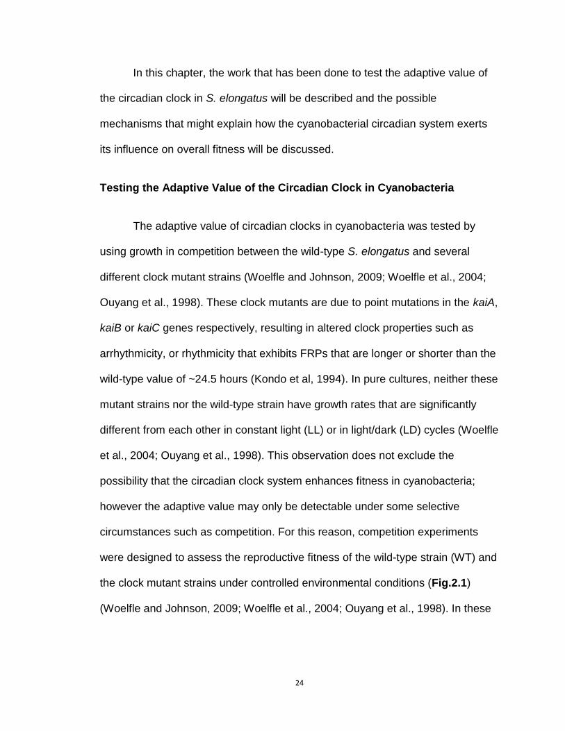
Figure 2.2 Competition of the WT strain with arhythmic strains (Woelfle et al., 2004). A, phenotypes of three strains used in the competition experiments. The WT strain (top) shows circadian rhythms with a ~25 h FRP. CLAb (middle), a clock-disrupted kaiC mutant, is arhythmic. Another kaiC mutant, CLAc (bottom), is also ultimately arhythmic but initially shows a rapidly damped oscillation. B, competitions between the WT strain and arhythmic mutants under LD 12:12 (upper) or LL conditions (lower). Data are plotted as the fraction of the mutant strain in mixed cultures (ordinate) versus the estimated number of generations (abscissa). Figure modified from Woelfle et al., 2004 and Woelfle and Johnson, 2009.

30
defeat the WT strain when these strains were co-cultured in LD 15:15 cycles (15
hours of light followed by 15 hours of darkness) (Fig. 2.3B, bottom panel).
Conversely, the WT strain was the predominant strain in mixed cultures with
short period mutants when grown in LD15:15 (Fig.2.3B, bottom panel) or when
grown in mixed cultures with long period mutants in LD11:11 cycles (Fig. 2.3B,
upper panel). Our analyses suggested that all of these mutants entrained to the
LD cycles, but they entrained with different phase relationships relative to WT
that were based on the difference between their FRP and the period of the LD
cycle (Ouyang et al., 1998). It appears to be clear from these results that the
cyanobacterial strain whose circadian clock was optimally entrained to the
environmental cycle was more fit than the strains whose clock was entrained to
the LD cycles in non-ideal phase relationships.
Furthermore, this fitness advantage is not dependent on which clock gene is
mutated, i.e., kaiA vs. kaiB vs. kaiC (Woelfle et al., 2004). This result indicates
that the difference in reproductive fitness is due to the clock phenotype itself,
rather than to a mutation in a particular clock gene. One of the most persuasive
features of these competition results is that mutants are able to out-compete WT
when the period of the LD cycle dovetailed better with the mutants’ FRP than
with WT’s FRP. When the period mutant were competed against the WT strain in
constant light, the proportions of the WT and clock mutant in the mixed culture
remained relatively constant, providing additional evidence that the adaptive
value of the circadian clock is extrinsic rather than intrinsic. In many other cases

31
Figure 2.3 Competition of the WT strain with period-altered mutants under LD 11:11 and LD 15:15 cycles (Ouyang et al., 1998). A, circadian phenotypes of the WT strain and period-altered mutants used in these competition experiments. The short period mutants (FRP ~ 22 h) include the kaiB mutant B22a and the kaiC mutant C22a, and the long period mutants (FRP ~ 30 h) include the kaiA mutant A30a and the kaiC mutant C28a. All strains have a luciferase construct that reports the clock-regulated promoter activity of the psbA1 gene by time-dependent luminescence intensity. B, competitions between the WT strain and the period-altered mutants under LD 11:11 cycles (upper) or LD 15:15 cycles (lower). Data are plotted as the fraction of the mutant strain in the mixed culture versus the estimated number of generations. Symbols for each strain are identified under the abscissa. Figure modified from Ouyang et al., 1998.

32
of tests of adaptive significance, mutant strains are uniformly out-competed by
WT strains. But these studies of the cyanobacterial clock provide an example
where mutants can out-compete the WT strain if their particular properties (e.g.,
FRP) resonant better than WT’s with an imposed environmental condition (e.g.,
the period of the environmental light/dark cycle).
Taken together, the competition experiments between cyanobacteria with
a normally functioning circadian clock and strains carrying mutations in clock
genes have demonstrated (i) that the circadian clock enhances the reproductive
fitness of cyanobacteria in cyclic environments but not in non-cyclic
environments, and (ii) that this enhancement is the greatest when the period of
the internal clock closely matches the period of the external cycle so that an ideal
phase angle is achieved under entrainment (Woelfle et al., 2004; Ouyang et al.,
1998).
Testing the adaptive value of cyanobacterial circadian clock in continuous
cultures and on solid medium
The competition experiments have clearly demonstrated the adaptive value
of circadian clocks in liquid batch cultures. Cyanobacteria, however, grow under
many other conditions in its natural environments, e.g., the solid surfaces in soil,
or in constantly mixed environments such as rivers. To test if circadian clocks
enhance fitness under these conditions, competition experiments were
performed on solid agar medium as well as in continuous cultures. As shown in

33
Figure 2.4 Competition experiments on solid agar medium and in continuous cultures. A, the competition experiment between the WT and ClAb on BG-11 agar plates. The WT cells was mixed with CLAb cells in the ratio of 1:1 and then plated on BG-11 agar plates. Cultures were incubated under LD 12:12 cycles or LL conditions. The fraction of each strain was quantified on 5th day. B, cell densities monitored in the continuous cultures. Red, 50ml cultures; Black, 100ml cultures. C, the fraction of CLAb in continuous co-cultures with the WT strain.

34
Fig.2.4A, after the WT was co-cultured with CLAb on agar medium for 5 days,
the WT dominated the cultures under LD condition but not under LL condition,
which is consistent with the results in batch cultures. Similarly, in continuous
cultures where cell densities were relatively low and stable (Fig. 2.4B), CLAb
was always out-competed by the WT under LD 12:12 cycles, regardless of the
volume of the cultures (Fig. 2.4C). Altogether, these results suggest that the
clock-mediated fitness enhancement can be extended to other growth conditions
and that it is not limited to batch cultures.
Some cell physiological properties under the competition conditions
From the competition experiments we conclude that strains with a
functioning clock out-compete strains without a functioning clock or with non-
ideally entrained clocks in cyclic environments. Nevertheless, some details are
missing. For instance, the decreasing proportion of the “loser” in mixed cultures
could be due to much slower growth rates than the “winner,” or it can be the
consequence of cell death. Understanding these details would help us uncover
the mechanism of the competition. To address the question if the “loser” is still
growing, a Yellow Fluorescence Protein (YFP) reporter (Chabot et al., 2007) was
transformed into the WT such that two strains in mixed cultures can be
distinguished by fluorescent microscopy and flow cytometry. The YFP strain
shows yellow fluorescence upon excitation, while strains without YFP display red

35
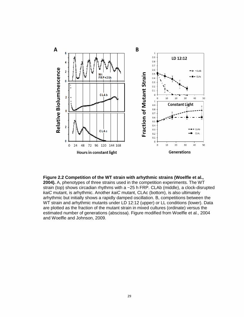
Figure 2.5 Competition experiments between the WTYFP strain and CLAb. A, flow cytometry plot used to quantify the ratio of WTYFP and CLAb in mixed cultures. The x-axis represents the fluorescence intensity around 650nm (red fluorescence), and the y-axis represents the fluorescence intensity around 540 nm (yellow fluorescence). Due to different fluorescence spectra, WTYFP and CLAb cells were located at different regions on the graph. By count the cell numbers through flow cytometer, the ratio of WTYFP and CLAb can be quantified in mixed cultures. B, image taken by a fluorescent microscope. WTFYP cells show yellow fluorescence due to the expression of yfp, and CLAb cells show red fluorescence due to chlorophyll. C, growth curves of the WT and WTYFP strains under LL conditions. D, growth curves of the WT and WTYFP strains under LD 12:12 cycles. E, fraction of the WTYFP (red) and CLAb (black) in co-cultures under LD 12:12 cycles quantified by flow cytometry.

36
fluorescence coming from the chlorophyll. These different fluorescence spectra
can be separated by flow cytometry, which is used for cell counting, as shown in
Fig. 2.5A. The cell morphology was measured and visualized by using
fluorescent microscopy, as shown in Fig. 2.5B. No growth defect was observed
in the YFP strain (Fig. 2.5C&D), and the WTYFP out-competed CLAb under LD
12:12 cycles (Fig. 2.5E), suggesting that the YFP reporter has no effect on the
fitness of the WT. The WTYFP was then co-cultured with CLAb under LD 12:12
condition, and the growth rates of each strain in the mixed cultures were
measured. As shown in Fig. 2.6A, CLAb kept growing in the first 5 days, but it
grew significantly slower than the WT. The overall growth of the mixed cultures
showed no significant difference from the pure cultures (Fig. 2.6B), suggesting
that only CLAb was impaired by the competition.
Following the growth experiments, cell lengths of each strain and cell division
rates were examined in pure and mixed cultures (Fig. 2.6C&D). Interestingly, in
mixed cultures, the lengths of CLAb cells decreased during the process of
culturing, and they are significantly shorter than cells in pure cultures after the 5th
day. Consistently, cell division rates of CLAb were also reduced in mixed
cultures, whereas in pure cultures both WTYFP and CLAb showed increasing
division rates on the 3rd day (the exponential phase). Overall, the cell length and
division rates of WTYFP were not influenced by co-culturing under LD 12:12
condition, whereas CLAb was weakened by co-culturing. Taken together, these
results indicate that some of the physiological properties of CLAb
37
Figure 2.6 Dynamics of the competition experiments between WTYFP and CLAb. A, growth curves of WTYFP (red) and CLAb (black) in co-cultures under LD 12:12 cycles. B, growth curves of pure cultures and mixed-cultures under LD 12:12 cycles. Red, pure cultures of WTYFP; black, pure cultures of CLAb; green, co-cultures of WTYFP and CLAb. C, cell length of each strain in pure cultures or mixed-cultures. D, cell division events of each strain in pure cultures or mixed-cultures.
38
were adversely affected by co-culturing with the WT. However, no clear evidence
showed that CLAb cells were undergoing cell death. Although with a significantly
slower growth rate, these “losers” could still grow, indicating that the competition
may be mediated by some growth inhibitors or limiting nutrients.
Another example: cyanobacterial circadian clock enhances fitness by non-
optimal codon usage
The competition experiments have clearly demonstrated that the circadian
clock enhances fitness of cyanobacteria in cyclic environments. Recently,
another study confirmed the adaptive value of cyanobacterial circadian clock
from a different perspective by studying the non-optimal codon usage of kaiBC
genes (Xu et al., 2013). The circadian system regulates nearly all expression of
the cyanobacterial genome (Liu et al, 1995; Ito et al., 2009), indicating the
importance of kai genes in these organisms. A general observation from many
organisms is that genes with high expression levels and functional importance
are usually encoded by optimized codons (Ikemura, 1981) that have higher
usage frequencies than other synonymous codons. Thus we might predict that
kai genes should be encoded by optimized codons. An examination of the codon
usage of kaiBC genes, however, revealed that the codon usage of kaiBC genes
is not as translationally efficient as genes with high functional importance, e.g.,
ribosomal genes (Xu et al., 2013). To understand why kaiBC is not encoded by
optimized codons, cyanobacterial strains expressing optimized codon kaiBC
(OptkaiBC) were generated by Dr. Yao Xu in the Johnson lab. As predicted, the

39
expression levels of kaiB and kaiC genes are increased in OptkaiBC due to the
optimization of codon usage. Interestingly, OptkaiBC showed no difference from
the WT in its circadian rhythms at warm temperatures, while it displayed robust
circadian rhythms at low temperatures where the wild-type strain tends to be
arrhythmic or highly damped (Xu et al., 2013).
This result is quite surprising since we expected that better rhythms
provide better fitness under rhythmic conditions, based on the conclusion of the
previous competition experiments. From this line of reasoning, natural selection
should prefer the OptkaiBC strain rather than the non-optimized codon strain. To
address this question, I measured the growth rates of the WT, OptkaiBC and two
arrhythmic strains (CLAb and CLAc) at different constant temperatures within the
physiological range of temperatures (18 °C to 37°C) for this cyanobacterial
species under LD 12:12 cycles. As shown in Fig.2.7 and Table2. 1, at warm
temperatures where both the WT and OptkaiBC show robust circadian rhythms,
no significant difference was observed among their growth rates, including the
two arrhythmic strains. However, as the temperature is reduced to 20 °C and
18 °C, OptkaiBC grew significantly slower than the other strains. Moreover, the
two arrhythmic strains grew even slightly better than the WT (Fig.2.7 and Table
2.1). From Fig.2.8 we can see that the warm temperatures around 30 °C allow
our cyanobacterial strains, both the WT and OptkaiBC, to grow at the fastest
rates, while at cooler temperatures the growth rates were significantly reduced
(Fig. 2.8C). Therefore we consider ~ 30 °C to be the optimal growth

40
Figure 2.7 Growth curves of cyanobacterial strains at different temperatures. WT, optKaiBC, CLAb (arrhythmic) and CLAc (damped oscillation) strains were grown in LD 12:12 cycles at 37°C, 30°C, 25°C, 20°C, or 18°C with constant air bubbling and shaking. Cell densities were monitored by measuring OD750 every two days. Data are averages ± SEM from 2 to 6 independent experiments for each strain and condition. For a better comparison at 18°C, 20°C, and 25°C, the insets are a magnified portion for the specified times.

41
Table 2.1 Doubling time of WT, optkaiBC, CLAb and CLAc strains at different temperatures under LD 12:12 cycles.

42
Figure 2.8 Growth of the WT and OptkaiBC strains at different temperatures under LD12:12 cycles. A, growth curves of WT at 37°C, 30°C, 25°C, 20°C, or 18°C . B, growth curves of OptkaiBC at 37°C, 30°C, 25°C, 20°C, or 18°C . C, growth rates of the WT (black) and OptkaiBC (red) strains was plotted as the function of temperatures.

43
Figure 2.9 Competition experiments between the WT and OptkaiBC strains at 30 oC or 20 oC under LD 12:12 cycles. The WT strains either with Cb resistance (left) or Kn resistance (right) was competed against the OptkaiBC strains with the opposite resistance at 30 oC (upper panels) or 20 oC (lower panels).

44
Figure 2.10 Same strains with different antibiotic resistances do not compete with each other.

45
temperatures for this particular species. These results indicate that a robust
circadian rhythm at cool temperatures actually impacts the fitness, and that
circadian rhythms provide an advantage only within a range of growth
temperatures.
Although the OptkaiBC strain showed reduced growth rates in pure
cultures at cool temperatures, perhaps the robust rhythms that it confers might
be an advantage under competition conditions. To test this hypothesis,
competition experiments between the WT and OptkaiBC strains were conducted
at 30 oC and 20 oC under LD 12:12 cycles. As shown in Fig.2.9, the results were
consistent with the observations of pure cultures (Fig.2.7): the OptkaiBC
displayed a slight advantage over the WT at 30 oC (Fig. 2.9 A&B), while it was
out-competed by the WT at 20 oC (Fig. 2.9C&D). To exclude the possibility that
the competition results were compromised by the different antibiotic resistance
genes incorporated in the genome of each strain, two sets of competition
experiments, WT(Cb) vs. OptkaiBC(Kn) and WT(Kn) vs. OptkaiBC(Cb), were
conducted, as shown in Fig. 2.9. No significant difference was observed between
these two experiments. Furthermore, when strains with the same genotype but
different antibiotic resistances {i.e., WT(Cb) vs. WT(Kn) and OptkaiBC (Kn) vs.
OptkaiBC (Cb)} were competed against each other (Fig. 2.10), no competition
was detected, thus indicating the competition resulted from the kai genotypes
and not the antibiotic markers.

46
Results from this study suggest that having a robust circadian rhythm is
not always an advantage. Under cold temperatures, cyanobacteria may face
more challenges from the environment and simply surviving these environmental
stresses might be a priority. Therefore, it is possible that running a robust timing
mechanism is burdensome. By adapting the non-optimal codons to kaiBC genes,
the circadian clock obtained the flexibility to guarantee the best fitness at different
temperatures, suggesting another way whereby circadian clock enhances fitness,
namely “conditionality” (Njus et al., 1977).
Potential Mechanisms of Clock-mediated Fitness Enhancement
While competition experiments have clearly demonstrated a clock-
mediated fitness enhancement in cyanobacteria, the cellular mechanism remains
unknown. Cyanobacterial strains with different clock properties all showed a
similar growth rate when cultured alone; however, the reproductive fitness was
negatively affected in mixed cultures in a way that is dependent on the light/dark
cycles (Woelfle et al., 2004; Ouyang et al., 1998). To explain these observations,
three models have been proposed: the “limiting resource model,” the “diffusible
inhibitor model” and the “cell-to-cell communication model” (Woelfle and
Johnson, 2009).
The Limiting Resource Model
The limiting resource model proposes that the circadian system enables
individual cyanobacterial cells to maximally utilize some limiting environmental

47
resource by phasing their metabolism to the environmental cycle (Woelfle and
Johnson, 2009). For instance, transcription of genes that encode components of
the photosynthetic machinery in cyanobacteria is up-regulated during the day
and down-regulated at night (Tomita et al., 2005), and this rhythmic gene
expression may facilitate cyanobacterial cells to perform photosynthesis more
efficiently and to consume less energy at night by limiting unnecessary
transcription and translation. In contrast, cyanobacterial cells without a
functioning circadian clock or with a non-optimally entrained clock may be less
efficient metabolically, thereby having an inherent disadvantage when competing
for a limited resource with cells that have a clock that is favorably entrained to the
environmental cycle.
Our published results that the growth rates of the various pure cultures
(WT and mutants) were experimentally indistinguishable led us to believe that the
Limiting Resource Model was incorrect (Woelfle et al., 2004; Ouyang et al.,
1998), but a more recent modeling paper from Hellweger has forced us to re-
evaluate the experimental evidence for and against this model (Hellweger, 2010)
(see below).
To show that the limiting resource model could be hypothetically true,
Hellweger’s mathematical modeling of the competition experiments attributed
small differences in the growth rates between the cyanobacterial strains as a key
determining factor in predicting the outcome of the competition (Hellweger,
2010). This model simulated the growth of the WT strain and period-altered

48
mutants in both pure and mixed cultures and was able to successfully reproduce
the experimental outcomes (Hellweger, 2010). After examining parameters in the
simulation, it was found that a circadian clock will have a higher amplitude when
its FRP matches the period of the LD cycle, and this higher amplitude leads to
greater expression of photosynthesis genes and ultimately to higher growth
rates. The difference in predicted gene expression and growth rates was
therefore suggested by this model to be the mechanism of the clock-mediated
fitness enhancement. Although our experimental measurements of overall growth
rates had detected no differences in pure cultures of the strains used in
competition (Woelfle et al., 2004; Ouyang et al., 1998), Hellweger’s model
suggested that small differences may exist between the strains which are difficult
to detect in the batch cultures used for the competition experiments; however,
these hypothetically small differences in growth rates between the strains might
be detectable when cyanobacteria are cultured using chemostats (Hellweger,
2010). Using a chemostat culturing method, cells can be grown in a physiological
steady state where they grow at a constant rate, and all culture parameters
remain constant (Harder and Kuenen, 1977). Previously, competition
experiments between the WT strain and the long period mutant, C28a,
conducted in chemostats produced results that are quite similar to the
competition results using the batch culture method (Ouyang et al., 1998), but a
re-evaluation of this approach was warranted based upon the modeling study of
Hellweger (Hellweger, 2010).

49
Another way to reconcile the Limiting Resource Model with our
experimental data is to consider the impact of transfers/dilutions in the
competition assay. In our previous studies, we did not detect any significant
differences in the growth rates between strains with different circadian
phenotypes during exponential growth (Woelfle et al., 2004; Ouyang et al., 1998).
We therefore considered the possibility that there could be small differences in
the latency of these strains to start growing at the time of each transfer to new
culture medium. If so, the initial growth rates might be different among the strains
and these differences might accumulate over successive transfers. Therefore, to
test the Limiting Resource Model experimentally, we focused on testing a
hypothesis that small differences in initial growth rates may be cumulative over
repeated dilutions of cell cultures and therefore are responsible for the result of
the competition studies (Dr. Tetsuya Mori, personal communication). By analogy
to a race, it is as if one runner gets started “off the blocks” from the starting line a
little earlier than the other runners. If one strain has the innate ability to adapt to
the new medium more quickly than the other such that it enters the exponential
phase of growth in a shorter period of time after each transfer to new medium,
small differences in this period of adaptation between strains with different
circadian phenotypes could give rise to large differences in the composition of
the mixed cultures after several generations (Woelfle and Johnson, 2009).
To test this hypothesis, we examined whether there were small
differences in initial growth rates over several serial dilutions/transfers of pure
cultures of the WT strain and of the arrhythmic mutant, CLAb. In previous

50
studies, the WT strain and clock mutants did not show any significant difference
in their overall growth rates in pure cultures (Woelfle et al., 2004; Ouyang et al.,
1998), however, the difference might have been too subtle to be observed with
the sampling techniques that were used. If there were small differences in the
ability of cells to adapt to new medium, we would expect that this difference could
be enhanced by a series of dilutions to new medium over many generations. To
experimentally test this version of the Limiting Resource Model, stationary phase
cultures of the WT strain and CLAb cells were diluted one thousand fold every 8
days, and the growth in constant light or in a light/dark cycle of each of the two
strains was monitored by measuring the optical density. As shown in Fig.2.11,
during a series of four dilutions, no obvious differences were observed by eye in
the growth curves of the WT and arrhythmic strains in pure cultures in either
constant light or in LD 12:12 cycles. However, some small differences were
detected when the initial growth rates were calculated (Table 2.2; the initial
growth rates were calculated as the doubling time in the first 24 hours after
dilution). In constant light, even though the initial doubling time of CLAb was
significantly greater (i.e., slower) than that of the WT strain after the second
dilution, no significant difference was observed after the third dilution, and after
the fourth dilution, the direction of the difference was the opposite to that in the

51
Figure 2.11 Growth curves of the WT strain and the clock mutant (CLAb) in pure cultures that were serially diluted four times. A, pure cultures of the WT strain (blue diamond) and CLAb (red squares) were set up under LL conditions. B, pure cultures of the WT strain and CLAb were set up under LD conditions. After the cells reached the stationary phase, they were diluted 1:1000 into fresh BG-11 medium. When the diluted cultures reached the stationary phase, they were diluted again. The cultures were serially diluted four times. Cell density was measured as OD750 value.

52
Table 2.2 Initial doubling time of the WT and CLAb under LL conditions or LD 12:12 cycles.

53
second dilution. Therefore, the end result was that there is no difference in the
initial growth rates between the WT and CLAb strains. In LD 12:12 cycles, the
CLAb strain grew significantly faster than the WT strain in the lag phase after the
first transfer to fresh medium, whereas in the subsequent dilutions, CLAb showed
slower initial growth rates than the WT strain (and the difference was not
significant after the fourth dilution).
Although these results indicate that the WT strain and the CLAb strain
may have different abilities to adapt to new medium, the different initial growth
rates in pure cultures were not amplified in a straightforward way by subsequent
dilutions (Fig.2.11). Therefore, whether the outcome of these competition
experiments is caused by small differences in the initial growth rates between
strains remains unclear, and indicates that a better experimental design is
needed to more rigorously test this model. Previous work utilizing chemostats to
culture cyanbacterial cells in competition yielded results that were substantially
the same as those obtained when competition is conducted in batch cultures
(Ouyang et al., 1998). This culture method may allow us to more accurately
determine whether there are differences between cyanobacterial strains in their
ability to adapt to the introduction of new medium.
The Limiting Resource Model is supported by mathematical modeling that
predicts there are differences in the physiological states between WT and clock
mutants when grown in pure cultures versus mixed cultures. However, the clock
mutants used in the competition experiments have not yet been found to differ

54
significantly from the WT strain in physiological properties that have been
measured experimentally (Woelfle and Johnson, 2009). Further research is
needed to address these questions.
The Diffusible Inhibitor Model
The Diffusible Inhibitor Model proposes that cyanobacterial cells
rhythmically secrete a diffusible molecule that acts to inhibit the growth of other
cells (i.e., cells of the same strain/species or cells of another strain/species) in
the same environment (Woelfle and Johnson, 2009). If cyanobacteria are
sensitive to their own inhibitory molecule, it would be advantageous for them to
possess some mechanism to be insensitive to or to inactivate this inhibitor. For
example, cyanobacterial cells could avoid inhibition by regulating both the timing
of secretion of the inhibitor and their own sensitivity to the inhibitor. This model
has two underlying assumptions: 1) the secretion of the inhibitor is light-
dependent and is therefore limited to the light phase and the subjective day in
constant conditions (clock and light dependent); 2) cyanobacterial cells are only
sensitive to the inhibitor during the dark phase and in the subjective night (clock
dependent) as depicted in Fig. 2.12A (Woelfle and Johnson, 2009). This
hypothetical phenomenon could allow cyanobacterial cells that are entrained with
the best phase angle to produce an inhibitor that retards the growth of
competitors (inter- or intra-species competitors) without inhibiting themselves.
The subjective day of the cyanobacterial strain whose clock is entrained to a
cycling environment in the most appropriate phase angle will coincide with the

55
Figure 2.12 Test of the Diffusible Inhibitor Model. A, Depiction of the Diffusible Inhibitor Model. Entrained phases of two strains are modeled in terms of their entrained phase relationship to an LD 12:12 cycle. For the optimally-phased cells, the subjective day overlaps with the daytime (white box), and the subjective night phase overlaps with the nighttime (black box). Therefore, their secretion phase (yellow box) coincides with the daytime, and the sensitive phase (red box) coincides with the nighttime. For non-optimally-phased cells, their subjective day starts from middle of the daytime, and their subjective night starts from the middle of the night and ends in the middle of the daytime. Therefore, the secretion phase (yellow box) of the non-optimal-phased cells is only from the middle of the day to the end of the day, while the secretion-competent phase that overlaps with the dark phase (yellow-black box) does not result in secretion because the secretion is postulated to be light-dependent. The sensitive phase (red box) of the non-optimally-phased cells starts in the middle of the night and ends in the middle of the day. B, the semi-co-culture apparatus used to test the existence of a diffusible inhibitor. Two
chambers (left and right) were separated by a membrane with 0.2 m pores. Cells of different strains could be cultured separately in these two chambers, but their media passes freely through this membrane such that the putative inhibitor could diffuse to the other side. The cultures were illuminated by white fluorescent light from the top, and the light intensity was 50 uE*m-2*s-1. C, the WT strain and CLAb were semi-co-cultured in this apparatus under LD 12:12 cycles. Cell densities (OD750) were measured on the fifth day. Bars which share the same color represent cultures in the same apparatus. Panel A modified from Woelfle and Johnson, 2009.

56
light phase,but strains that are entrained to a non-optimal phase relationship
would have their subjective day in a different temporal regime (e.g., one
hypothetical non-optimal phase relationship could be the subjective day starting
in the middle of the light phase and ending in the middle of dark phase). In this
scenario, the secretion of the inhibitor from these poorly entrained cells occurs
during only part of the light phase and the secretion phase that overlaps with the
dark phase does not result in active secretion (because the secretion is light-
dependent, see underlying assumption # 1 above). However, the sensitive phase
spans part of the subjective night starting in the middle of the night and ending
during the middle of the day (Fig.2.12A) (Woelfle and Johnson, 2009).
Using this line of reasoning, in a mixed culture of an optimally entrained
strain with a non-optimally entrained strain, one would expect the growth of the
poorly entrained cells to be inhibited because their sensitive phase overlaps with
the secretion phase of the optimally entrained strain. Therefore, the growth of the
poorly entrained cells would be inhibited by the secretions from the optimally
entrained cells (but not vice versa). Similarly, if an arrhythmic strain is grown in
mixed culture with the WT cells in a LD 12:12 cycle, arrhythmic cells might be
sensitive to the inhibitor all the time due to the lack of a functioning clock. There
have been reports suggesting that cyanobacteria secrete secondary metabolites
that are toxic to other species (Gleason and Paulson, 1984; Zaccaro et al., 2006)
(and even to themselves). For example, a secreted secondary metabolite of the
cyanobacterium Scytonema hofmanni, cyanobacterin, inhibits the growth of
several other cyanobacterial species (Gleason and Paulson, 1984). A proteomic

57
study demonstrates that some proteins in S. elongatus are secreted into medium,
but the function of these secreted proteins remains unknown (Koksharova et al.,
2005). Therefore, it is possible that the competition between the WT and clock
mutants could be mediated by secreted metabolites or proteins.
In contrast to Hellweger’s mathematical modeling that best supports the
Limiting Resource Model, an earlier modeling of the competition experiments
provided support for the Diffusible Inhibitor Model (Roussel et al., 2000; Gonze et
al., 2002). By postulating the existence of a diffusible inhibitor, Roussel et al.
successfully reproduced all the experimental observations, whereas the
theoretical model based on differences in resource exploitation did not lead to a
satisfactory result (Roussel et al., 2000). Gonze et al. proposed and tested a
more sophisticated model that supported the Diffusible Inhibitor Model and
predicted that the outcome of the competition depends on the initial proportions
of cells and on the FRPs of the different cyanobacterial strains (Gonze et al.,
2002). However, this prediction was not supported by experimental testing–in
contrast to the model predictions, competition experiments between the WT and
CLAb strains found that CLAb was defeated by the WT strain even though the
starting proportion of CLAb was as high as 90% of the whole culture (Woelfle et
al., 2004).
As a different strategy to test the Diffusible Inhibitor Model experimentally,
we designed a growth chamber in which two cyanobacterial strains are
separated by a permeable membrane that allows the exchange of medium and
58
small molecules, but prevents the two strains from directly contacting each other
(Fig.2.12B). Thus, if a diffusible inhibitor is secreted by one strain, it could pass
through the membrane and affect the growth of the strain on the other side of the
membrane. As discussed earlier, the WT strain rapidly becomes the predominant
strain in mixed cultures with the arhythmic strain CLAb in LD 12:12 cycles
(Woelfle et al., 2004); therefore, we conducted a series of experiments by using
these two strains separated by a membrane. The pore size of the membrane was
0.2 m that allows most molecules and small proteins to pass through but not the
cyanobacterial cells themselves. If the Diffusible Inhibitor Model is correct, we
would expect that the arhythmic strain would show a significantly slower growth
rate; however, CLAb displayed the same growth rate as WT in LD 12:12 cycles
(Fig.2.12C). One possible explanation for this result is that the putative diffusing
molecule is too large to pass through the pores of the membrane; therefore,
membranes with varying pore sizes could be tested (a pore size of 0.2 m was
used in the experiments whose results are depicted in Fig. 2.12C). Alternatively,
the inhibitor could be a cell-surface molecule rather than a diffusible factor.
Contact-dependent inhibition (CDI) has been reported in E.coli (Aoki et al., 2008;
Aoki et al., 2005) and interestingly, a few potential homologs of genes involved in
CDI in E.coli have been identified in S. elongatus. Knock-out mutants of these
potential homologs could be constructed in order to address whether a contact-
dependent inhibition mechanism is involved in the competitions between
cyanobacterial strains.
59
The Cell-to-Cell Communication Model
The Cell-to-Cell Communication Model, which is a combination of the two
hypotheses discussed above, postulates that circadian clocks regulate some
pathways involved in cell to cell communication in cyanobacteria such that
individual cells can cooperate as a group in order to adapt to the environment
and/or best utilize a limiting resource (Woelfle and Johnson, 2009). This
hypothesis postulates that when the circadian clock is disrupted or is not ideally
entrained to the environment, cells do not effectively communicate due to the
absence of proper circadian regulation. Under these conditions, cells with
mutations in the circadian clock compete as individuals with WT cells that can act
as a group, and thus are at a competitive disadvantage in utilizing a limited
resource.
Quorum sensing is recognized as a mechanism by which many bacterial
species communicate and cooperate (Ng and Bassler, 2009). Bacterial species
that engage in quorum sensing secrete signaling molecules called autoinducers
(AI), and the detection of the AI allows cells to switch between two distinct
patterns of gene expression, depending upon cell density. When the cell density
is high and AI reaches a threshold concentration, individual cells cooperate with
others such that the entire population turns on a gene expression mode that
triggers biological activity, for example the formation of a biofilm or virulence
factor production (Ng and Bassler, 2009).

60
To date, there are only a few cyanobacterial species in which quorum-
sensing has been reported (Sharif et al., 2008; Mooy et al., 2012). One of the
AIs, N-octanoyl homoserine lactone (C8-AHL), was found in Gloeothece sp.
PCC6909 cultures, and 43 genes were expressed differently in response to C8-
AHL treatment. It was suggested that this quorum sensing may mediate the
formation of a biofilm (Mooy et al., 2012). Another cyanobacterium,
Trichodesmium consortia, was reported to respond to quorum-sensing molecules
called acylated homoserine lactones (AHLs) that are produced by epibionts
attached to its surface, and colonies of Trichodesmium that were treated by
AHLs doubled their activity of alkaline phosphatases, which are enzymes used
by epibionts in the acquisition of phosphate from dissolved-organic phosphorus
molecules (Mooy et al., 2012). Homologs of luxO and luxU, genes that encode
components of the quorum sensing pathway, are present in the cyanobacterial
species Synechocystis sp. PCC 6803 (Sun et al., 2004). When quorum sensing
genes from Vibrio harveyi (Ng and Bassler, 2009) were used to search the
genome sequence of S. elongatus, potential homologs were identified; the
homologs identified included the AI receptor cqsS (Ng et al., 2011), and cqsA (an
enzyme involved in AI synthesis), as well as the signal transduction pathway
component, luxO (Ng and Bassler, 2009) (Table 2.3). In addition, a homolog of
aphA, the gene that encodes the transcription factor that is a master regulator of
the quorum sensing pathway and is active at low cell density (LCD) in V. harveyi
(Rutherford et al., 2011), is also present in the S. elongatus genome. Taken

61
Table 2.3 Homologs of quorum sensing genes of V. harveyi in S. elongatus.

62
together, the data from this bioinformatics approach suggest that S. elongatus
may be capable of quorum sensing with clock-enhanced reproductive fitness
mediated by this form of cell-to-cell communication. This possibility could be
tested by knocking out these potential homologs.
Future Directions
Competition experiments have demonstrated the adaptive value of the
circadian clock in cyanobacteria. It is clear that the circadian clock enhances the
fitness of cyanobacterial cells in light/dark cycles, and that this enhancement in
reproductive fitness is greatest when the circadian clock resonates with the
environmental cycle. In contrast, the circadian clock in cyanobacteria provides
little or no reproductive advantage under constant conditions, indicating that the
adaptive value conferred by circadian clock is an extrinsic rather than intrinsic
property (Woelfle et al., 2004; Ouyang et al., 1998).
To date, the underlying mechanism by which the circadian clock enhances
reproductive fitness remains elusive. Although several models have been
proposed and tested, each has some evidence that supports it and none can be
excluded at this time. Each of the models discussed here will be examined
further using a complementary approach. These include alternative culture
methods aimed at accurately detecting small (but significant) differences in
growth rates between wild-type and clock mutant strains. These small differences
in growth rate could explain the competition outcomes. In addition, a genetic
approach guided by bioinformatics and mathematical modeling could also be

63
used to identify genes involved in the pathway and define the mechanism of
fitness enhancement.
Experimental Procedures
Bacterial strains and culture conditions
All cyanobacterial strains used in this study is listed in Table 2.4 The
cyanobacterial strains were grown in BG-11 medium (Bustos and Golden, 1991)
at 30 °C, and illuminated by cool-white fluorescence bulbs (40-50 µE m−2 s−1)
with air bubbling. Spectinomycin (25µg/ml) and/or kanamycin (25µl/ml) were
supplemented to the medium when necessary. For growth on solidified agar
plates, BG-11 medium was supplemented with 1.5% agar and appropriate
antibiotics.
Competition experiment
The clock phenotypes of cyanobacterial strains were determined by their
luminescence rhythms that report the promoter activity of psbAI. Strains with
different clock phenotypes and different antibiotic resistance, e.g., wild type vs.
arhythmic, were selected for the competition experiments. Different antibiotics
enable us to track their fractions in mixed cultures by plating on selective media.

64
Table 2.4 Cyanobacterial strains used in this chapter.

65
Pure cultures of two strains were set up under LL condition, and when they
reached log phase (OD750 ~ 0.6), equal numbers of cells were mixed and
cultured under different LD cycles or LL condition. Aliquots were taken from the
mixed cultures every ~8 generations in LD and every ~16 generations in LL, and
they were plated on selective media to count the number of colony-forming units
(CFU) of each strain. Meanwhile, the mixed culture was diluted into fresh
medium and grown for another ~8 generations (LD) and ~16 generations (LL).
This process was repeated for 4 cycles to allow cells to grow for 40-50
generations. The fraction of each strain in the mixed culture was calculated by
the number of colonies of each strain growing on selective media. Circadian
phenotypes were confirmed by monitoring the luminescence rhythms of colonies
of each strain at different sampling times. Similar procedure was applied in
competition experiments on solid agar medium and in continuous cultures, and
the continuous cultures were conducted in home-made apparatus with lamination
and air bubbling.
Growth curves and calculation of doubling time
To generate growth curves, seed cultures of cyanobacterial strains were
grown under LL conditions at 30°C until exponential phase before inoculation. To
measure the initial growth rates of WT and CLAb, pure cultures of WT and CLAb
strains were grown under either LD 12:12 cycles or LL conditions at 30°C with air
bubbling. When cells reached stationary phase, cultures were diluted at the ratio
of 1:1000 to fresh BG-11 medium, and this process was repeated for 4 times. To

66
compare the growth rates of WT, OptkaiBC, CLAb and CLAc strains at different
temperatures, pure cultures of these strains were grown under LD 12:12 cycles
at 37°C, 30°C, 25°C, 20°C and 18°C with air bubbling and shaking. Cell densities
were monitored by measuring the optical density at 750nm (OD750). OD750 was
then plotted against time for a comparison of growth among strains. The growth
constant k was generated by fitting exponential curves to the growth curves at
log scale, and doubling time was calculated using this equation: doubling time (h)
= (ln(2)/k) × 24. For initial growth rates calculation, the same equation was
applied to the first 2-3 days of the data.
Membrane experiment
A semi-co-culture apparatus was used to test the existence of a diffusible
inhibitor. Two chambers (left and right) were separated by a polyethersulfone
membrane (Sterlitech, WA; Cat. # PES022005) with 0.2 m pores. Cells of
different strains can be cultured separately in these two chambers, but the
medium passes freely through this membrane such that the putative inhibitor
could diffuse to the other side. The cultures were illuminated by white fluorescent
light from the top, and the light intensity was 50 uE*m-2*s-1. The WT strain and
CLAb were semi-co-cultured in this apparatus under LD 12:12 cycles. Cell
densities (OD750) were measured on the fifth day.

67
Dynamics of the competition
A copy of the yfp gene (Chabot et al., 2007) driven by the ptrc promoter
was incorporated into the Neutral Site II of the WT strain, generating an YFP
reporter strain WTYFP. No effect was observed in the growth rates and FRP of
the WTYFP, comparing to the wild-type strain, thus suggesting that WTYFP can
be used to monitor the dynamic of the competition experiments. WTYFP was co-
cultured with CLAb under LD12:12 cycles, and aliquots of cells was taken every
two days. The growth of pure and mixed cultures was recorded by monitoring
OD750. Cell lengths and cell division rates were observed and quantified by using
fluorescence microscopy. Fractions of each strain in mixed cultures were
quantified by flow cytometry (Fig. 2.5A), and the OD750 value of the mixed
cultures was converted to cell numbers by fitting to a standard curve. Cell
numbers of each strain in mixed cultures were calculated by multiplying the
fraction by the total cell numbers in the mixed cultures.
Bioinformatics analysis
Amino acid sequences of quorum sensing genes in V. harveyi were
retrieved from the NCBI database
(http://www.ncbi.nlm.nih.gov/bioproject/?term=19857). Homology search of these
protein sequences was performed against the S. elongatus genome by using the
BLASTP program (Altschul et al., 1997). The threshold of expected value is set
up at 1e-10, and BLOSUM80 matrix and Low Complexity Filter were applied.

68
Chapter III
The global metabolic profiles of the wild-type cyanobacterium Synechococcus elongatus PCC 7942 and adaptive-fitness mutants
Abstract
Although numerous experiments have been done to test the potential
mechanisms of the clock-mediated fitness enhancement, it is difficult to draw
conclusions from these results. Recently, several studies have reported
connections between metabolism and the circadian clock in Synechococcus
elongatus PCC 7942. In addition, some of my preliminary results also revealed
that the steady-state levels of metabolites in the clock mutant are different from
the metabolite levels in WT. Therefore, in this study, I hypothesize that the clock-
mediated fitness enhancement results from altered metabolism in the mutant
strains. To test this hypothesis, I took advantage of the rapid development of
metabolomics techniques and established the metabolic profiles for the WT strain
and two clock mutant strains under light-dark cycles and in constant light. My
results demonstrated that the global metabolic profiles were altered in the clock
mutants, and that the metabolism of WT is dramatically different in light-dark
cycles vs. constant light condition.
Introduction
Circadian clocks are endogenous timing mechanisms that regulate many
physiological activities of organisms. The regulation of metabolic processes by

69
circadian clocks has been reported in a broad range of organisms ranging from
bacteria to mammals (Bailey et al., 2014). In mice and humans, the blood
pressure, body temperatures and many other metabolic events are regulated by
circadian clocks. In plants, circadian clocks control the secretion of selected
hormones involved in the defense against herbivores (Goodspeed et al., 2012).
In the prokaryotic cyanobacteria, nitrogen fixation activities are separated from
photosynthesis by circadian regulation to avoid the toxicity of oxygen to
nitrogenase (Grobbelaar et al., 1986; Mitsui et al., 1986). On one hand, circadian
clocks regulate metabolism; on the other hand, metabolism can also affect
circadian clocks. This relationship implies that clocks and metabolism could form
an autoregulatory feedback network. Recently, researchers proposed that the
coupling of cyanobacterial circadian clocks and metabolism might be the
mechanism through which circadian clocks receive entraining information from
the environment (Rust et al., 2011; Kim et al., 2012). Kim et al. (2012) proposed
that the redox state of quinone and the ATP/ADP ratio in the cell could work
together to reset the phase of the clock, suggesting the importance of the
coupling of circadian clocks and metabolism.
As one of the simplest circadian machineries, the cyanobacteria circadian
clock confers adaptive significance in rhythmic environments, as indicated by a
series of competition experiments in the cyanobacterium Synechococcus
elongatus PCC 7942 (S. elongatus) (Ouyang, et al., 1998; Woelfle et al., 2004).
We have been trying to understand for long time the mechanism by which the
circadian clock enhances fitness under the competition conditions. Although

70
several models have been proposed and numerous experiments have been done
to test these models, no definite answer is known at this time (Ma et al., 2013).
However, because there is increasing evidence showing that the connection
between circadian clocks and metabolism is important, I wish to test if
metabolism can be altered by the disruption of the circadian clock, and whether
the metabolic status under the competition conditions can help us to understand
clock-conferred fitness. Answering these questions will not only help us to further
understand the coupling of metabolism and circadian clocks, but may also
provide clues to the underlying mechanism of the competition results. In
particular, an experiment to study the metabolites in the cyanobacterial culture
medium revealed differences between the wild type (WT) and the clock mutants,
leading us to hypothesize that the metabolic profiles of clock mutants are altered,
which could be the potential mechanism of the clock-mediated fitness
enhancement. In this chapter, global metabolic profiles of the WT and two clock
mutants, CLAb and CLAc, were examined by metabolomics techniques. From
this study I demonstrated that metabolism varies in these three strains under
light-dark (LD) conditions, while the regulation of metabolism by circadian clock is
dominated by light conditions {LD vs. constant light (LL)}.
Results
The metabolic profiling is influenced by both clock phenotypes and light
conditions
The clock phenotypes of three strains used in this study, WT, CLAb and

71
CLAc, are shown in Fig. 3.1. To establish the global metabolic profiles, cells of
these three strains were collected every 6 hours under LD 12:12 cycles from
Zeitgeber Time (ZT) 0 to ZT24. For the WT, cells under LL condition were also
collected every 6 hours from Circadian Time (CT) 12 to CT36. After sample
collection, cells were immediately frozen by liquid nitrogen and sent to
Metabolon, Inc., which is a biotech company specialized in metabolomics, for
metabolomics analysis. At Metabolon, samples were prepared by using a
proprietary series of organic and aqueous extractions to remove the protein
fraction while allowing maximum recovery of small molecules. Then intracellular
metabolites were identified and quantified by liquid chromatography–mass
spectrometry (LC-MS) and gas chromatography–mass spectrometry (GC-MS).
Raw and normalized data was obtained from Metabolon after sample processing.
I then analyzed these data by using R programming language (http://www.R-
project.org). After removing metabolites with missing values during the 24-hour
time course, 173 metabolites were included to build the metabolic profiles. These
173 metabolites are categorized into 6 super pathways and 37 sub pathways
(Fig. 3.2).
To test if the metabolic profiles from the 4 groups (WTLD, CLAbLD,
CLAcLD and WTLL) are statistically different from each other, the whole dataset
was transformedg by natural logarithm and principal component analysis (PCA)
was applied. PCA is a statistical procedure that reduces the dimensionality of the

72
Figure 3.1 Clock phenotypes of three strains used in this study. Upper panel, WT whose FRP is about 25 hours; middle panel, the arrhythmic strain CLAb; bottom panel, CLAc with a damped rhythm.

73
Figure 3.2 173 metabolites were included to establish the metabolic profiles. And they are categorized to 6 super pathways (A), and 37 sub pathways (B).

74

75
Figure 3.3 Principal component analysis (PCA) indicates that the metabolic profiling is associated with genotypes and light conditions. Green circle, WT samples under LL condition; orange circle, WT samples under LD condition; blue circle, CLAb samples under LD condition; purple circle, CLAc samples under LD condition.

76
data but the variations among samples can be preserved (Ringner, 2008). The
first principal component (PC1) represents the direction that samples shows the
largest variation of those metabolites levels. The second component (PC2) is
uncorrelated with PC1, and it is the direction that samples show the second
largest variation (Ringner, 2008). PCA enables the visualization of multi-
dimensional data.
Our whole data set contains 40 samples, and each of these samples has
173 variables (173 metabolites) that define the property of this particular sample.
By PCA, the property of one sample can be simply represented by two principal
components with the largest variations among samples, and visualized by a two-
dimensional plot, as shown in Fig. 3.3. For each strain and each condition, five
data points are plotted to represent the five time points. As can be seen in Fig.
3.3, the clustering indicates that the metabolic profile is determined by both clock
phenotypes and the light/dark conditions. For instance, samples from the WT
under LD condition (orange circle) were clustered together, while samples from
the WT under LL condition (green circle) were grouped together in another
coordinate, suggesting that there are significant differences in their metabolic
profiles when light conditions are different. When the samples were collected
from different strains but under the same light condition, two situations were
observed. The WT and CLAb displayed significant differences, as can be seen
from Fig. 3.3. Interestingly, the five time points of CLAb (blue circle) did not show
large variations among each other, while the five time points from other samples
were spread out on the PCA plot, suggesting that the metabolic profile of CLAb

77
did not vary significantly at different time points. In contrast, samples from CLAc
(purple circle) were not separated with the WT samples, but more variations were
observed among the five time points of CLAc, indicating some small temporal
differences between these two groups.
Global metabolic profiling was altered in CLAb and CLAc under LD 12:12
cycles
To visualize the metabolic profiles of these four groups, a heatmap was
generated by hierarchical clustering, which shows the temporal patterns and the
production levels of each metabolite in each group (Fig. 3.4). As can be seen in
Fig. 3.4, the metabolic profiling was dramatically altered in the arrhythmic mutant
CLAb. On one hand, production of most of the metabolites was down-regulated
in CLAb (Fig. 3.4); on the other hand, it is apparent that some of the metabolites
that display daily oscillation in the WT strain do not cycle in CLAb (Fig. 3.4).
I then quantified the number of cycling metabolites in these strains by
using the modified cosiner method (Kucho et al., 2005; see methods). By fitting
the detrended data to a 24-h cosine curve, an error factor (Ef) and the amplitude
(Amp) was calculated to evaluate the fitting quality. For each metabolite, if its Ef
is less than 0.2 and its Amp is greater than 1.2, it will be considered as a cycling
metabolites (Kucho et al., 2005). As shown in Fig. 3.4, in WT, almost 40% of the
metabolites were cycling in LD 12:12. In CLAb, however, only about 5% of the
metabolites were oscillating under the same conditions. Besides the reduced
numbers of cycling metabolites in CLAb, the peak time of these cycling

78
metabolites also differed from the WT (Fig. 3.6 A&C). In WT, about 70% of the
cycling metabolites peaked around dusk (ZT12), whereas only 30% of the cycling
metabolites peaked at this time in CLAb. In contrast, the majority of metabolites
of CLAb peaked in the dark phase or near dawn, as shown in Fig. 3.6A&C.
Interestingly, some of the cycling metabolites of CLAb are different from those in
WT (Table 3.1). For instance, I did not observe glycerol oscillating in the WT,
whereas it was cycling with the peak time at ZT22 in CLAb. In addition, although
the majority of metabolites were down-regulated in CLAb, I detected a group of
metabolites that were up-regulated and peaked in the dark phase (Fig. 3.4 and
Table 3.2). These up-regulated metabolites were highlighted in Table 3.2, and
they belong to the free fatty acid pathway, indicating that the free fatty acid
pathway was perturbed in CLAb.
In CLAc, which shows a damped rhythm under LL conditions and is also
outcompeted by WT in LD but more slowly than is CLAb (Woelfle et al., 2004),
the global metabolic profile was also affected. Unlike CLAb, I did not observe
significant down-regulation of metabolite levels in CLAc (Fig. 3.4). However,
some metabolites showed higher production levels during the dark phase in
CLAc and were practically in antiphase to their pattern in WT, (i.e., at higher
concentrations during the light phase in WT), as shown in the orange box in Fig.

79

80
Figure 3.4 Global metabolic profiling of 173 metabolites in WT and arrhythmic mutants strains under LD 12:12 conditions and LL condition (only for WT) for 24 hours. The white bars under the heatmap represent 12-hour light conditions, and the black bars represent 12-hour dark conditions. Metabolites are sorted by hierarchical clustering. The yellow and orange boxes highlight differences between the WT and CLAb (yellow) or CLAc (orange).

81
3.4. Furthermore, 15% of the metabolites displayed a 24-hour oscillation in CLAc,
which is lower than the WT (40%), but is higher than CLAb (5%) (Fig. 3.5).
Interestingly, almost all of the cycling metabolites of CLAc were also oscillating in
the WT (Table 3.1), suggesting that the metabolic profile of CLAc preserved
features of the WT profile. Even though the number of cycling metabolites was
decreased in CLAc (Fig. 3.5), their peak time was not dramatically altered. As
can be seen in Fig. 3.6A &D, the distribution of peak time in CLAc is quite similar
to that of the WT, further confirming that CLAc is more similar to the WT than
CLAb. These patterns are consistent with the observation that the “damped”
strain CLAc is out-competed by WT more slowly than CLAb in the competition
experiment (Woelfle et al., 2004). The probable explanation for that result is that
the daily regulation of metabolism in CLAc is more “WT-like” than is that of CLAb,
and therefore is able to compete more efficiently against WT than is the
arrhythmic CLAb strain.
Taken together, the global profiles of these three strains, WT, CLAb and
CLAc, revealed that their metabolic “signatures” are altered under LD 12:12
conditions when the clock phenotypes are changed, suggesting that the circadian
clock plays a critical role for regulating metabolism of cyanobacteria over the
daily cycle to enhance adaptation to the environment. It is likely that the altered
metabolism may be a potential explanation for the results of the competition
experiments (Woelfle et al., 2004).

82
Table 3.1 Metabolites that showed 24-hour rhythms in at least one group.
Metabolites WTLD WTLL CLAbLD CLAcLD
1,3-dihydroxyacetone + _ _ _
1-oleoylglycerophosphoglycerol* + _ _ _
1-palmitoylglycerol (1-monopalmitin) _ _ _ +
1-palmitoylglycerophosphoglycerol* + _ _ _
2,3-dihydroxyisovalerate + _ + _
2'-deoxyadenosine _ _ _ +
2-palmitoylglycerophosphoglycerol* + _ _ _
2-phenoxyethanol + _ _ +
3-dehydrocarnitine* _ _ + _
3-hydroxymyristate + _ _ _
3-hydroxypalmitic acid methyl ester + _ _ _
3-methyl-2-oxovalerate + _ _ _
4-methyl-2-oxopentanoate + _ _ _
5-methylthioadenosine (MTA) + _ _ _
5-oxoproline + _ _ _
7-methylguanosine + _ _ +
8-hydroxyguanine + _ _ _
acisoga + + _ +
adenosine _ _ _ +
alanine + _ _ _
alpha-ketoglutarate + _ _ +
asparagine + _ _ _
benzoate _ + + _
biopterin _ _ _ +
citrulline + _ + _
cytidine + _ _ +
cytidine 5'-monophosphate (5'-CMP) _ _ _ + dihydroxyacetone phosphate (DHAP) + _ _ _
erythronate* + _ _ +
flavin mononucleotide (FMN) + _ _ _
fructose + _ _ _
fumarate _ + _ _
gamma-glutamylalanine + _ _ _
gamma-glutamylglutamine _ + _ _
gamma-glutamylisoleucine* + _ _ +

83
gamma-glutamylmethionine + _ _ _
gamma-glutamylphenylalanine _ + _ _
gamma-glutamylthreonine* + _ _ _
gamma-glutamyltyrosine _ + _ _
glucose-6-phosphate (G6P) _ _ + _
glutamate + _ + _
glutathione, oxidized (GSSG) + _ _ _
glycerol _ _ + _
glycolate (hydroxyacetate) + _ _ _
guanidine + _ _ _
guanine + + _ +
guanosine + _ _ _
histidine + _ _ _
hypoxanthine _ _ _ +
inosine + _ _ _ Isobar: UDP-acetylglucosamine, UDP-acetylgalactosamine _ + _ _
isoleucine + _ _ _
leucine + _ _ _
malate + _ _ _
maltose + _ _ +
maltotetraose _ _ _ +
margarate (17:0) _ _ _ +
methionine + _ + _
methionine sulfoxide _ _ _ +
myristate (14:0) + _ _ _
N2-acetyllysine + _ _ _
N6,N6-dimethyladenosine + _ + _
N6-acetyllysine + + + +
N6-carbamoylthreonyladenosine + _ _ +
N-6-trimethyllysine _ _ _ +
N-acetylaspartate (NAA) + _ _ +
N-acetylglucosamine + _ _ _
N-acetylglutamate + _ _ _
N-formylmethionine + _ _ _
nicotinamide + _ _ _
nicotinamide riboside* + _ _ _
norophthalmate* + _ _ _
ophthalmate + _ _ _
palmitate, methyl ester + _ _ _

84
palmitic amide _ _ _ +
pentadecanoate (15:0) + _ _ _
phenylpyruvate _ _ _ +
pheophorbide A + _ _ _
phosphate + _ _ _
proline + _ _ _
S-adenosylhomocysteine (SAH) + _ _ +
serine + _ _ +
succinate + _ _ _
symmetric dimethylarginine (SDMA) + _ + _
threonate + _ _ +
threonine + _ _ _
thymidine 5'-monophosphate + _ _ _
uracil + _ _ _
uridine + + _ _
valine + _ _ _
xanthine _ + _ _
xanthosine + + _ _
Total cycling metabolites 70 12 11 27

85
Figure 3.5 Percentage of cycling metabolites in total 173 metabolites detected. Criteria for distinguishing cycling vs. non-cycling metabolites are explained in the text and methods section.

86
Figure 3.6 Peak time of cycling metabolites. A, WT under LD 12:12 cycles; B, WT under LL condition; C, CLAb under LD 12:12 cycles; D, CLAc under LD 12:12 cycles.

87
The metabolic profiles of WT are significantly different in LD vs. LL
For photosynthetic cyanobacteria, light plays an essential role in
regulating metabolism through photosynthesis. My results from the clock mutants
CLAb and CLAc suggest that circadian clock also regulates metabolic patterns
beyond simple lights-on and light-off switches. Therefore I would like to know if
circadian clock cycling is able to dominate the regulation of metabolism. If yes, I
would expect that many metabolites that oscillate under LD cycles should also
show circadian rhythms under LL conditions. In S. elongatus, the global gene
expression continues oscillating under LL conditions (Liu et al., 1995; Ito et al.,
2009). To investigate if the metabolism shows similar patterns as the gene
expression under LD and LL conditions, I collected cells from the WT cultures
every 6 hours in LL from CT12 to CT36. As shown in Fig. 3.4, some differences
were observed when the metabolic profile of the WT under LL condition was
compared with it under LD condition. For instance, some of the cycling
metabolites under LD condition exhibited constant high levels under LL condition
(blue boxes in Fig. 3.4). Moreover, the number of metabolites that cycle in LD
was reduced to only 5% under LL conditions (Fig. 3.5), indicating that constant
light over-rides the circadian control of gene expression as far as metabolite
levels are concerned. In addition, under LL conditions, nearly 70% of the cycling
metabolites reached the peak around CT0 or CT24, whereas under LD condition
most of the cycling metabolites peaked around dusk (Fig. 3.6 A&B), once again
demonstrating that environmental light conditions (i.e., cyclic vs. constant)
mediate the production of cycling metabolites.

88
In summary, by comparing the metabolic profiles of the WT strain under LL
conditions and LD conditions, I demonstrated that most of the metabolites were
not oscillating when the cells were grown under LL conditions. This result is not
totally unexpected. On one hand, photosynthesis is always activated when light
is present, and the persistence of photosynthesis may not require simultaneous
gene expression. On the other hand, the production of metabolites reflects
enzyme activities, while microarray and promoter activity assays only detect
gene expression. The metabolic profiles revealed in this study is more likely to
predict the proteomics, therefore it is reasonable that the production of
metabolites exhibits different daily patterns from the global gene expression.
In the competition experiments, the WT was only able to defeat CLAb and
CLAc under LD conditions but not under LL conditions. Based on the metabolic
profiles of the WT, it is likely that light also plays an important role on the
metabolism of CLAb and CLAc under LL conditions. In this case, the defects of
the circadian clocks in these two mutant strains might not be sufficient to cause
any disadvantage to the growth of these two strains under LL conditions, which
could be the possible reason that the competition was only observed under LD
conditions. In particular, I hypothesize that the metabolic patterns of WT, CLAb,
and CLAc are very similar in LL.

89
Metabolites of CLAb and CLAc that show significant differences from the
WT are from a broad range of pathways
One of the aims of this study is to test the hypothesis that some changes
in the metabolism of the clock mutants caused the competition results. My results
have demonstrated that metabolism is altered in these clock mutants, providing
preliminary evidence to support this hypothesis. By tracking the production levels
and daily patterns of these metabolites, I expect to identify pathways that involve
the majority of these altered metabolites. In this ideal situation, I would be able to
target these pathways and eventually uncover the mechanism of the competition
experiments. From this line of reasoning, I identified some metabolites that
mainly contribute to the differences between the WT and CLAb or CLAc by
examining their principal component scores. As presented in Fig. 3.7 and Fig.
3.8, the PCA loading plots plot every metabolite according to their principal
component scores, and metabolites with high scores were selected and listed in
Table 3.2 (CLAb) and Table 3.3 (CLAc). For CLAb, 17 candidates from 5 super
pathways and 9 sub-pathways were identified (Fig. 3.9A and Table 3.2), and for
CLAc, 28 candidates were identified from 6 super pathways and 10 sub-
pathways (Fig. 3.9B and Table 3.3).
As shown in Fig. 3.9A, nearly 30% of the candidates of CLAb are from
the free fatty acid pathway. As I mentioned previously, these free fatty acids
exhibited elevated concentrations in the dark phase in CLAb, whereas they were
only produced in moderate levels and mostly in the light phase in WT (Fig. 3.4).

90
WT vs. CLAb under LD 12:12

91
Figure 3.7 Loading plot of PCA based on the comparison between samples of WT and CLAb under LD 12:12 condition. Each dot represents a metabolite and the position of each metabolite is determined by the values of 5 time points of the metabolites.

92
WT vs. CLAc under LD 12:12

93
Figure 3.8 Loading plot of PCA based on the comparison between samples of WT and CLAc under LD 12:12 condition. Each dot represents a metabolite and the position of each metabolite is determined by the values of 5 time points of the metabolites.

94
Figure 3.9 Metabolic candidates that showed significant difference from the WT. A, Percentage of metabolic candidates from CLAb. 17 metabolites were identified from 10 sub pathways. B, Percentage of metabolic candidates from CLAc. 28 metabolites were identified from 11 sub pathways.
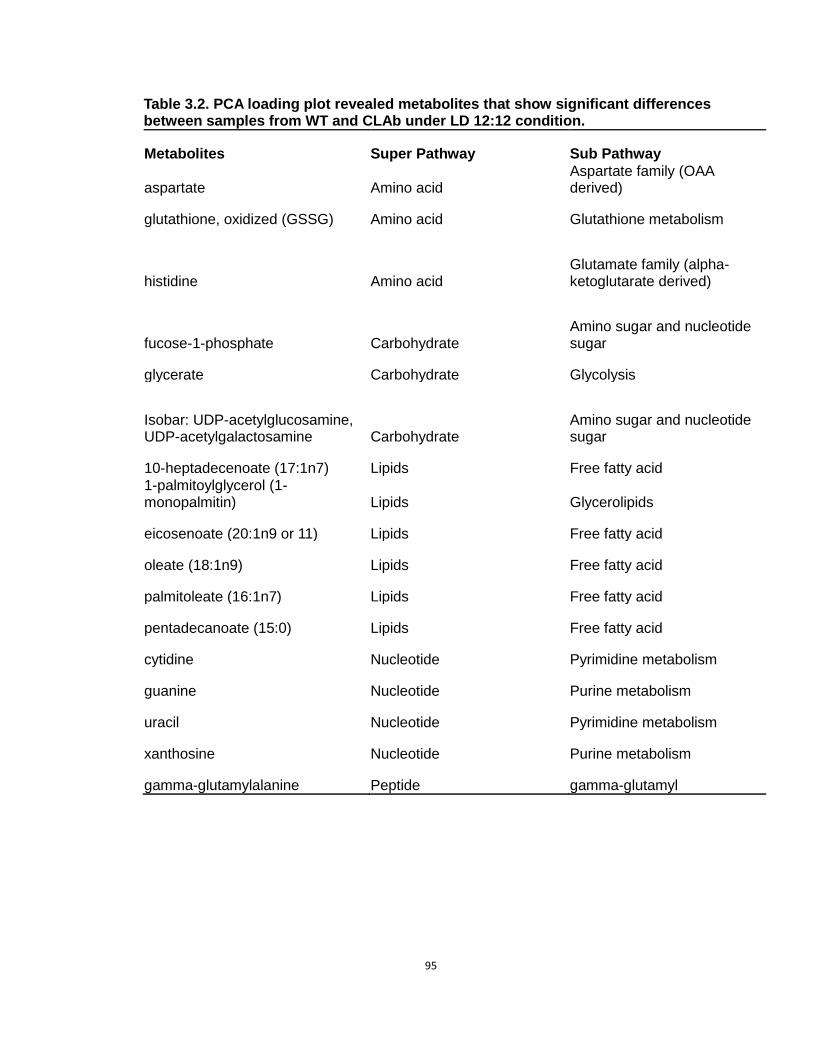
95
Table 3.2. PCA loading plot revealed metabolites that show significant differences between samples from WT and CLAb under LD 12:12 condition.
Metabolites Super Pathway Sub Pathway
aspartate Amino acid Aspartate family (OAA derived)
glutathione, oxidized (GSSG) Amino acid Glutathione metabolism
histidine Amino acid Glutamate family (alpha-ketoglutarate derived)
fucose-1-phosphate Carbohydrate Amino sugar and nucleotide sugar
glycerate Carbohydrate Glycolysis
Isobar: UDP-acetylglucosamine, UDP-acetylgalactosamine Carbohydrate
Amino sugar and nucleotide sugar
10-heptadecenoate (17:1n7) Lipids Free fatty acid
1-palmitoylglycerol (1-monopalmitin) Lipids Glycerolipids
eicosenoate (20:1n9 or 11) Lipids Free fatty acid
oleate (18:1n9) Lipids Free fatty acid
palmitoleate (16:1n7) Lipids Free fatty acid
pentadecanoate (15:0) Lipids Free fatty acid
cytidine Nucleotide Pyrimidine metabolism
guanine Nucleotide Purine metabolism
uracil Nucleotide Pyrimidine metabolism
xanthosine Nucleotide Purine metabolism
gamma-glutamylalanine Peptide gamma-glutamyl

96
Table 3.3 PCA loading plot revealed metabolites that show significant differences between samples from WT and CLAc under LD 12:12 condition.
Metabolites Super Pathway Sub Pathway
asparagine Amino acid Aspartate family (OAA derived)
aspartate Amino acid Aspartate family (OAA derived)
gamma-glutamyl-2-aminobutyrate Amino acid Glutathione metabolism
glutamate Amino acid Glutamate family (alpha-ketoglutarate derived)
glycine Amino acid Serine family (phosphoglycerate derived)
alpha-ketoglutarate Carbohydrate TCA cycle
fructose Carbohydrate Sucrose, glucose,fructose metabolism
phytol Cofactors, Prosthetic Groups, Electron Carriers
Chlorophyll and heme metabolism
cis-vaccenate (18:1n7) Lipids Free fatty acid
heptanoate (7:0) Lipids Free fatty acid
margarate (17:0) Lipids Free fatty acid
palmitate (16:0) Lipids Free fatty acid palmitate, methyl ester Lipids Free fatty acid
adenosine Nucleotide Purine metabolism
guanine Nucleotide Purine metabolism
inosine Nucleotide Purine metabolism
N6-carbamoylthreonyladenosine Nucleotide Purine metabolism
uracil Nucleotide Pyrimidine metabolism
xanthosine Nucleotide Purine metabolism
gamma-glutamylglutamine Peptide gamma-glutamyl
gamma-glutamylisoleucine* Peptide gamma-glutamyl
gamma-glutamylleucine Peptide gamma-glutamyl
gamma-glutamylmethionine Peptide gamma-glutamyl gamma-glutamylphenylalanine Peptide gamma-glutamyl
gamma-glutamylthreonine* Peptide gamma-glutamyl
gamma-glutamyltryptophan Peptide gamma-glutamyl
gamma-glutamyltyrosine Peptide gamma-glutamyl
gamma-glutamylvaline Peptide gamma-glutamyl

97
In cyanobacteria, free fatty acids are mainly involved in lipid and membrane
synthesis (Ruffing and Jones, 2012). Ruffing and coworkers reported that extra
free fatty acids could damage the cell physiology in S. elongatus (Ruffing and
Jones, 2012; Ruffing, 2013). For example, when S. elongatus was engineered to
produce more free fatty acids, the permeability of cell membrane was increased,
and photosynthesis was impaired (Ruffing and Jones, 2012). Furthermore,
Ruffing also found that the level of reactive oxygen species (ROS) was
significantly increased in strains that over-produced free fatty acids, indicating
that these cells were under stress conditions (Ruffing, 2013). Although the
production level of free fatty acids in CLAb may not be as high as that in the
engineered strains, these over-produced molecules may still result in a stressful
environment for CLAb cells.
Similarly to CLAb, some metabolites in the free fatty acid pathway were
also over-expressed in CLAc (Table 3.3 and Fig. 3.9B). In addition, 30% of the
“aberrantly expressed” candidates of CLAc are gamma-glutamyl amino acids that
belong to the peptide super pathway. Gamma-glutamyl amino acids are the
products of amino acid gamma-glutamylation which is a process catalyzed by
gamma-glutamyltranspeptidase (GGT). GGT is involved in glutathione (GSH)
synthesis, and its function is conserved in a wide range of organisms from
bacteria to mammals (Suzuki et al., 2007). GSH is an important antioxidant which
prevents damages from ROS. Consistently, some metabolites involved in GSH
metabolism were also among the candidates in both CLAb and CLAc (Fig. 3.9),
indicating that GSH metabolism is affected by the disruption of the circadian

98
clock.
Besides the metabolites identified from the free fatty acid pathway and
pathways involved in glutathione metabolism, candidates were also identified
from many other pathways, but in relatively smaller degrees of difference (Table
3.2, Table 3.3 and Fig. 3.9). It is possible that these pathways also contribute to
the competition results, either directly or indirectly. Because the global
metabolism is composed of numerous pathways and these pathways are all
inter-connected, it is difficult to conclude whether or not specific pathways are
critical for the competition at this moment. Experiments targeted to these
pathways are in the planning stage.
Discussion
In this study, two questions are addressed. First, the two clock mutants,
CLAb and CLAc that were out-competed by the WT under LD conditions,
displayed different metabolic profiles from the WT, suggesting that the disruption
of circadian clocks affected the metabolism, and that maladaptive metabolism of
the clock mutants could be the potential mechanism of the competition. Second,
the WT showed different metabolic profiles under LD vs. LL, indicating that
constant light can over-ride the regulation of metabolism that is normally
controlled by the circadian clock entrained to LD.
It has been more than a decade since scientists discovered that circadian
clocks enhance the fitness of cyanobacteria in cyclic environments. The

99
underlying mechanism, however, remains elusive. Although several hypotheses
have been proposed, none of them has been experimentally proven (Ma et al.,
2013). Considering the fact that the cyanobacterial circadian clock regulates the
transcription of almost all of the genes in the genome (Liu et al., 1995; Ito et al.,
2009), we gradually realize that this competition phenomenon may be a global
effect of the disruption of the circadian clock. In addition, an experiment designed
to identify metabolites in the cyanobacterial culture medium discovered some
differences between the WT and a clock mutant (data not shown), leading us to
consider the global metabolic profiles of these strains.
As an ongoing project, results presented in this chapter open the window
for us to reveal the mystery of the competition experiments. However, more work
needs to done to fully address this question. For instance, in CLAb, the free fatty
acid pathway displayed some abnormal production. Based on this observation, I
hypothesize that the malfunction of the free fatty acid pathway in CLAb affects
the metabolism of CLAb and reduces its fitness in under LD conditions. To test
this hypothesis, first, I need to do a more specific experiment to confirm that this
pathway is not functioning properly. An experiment targeted to specifically
measure the metabolites in this pathway should be designed and conducted.
Second, I would like to find a way to restore the normal metabolism back to the
clock mutants, and then test if the competition effect can be eliminated or
reversed. While the first step is relatively easy to conduct, the second step is
challenging. Alternatively, I can test if the WT strain can display CLAb-like
phenotypes when free fatty acid pathways are over-expressed.

100
In addition, studying period mutants could be another option. In the
competition experiments, the WT out-competed not only the arrhythmic mutants,
but also a long period mutant, (C28a, free running period ~ 30 hours) under LD
12:12 cycles. But when C28a was co-cultured with the WT under LD 15:15
cycles, it defeated the WT (Ouyang et al., 1998). Therefore, comparing to the
arrhythmic mutants (CLAb and CLAc), it might be easier to manipulate the
metabolism of WT vs. C28a by changing the light conditions between LD 12:12
(optimal for WT, non-optimal for C28a) and LD 15:15 (optimal for C28a, non-
optimal for WT) to compare their metabolic profiles.
Methods
Sample collection
Samples were collected from the WT strain and two arrhythmic mutants
CLAb (arrhythmic in LL from LL onset) and CLAc (damps in LL to arrhythmicity in
2-3 days). Cells were grown at LD 12:12 cycles for 4 days or until the OD750
value reached 0.5, then cultures were either kept in LD12:12 or released into LL
conditions. 100ml cultures were collected at ZT0, ZT6, ZT12, ZT18 and ZT24
under LD 12:12 conditions or at CT12, CT18, CT24, CT30 and CT36 under LL
conditions (only for WT). For each strain, samples from two parallel cultures were
collected as duplicates. After sample collection, cells were immediately
centrifuged at 4 oC in darkness for 15 mins at 4000 rpm, and cell pellets were
immediately frozen by using liquid nitrogen and kept at -80 oC until the samples
were processed.
101
Metabolomics measurement
Metabolites were measured in 40 samples including the WT strain, CLAb
and CLAc by Metabolon, Inc. (Durham, NC, USA). To identify metabolites from
samples, LC/MS and GC/MS were used by Metabolon. A total number of 182
metabolites were identified. Among these metabolites, 9 were excluded for
further analysis due to missing values for more than 20% of the time points.
Statistical analysis and clustering
Raw and normalized LC/MS and GC/MS data was obtained from
Metabolon. Statistical analysis and data visualization was performed by using R
programming language with the packages “metabolomics” (De Livera and
Bowne, 2013) and “gplots” (Warnes et al., 2014). ANOVA was applied to assess
the statistical significance between duplicates or among samples from different
genotypes and/or light conditions. To establish the metabolic profiling, PCA and
hierarchical clustering were performed.
Identification of cycling metabolites
Cycling metabolites were identified by using the modified cosiner method
(Kucho et al., 2005). In short, the normalized and natural log transformed LC/MS
and GC/MS data was detrended linearly as described by Kucho et al.. Because I
only have data in 24 hours, only metabolites cycling with a 24-hmy period were
identified by fitting to the 24-hour cosine curve. Along with curve fitting, the peak
time and amplitude were calculated as described (Kucho et al., 2005). I then

102
filtered the metabolites by two criteria: first, the cycling metabolites should have
an error factor less than 0.2; second, the amplitude should be greater than 1.2.
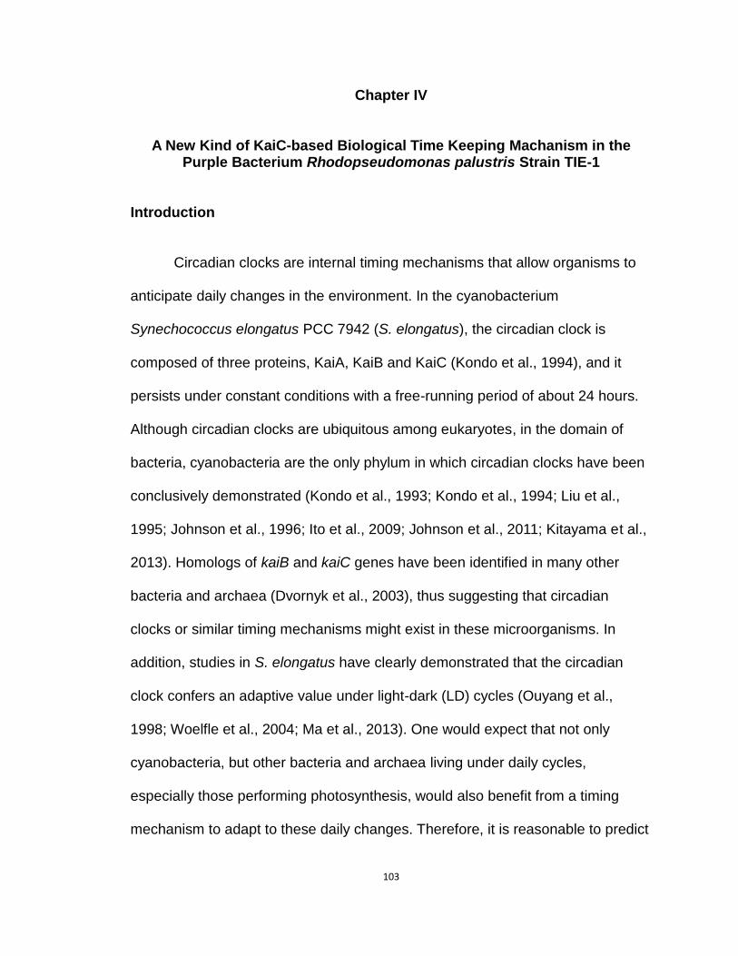
103
Chapter IV
A New Kind of KaiC-based Biological Time Keeping Machanism in the Purple Bacterium Rhodopseudomonas palustris Strain TIE-1
Introduction
Circadian clocks are internal timing mechanisms that allow organisms to
anticipate daily changes in the environment. In the cyanobacterium
Synechococcus elongatus PCC 7942 (S. elongatus), the circadian clock is
composed of three proteins, KaiA, KaiB and KaiC (Kondo et al., 1994), and it
persists under constant conditions with a free-running period of about 24 hours.
Although circadian clocks are ubiquitous among eukaryotes, in the domain of
bacteria, cyanobacteria are the only phylum in which circadian clocks have been
conclusively demonstrated (Kondo et al., 1993; Kondo et al., 1994; Liu et al.,
1995; Johnson et al., 1996; Ito et al., 2009; Johnson et al., 2011; Kitayama et al.,
2013). Homologs of kaiB and kaiC genes have been identified in many other
bacteria and archaea (Dvornyk et al., 2003), thus suggesting that circadian
clocks or similar timing mechanisms might exist in these microorganisms. In
addition, studies in S. elongatus have clearly demonstrated that the circadian
clock confers an adaptive value under light-dark (LD) cycles (Ouyang et al.,
1998; Woelfle et al., 2004; Ma et al., 2013). One would expect that not only
cyanobacteria, but other bacteria and archaea living under daily cycles,
especially those performing photosynthesis, would also benefit from a timing
mechanism to adapt to these daily changes. Therefore, it is reasonable to predict

104
that there should be other bacteria and/or archaea possessing a timing
mechanism besides cyanobacteria.
Among non-cyanobacteria prokaryotes whose genome include kaiB and
kaiC homologs, the purple non-sulfur bacterium Rhodopseudomonas palustris
(R. palustris) attracted our attention due to its ability to perform photosynthesis.
Unlike cyanobacteria, R. palustris is an anoxygenic phototrophic bacterium that
belongs to alpha-proteobacteria, and it uses different electron donors during
photosynthesis (Larimer et al., 2004; Jiao et al., 2005). Oxygen actually inhibits
its growth and many other activities such as photosynthesis and nitrogen fixation
(Larimer et al., 2004; Jiao et al., 2005). Despite this difference, R. palustris
shares many similarities with cyanobacteria. First, it is as widely distributed as
cyanobacteria, and in nature, it frequently shares the same locations with some
cyanobacteria, but at a different depth of water or sand (Proctor, 1997; Larimer et
al., 2004). A case where purple bacteria are located together with cyanobacteria
is the photosynthetic mats near coastal areas, as can be seen in Fig.4.1A.
Cyanobacteria reside in the top layer of the mat underneath a layer of sands to
avoid high light intensities. Purple bacteria can be found under the cyanobacteria
where concentrations of oxygen are relatively low (Stal, 1995). Second, R.
palustris uses sunlight as the energy source, indicating that some of its
physiological activities are regulated by light. Therefore, being able to anticipate
light/dark changes could be adaptive.
Along with the presence of kaiB and kaiC homologs in R. palustris (kaiBRp

105
and kaiCRp thereafter) (Dvornyk et al., 2003; Larimer et al., 2004; Jiao et al.,
2005), these lines of evidence suggest that R. palustris might have evolved a
timing mechanism. In this study, I explored this possibility by measuring the
nitrogen fixation activities of R. palustris under LD cycles and in constant light
(LL) conditions. Furthermore, the function of kaiCRp was investigated in both S.
elongatus and R. palustris. I report here that R. palustris possesses a timing
mechanism regulated by kaiCRp, and that this timing mechanism might be an
“hour glass” timer rather than a circadian clock. Consistent with the results in S.
elongatus, this timing mechanism enhances the fitness of R. palustris under LD
cycles but not LL conditions.
Results
Homologs of kaiB and kaiC were identified in R. palustris
The three clock genes of S. elongatus, kaiA, kaiB and kaiC, (kaiASe, kaiBSe
and kaiCSe thereafter) are essential for generating circadian rhythms in
cyanobacteria (Kondo et al., 1994). In R. palustris, however, only kaiBRp and kaiCRp
were identified, as depicted in Fig.4.1B. Instead of kaiA, a histindine kinase (hkRp)
containing a PAS domain is closely located downstream of kaiBRp (Fig. 4.1B). It is
not surprising that no kaiA exists in R. palustris; although kaiC and kaiB have been
found across the bacteria and archaea domains, kaiA has only been identified in
cyanobacteria (Dvornyk et al., 2003). The presence of hkRp
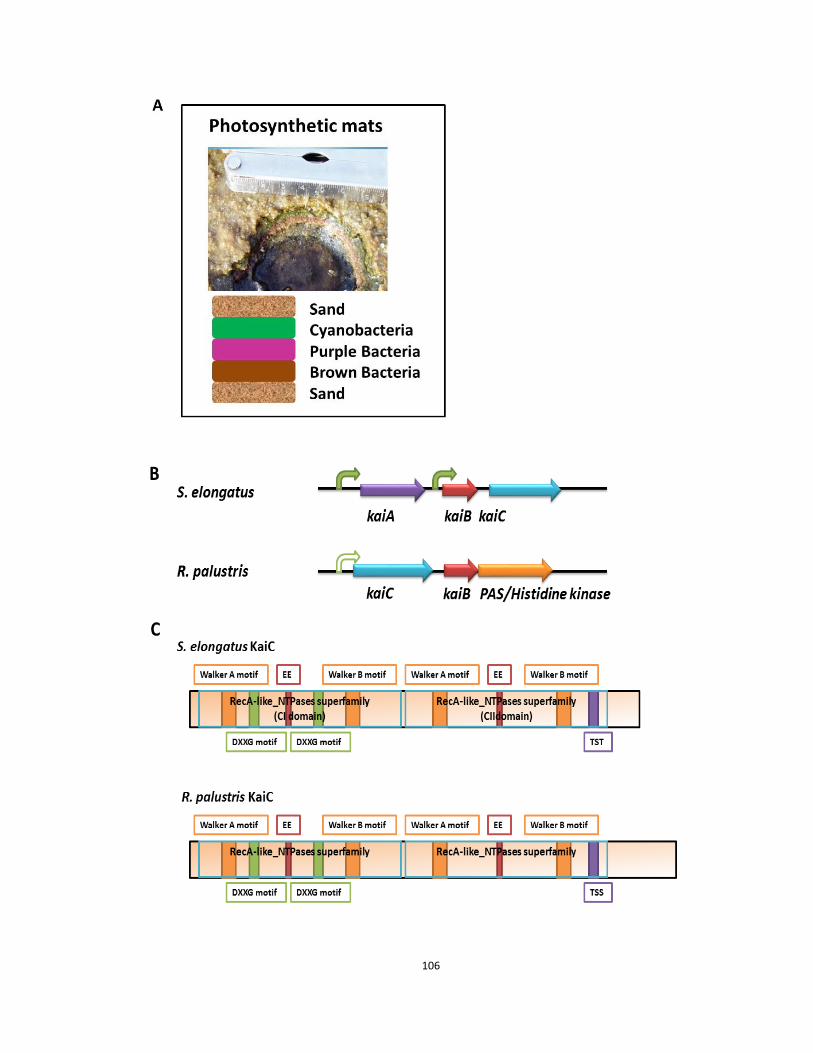
106
107
Figure 4.1 Homologs of kaiC and kaiB are identified in R. palustris. A, comparison of the kaiABC locus in S. elongatus with the kaiBC locus in R. palustris. In S. elongatus, kaiB and kaiC are co-transcribed by the same promoter. KaiA is located upstream of kaiB and is separately transcribed by another promoter (Ishiura et al., 1998). No kaiA was identified in R. palustris. Instead of kaiA, a histidine kinase containing PAS domain is located downstream of kaiB. Sequences for the primary sigma factor RpoD binding (rpoD promoter) were predicted to be the upstream of kaiC in R. palustris. B, comparison of predicted secondary structures of S. elongatus KaiC and R. palustris KaiC. Both of the KaiC proteins contain two RecA-like NTPase superfamily domains. In each of these domains, a Walker A and a Walker B motif is included as well as the EE residues that are the ATPase activity sites. In the first RecA-like NTPase superfamily domain (CI domain for S. elongatus), two DXXG motifs are present. Phosphorylation sites are located in the second RecA-like NTPase superfamily domain (CII for S. elongatus). In S. elongatus, these three phosphorylation sites are present as TST, while in R. palustris they are TSS. The C-terminus of R. palustris KaiC is about 50 amino acids longer than the S. elongatus KaiC.

108
suggests the possibility that kaiCRp functions through interactions with kaiBRp and
hkRp. Promoter prediction suggested that a binding site of the primary sigma
factor RpoD (Gruber and Gross, 2003) is present 17 bp upstream of kaiCRp (Fig.
4.1B), while no promoter was predicted upstream of kaiBRp or hkRp, indicating
that these three genes might be coordinately transcribed.
The KaiC protein of S. elongatus (KaiCSe) contains two RecA-like NTPase
superfamily domains that are named the CI domain and the CII domain (Johnson
et al, 2011). Each of these domains has an ATP/GTP-binding site known as the
Walker motif, as well as catalytic carboxylates (EE) that are recognized to be the
basis of autokinase and ATPase activity (Egli, et al., 2012). In addition, the CI
domain possesses two DXXG motifs for GTP binding (Nishiwaki, et al., 2000).
The sequence of the KaiC protein of R. palustris (KaiCRp) preserves all of the
conserved domains of S. elongatus KaiC, as illustrated in Fig.4.1C and Fig.4.2.
The C-terminus of KaiCRp, however, is about 50 amino acids longer than the
KaiCSe, and one of the phosphorylation sites of KaiCSe, Thr432 (Xu et al., 2004),
is present as a serine (Fig. 4.1C & Fig.4.2). Previous studies in cyanobacteria
have shown that the C-terminal region of KaiC is essential for KaiA binding
(Pattanayek et al., 2006; Vakonakis and LiWang, 2004). Given the fact that there
is no kaiA identified in R. palustris, it is not surprising that the C-terminus is not
conserved between KaiCSe and KaiCRp.

109

110

111
Figure 4.2 Alignment of KaiC proteins in some cyanobacteria species and two purple bacteria species. The Walker motifs are highlighted with black boxes. The DXXG motifs are highlighted with blue boxes. Red arrows indicate the phosphorylation sites, and the EE sites are labeled with red stars.

112
In addition to S. elongatus, kaiABC - driven timing mechanisms have also been
described in other cyanobacteria (Kucho et al., 2005; Wiegard et al., 2013;
Axmann et al., 2009; Holtzendorff et al., 2008; Gaudana et al., 2013; Cerveny et
al., 2013). Synechocystis sp. Strain PCC 6803 (Synechocystis), for instance, has
a circadian clock that regulates 2-9% of the genes in the genome under both
cyclic and constant conditions (Kucho et al., 2005).Unlike S. elongatus,
Synechocystis possesses three kaiC, two kaiB and one kaiA genes (Kucho et al.,
2005; Wiegard et al., 2013). Interestingly, as shown in Fig.4.2 and Fig.4.3,
protein sequence alignment and phylogenetic analysis of KaiC showed that the
KaiC (Synechocystis1) located together with KaiB and KaiA is grouped with KaiC
of S. elongatus, and that the KaiC (Synechocystis3) located with KaiB alone is
phylogenetically closer to KaiCRp than any other cyanobacterial KaiC. Moreover,
this KaiC (Synechocystis3) shares similar sequence and length with KaiCRp in the
C-terminus as well as the same phosphorylation sites (Fig. 4.2), indicating that
this version of kaiC experienced horizontal gene transfer events and that it may
contribute to the timing mechanism in a similar way to KaiCRp. In contrast,
Prochlorococcus, a marine cyanobacterium with only kaiB and kaiC, and
Cyanothece sp. ATCC 51142 with two kaiC, two kaiB and one kaiA, are clustered
within the cyanobacteria group (Fig.4.3). The PAS domain histidine kinase is only
identified in the R. palustris group (Fig.4.3), suggesting that hkRp may have a
specific function in purple bacteria.

113
Figure 4.3 Phylogenetic tree of KaiC proteins in some cyanobacteria species and two purple bacteria species. Among these KaiC proteins, three copies are from Synechocystis sp. PCC 6803 (Synechocystis 1, 2 & 3), and two copies are from Cyanothece sp. ATCC 51142 (Cyanothece 1 & 2). The others have only one copy of kaiC. Along with kaiC, kaiB, kaiA or hk genes are illustrated in the blue arrows to compare the difference among these species. The hk genes are only identified in the purple bacteria, while kaiA are only present among cyanobacteria.
114
KaiCRp can influence the circadian clock of S. elongatus, but it is not
sufficient to compensate the function of KaiC of S. elongatus
To investigate the function of KaiCRp, I first tested whether KaiCRp can
influence the molecular clock of S. elongatus by expressing kaiCRp in the wild-
type S. elongatus strain (AMC149) as well as the kaiC-deletion strain (DeltaC).
AMC149 and DeltaC harbor luminescence reporters that allow clock-controlled
promoter activities to be represented by luminescence levels (Kondo et al., 1993;
Xu et al., 2000). AMC149 displays roughly 24-hour circadian rhythms under LL
conditions, while DeltaC is arrhythmic due to the lack of kaiC (Kondo et al., 1993;
Xu et al., 2000). When kaiCRp was expressed in AMC149 under the control of the
trc promoter (Xu et al., 2003) by IPTG induction, the free running period of the
luminescence rhythm was lengthened by about 1 hour, and the amplitude was
reduced(Fig. 4.4A & B), suggesting some interference to the cyanobacterial
circadian clock by KaiCRp. The over-expressing strain exhibited a slightly longer
free running period even without IPTG induction, probably due to leaky
expression from the trc promoter.
Moreover, an interesting phenomenon was observed when kaiCRp was
overexpressed in the DeltaC strain. As can be seen in Fig. 4.4C, a transient peak
was elicited in DeltaC by expressing kaiCRp, whereas DeltaC alone only showed
declining luminescence over time. This peak was not observed in the absence of
IPTG, even though a stronger reporter activity was detected (Fig. 4.4C), possibly
due to leaky expression of kaiCRp.
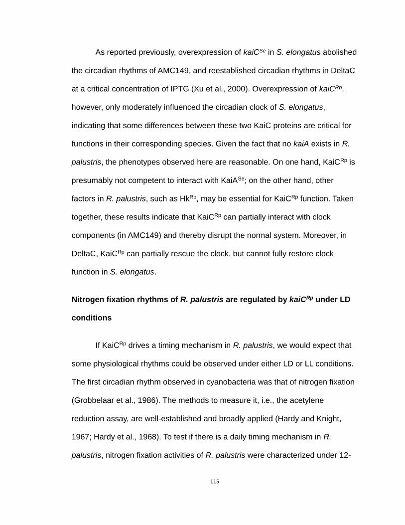
115
As reported previously, overexpression of kaiCSe in S. elongatus abolished
the circadian rhythms of AMC149, and reestablished circadian rhythms in DeltaC
at a critical concentration of IPTG (Xu et al., 2000). Overexpression of kaiCRp,
however, only moderately influenced the circadian clock of S. elongatus,
indicating that some differences between these two KaiC proteins are critical for
functions in their corresponding species. Given the fact that no kaiA exists in R.
palustris, the phenotypes observed here are reasonable. On one hand, KaiCRp is
presumably not competent to interact with KaiASe; on the other hand, other
factors in R. palustris, such as HkRp, may be essential for KaiCRp function. Taken
together, these results indicate that KaiCRp can partially interact with clock
components (in AMC149) and thereby disrupt the normal system. Moreover, in
DeltaC, KaiCRp can partially rescue the clock, but cannot fully restore clock
function in S. elongatus.
Nitrogen fixation rhythms of R. palustris are regulated by kaiCRp under LD
conditions
If KaiCRp drives a timing mechanism in R. palustris, we would expect that
some physiological rhythms could be observed under either LD or LL conditions.
The first circadian rhythm observed in cyanobacteria was that of nitrogen fixation
(Grobbelaar et al., 1986). The methods to measure it, i.e., the acetylene
reduction assay, are well-established and broadly applied (Hardy and Knight,
1967; Hardy et al., 1968). To test if there is a daily timing mechanism in R.
palustris, nitrogen fixation activities of R. palustris were characterized under 12-

116
Figure 4.4 Luminescence rhythms of S.elongatus strains overexpressing of kaiCRp. A, kaiCRp was expressed in the wild type AMC149. Upper panel, no IPTG induction; lower panel, IPTG was applied to the cultures after entrainment. Comparing to the rhythms of AMC149 (blue dots), the luminescence rhythm was suppressed and the FRP was lengthened when kaiCRp was overexpressed (red dots). B, the quantification of FRPs in AMC149 (blue) and AMC149oxkaiCRp (red) strains with and without IPTG induction. C, kaiCRp was overexpressed in the arrhythmic strain DeltaC. Upper panel, no IPTG induction; lower panel, IPTG was applied to the cultures after entrainment. Red arrows indicate the time that IPTG was applied to the cultures.
117
hour-light-12-hour-dark cycles (LD 12:12). As shown in Fig. 4.5A, nitrogen
fixation of the wild-type R. palustris (WT) exhibited an oscillating pattern under
LD cycles at 30 °C. Each trace represents an individual culture. Nitrogen was
mainly fixed during the day time, and this activity was shut down at night. At
around Zeitgeber Time (ZT) 8, all of the individual wild-type cultures reached their
maximal nitrogen fixation rates.
The observed oscillation could be one of the physiological activities
controlled by a timing mechanism, whereas it is also possible that it is merely a
response to light-dark signals. To distinguish between these two possibilities, a
kaiCRp-deletion strain of R. palustris (RCKO) was generated. Surprisingly, the
nitrogen fixation of RCKO showed similar fluctuations as the WT under LD
condition except that the timing of the peaks varied dramatically among individual
cultures (Fig. 4.5B). Quantitative RT-PCR further confirmed that no mRNA of
kaiCRp was detected in the RCKO strain, and kaiBRp was transcribed normally as
in WT (Fig. 4.6A). Furthermore, restoring a FLAG –tagged kaiCRp back to the
RCKO genome rescued the wild-type phenotype (Fig. 4.6B&C). Taken together,
these results indicate that deletion of kaiCRp resulted in a large increase in the
variability of the nitrogen fixation peak of RCKO.
While R. palustris is usually cultured at 30 °C under laboratory conditions,
it sometimes lives at lower temperatures in its natural environment (Jiao et al.,
2005). To test if nitrogen fixation rhythms can be observed at lower temperatures,
nitrogen fixation assays of the WT and RCKO strains were performed at 23°C

118
Figure 4.5 Nitrogen fixation rhythms of R. palustris under LD cycles. Samples were taken every 3 hours and incubated for 3 hours with acetylene under the same culture conditions. After 3 hours incubation, they were injected into GC to measure the acetylene reduction rates. A and C, nitrogen fixation rhythms of the wild-type R. palustris (WT) at 30 oC and 23 oC. Three traces represent three individual cultures. B and D, nitrogen fixation rhythms of the kaiCRp-deletion strain (RCKO) at 30 oC. The black and white bars underneath represent the light conditions.

119
Figure 4.6 A, Quantitative PRC to confirm the deletion of kaiCRp and the expression of kaiBRp. The mRNA levels of clpX were included as the internal control. The mRNA levels of kaiBRp and kaiCRp were quantified in the WT (red) and RCKO (black) strains. B, western blot by anti-FLAG antibody to confirm the expression of KaiCRp in the rescue strain (RCKO+ kaiCRp). C, nitrogen fixation of the kaiCRp rescue strain under LD 12:12 cycles at 30 oC. Three traces represent three individual cultures.

120
under LD 12:12 conditions. Similar to the results obtained at 30°C, at 23°C
nitrogen fixation rhythms of several WT cultures were in-phase with each other
(Fig. 4.5C), whereas the individual RCKO cultures differed from each other(Fig.
4.5D). Furthermore, the total nitrogen fixation activities of the WT strain were two
times higher than that of the RCKO strain at 23°C, suggesting some overall
impact to the physiology of RCKO at low temperatures.
One of the fundamental properties of circadian clocks is that they can be
entrained by environmental cues, whereby physiological processes adjust their
periods and phases according to the external cycles (Dunlap et al., 2004). The
stable phase angle observed in the WT strain suggests that WT is entrained to
the environmental cycles, thus indicating that a timing mechanism exists in R.
palustris, and that kaiCRp plays an important role in it.
Nitrogen fixation of R. palustris does not show clear rhythms under LL
conditions
A timing mechanism could be a sophisticated circadian clock that
persistently oscillates under constant conditions, or it could be an hour glass
operating only under cyclic environments and which damps rapidly in constant
conditions. As a continuation of the assays performed under LD cycles, the
nitrogen fixation rates were measured from cells cultured under LL conditions to
test if the rhythm persists without environmental signals. As shown in Fig. 4.7A,
at 30°C, the apparent nitrogen fixation rhythm of the WT continued for two short-
period cycles and then it damped out. The modified cosiner method (Kucho et al.,

121
2005) was applied to the data to statistically evaluate whether they conform to
being an oscillation. Usually two criteria have to be satisfied for a rhythm: first,
the error factor (Ef) of the curve fitting needs to be less than 0.2; second, the
relative amplitude should be greater than 1.2. As can been seen in Table 4.1, the
best fitting curve with the smallest Ef value (Kucho et al., 2005; see methods) of
the WT data was the curve with a 19-hour cycle and the relative amplitude is
15.33. However, this Ef value is significantly greater than 0.2, suggesting that
these data may not fit the criteria to qualify as a rhythm.
For the RCKO strain, possible oscillations with large variations were also
observed at 30 °C. Similar to the WT, these data do not fulfill both of the two
criteria, suggesting that no rhythm was detected in the RCKO strain at 30 °C. At
23°C, apparently no rhythm was detected in the RCKO strain (Fig. 4.7B and
Table 4.1). In the WT strain, the nitrogen was fixed cyclically, but these data also
failed the statistical test (Fig. 4.7B and Table 4.1).
Circadian rhythms usually exhibit periods that are nearly 24 hours in
constant conditions. The nitrogen fixation rhythm of the WT strain, however, is
not only significantly shorter than 24 hours, but also damped out after two cycles.
These results indicate that the timing mechanism of R. palustris could be a highly
damped circadian oscillator or possibly an hour glass that does not keep the
rhythms running under constant conditions. However, it is also possible that the

122
Figure 4.7 Nitrogen fixation activities of R. palustris under LL conditions. The WT strain and the RCKO strain were cultured under LD 12:12 cycles for 3 to 4 days, then they were released to LL condition. Under LL condition, samples were taken every 3 hours and incubated for 3 hours with acetylene under the same culture conditions before they were injected into GC to measure the acetylene reduction rates. A, nitrogen fixation activities of the WT (red) and RCKO (green) strains at 30 oC. B, nitrogen fixation activities of the WT (red) and RCKO (green) strains at 23 oC. Error bars were plotted as the standard deviation from three individual cultures (n=3).

123
Table 4.1 Statistical analysis of the nitrogen fixation data under LL conditions. Ef, error factor of the cosine curve fitting; Amp, relative amplitude; Peak, peak time calculated by curve fitting. The number after each category, e.g, Ef (24h), means the period of the cosine curve used for the fitting.
Ef
(24h)
Ef (22h) Ef (20h) Ef (19h) Ef (18h) Ef (17h) Ef (16h) Ef (14h) Ef (12h)
WT(30
oC)
4.19 3.45 2.85 2.69 2.75 3.11 3.74 4.99 4.54
RCKO(3
0 oC)
0.04 0.04 0.04 0.03 0.03 0.03 0.03 0.03 0.03
WT(23
oC)
0.04 0.04 0.04 0.04 0.04 0.04 0.04 0.05 0.04
RCKO(2
3 oC)
0.13 0.12 0.11 0.11 0.10 0.09 0.09 0.08 0.07
Amp
(24h)
Amp
(22h)
Amp
(20h)
Amp
(19h)
Amp
(18h)
Amp
(17h)
Amp
(16h)
Amp
(14h)
Amp
(12h)
WT(30
oC)
16.00 15.84 15.91 15.33 14.08 12.29 10.37 8.36 9.19
RCKO(3
0 oC)
1.06 1.06 1.05 1.04 1.04 1.04 1.04 1.04 1.05
WT(23
oC)
1.06 1.06 1.07 1.06 1.06 1.06 1.05 1.05 1.04
RCKO(2
3 oC)
1.11 1.07 1.06 1.09 1.12 1.15 1.18 1.12 1.12
Peak
(24h)
Peak
(22h)
Peak
(20h)
Peak
(19h)
Peak
(18h)
Peak
(17h)
Peak
(16h)
Peak
(14h)
Peak
(12h)
WT(30
oC)
21.54 0.09 2.43 3.54 4.65 5.78 6.97 19.58 24.00
RCKO(3
0 oC)
13.43 14.49 15.38 15.72 15.98 16.17 16.33 6.91 6.00
WT(23
oC)
15.65 17.56 19.56 20.62 21.72 22.88 0.12 2.90 6.00
RCKO(2
3 oC)
14.78 14.69 9.07 7.91 7.32 7.00 6.82 6.51 6.00

124
damping rhythms are caused by some technical issues. The exponentially
growing cells, for instance, may be entering the stationary phase where only a
small amount of nitrogen is required, or the nitrogenase could be inhibited after
fixing adequate nitrogen to the medium. Additionally, no clear patterns were
observed in the RCKO strain, although some fluctuations were detected. These
uncertainties leave this question unsolved.
The timing mechanism driven by KaiCRp enhances fitness of R. palustris
under LD cycles.
Several studies in cyanobacteria have demonstrated that the kaiC-driven
circadian clock enhances fitness under cyclic environments (Ouyang et al., 1998;
Woelfle et al., 2004; Ma et al., 2013). To test whether this adaptive value of kaiC
extends to R. palustris, growth rates of the WT and RCKO strains were
measured under LD 12:12 cycles and LL conditions at 30°C and 23°C. As shown
in Fig. 4.8A&C, at both temperatures, the RCKO strain grew in LL at about the
same rate as did WT. However, a dramatic difference in their growth rates was
observed when they were cultured under LD conditions (Fig.4.8B&D); the WT
strain grew significantly faster than the RCKO strain. Furthermore, when a FLAG-
tagged kaiCRp was restored into the RCKO genome, the rescued strain showed
similar growth rates as the WT strain under both LL and LD conditions (Fig. 4.8),
confirming that the reduced growth rate of RCKO is caused by the absence of
kaiCRp. Taken together, these results suggest that a KaiCRp-dependent timing

125
Figure 4.8 Growth curves of R. palustris under LL or LD 12:12 conditions. A, cell densities (OD600) of the WT (red), RCKO (black) and the kaiCRp rescue strain (green) were measured every two days under LL condition at 30 oC. B, cell densities of the WT (red), RCKO (black) and the kaiCRp rescue strain (green) were measured every two days under LD 12:12 cycles at 30 oC. The OD600 value was plotted as the function of time (days).

126
Figure 4.9 ATPase activity of KaiCRp in vitro. Purified KaiCRp was incubated with ATP at 0 oC, 23 oC, 30 oC and 37 oC for 24 hours. Then the amount of ATP/ADP/AMP was quantified by HPLC. Blue, ATP; red, ADP; green, AMP.

127
mechanism confers an adaptive value under cyclic conditions but not constant
conditions.
KaiCRp displays ATPase activity in vitro
It is believed that the phosphorylation rhythm of KaiC along with the
interactions of KaiA and KaiB is the basic mechanism of circadian clock in S.
elongatus (Nakajima et al., 2005; Kageyama et al., 2006). Additionally, the
ATPase acitivities of KaiCSe was reported (Terauchi et al., 2007), and several
studies suggested that three activities of KaiCSe (ATPase, autokinase and
autophosphatase) are coupled and that they are essential for generating
circadian rhythms in S. elongatus (Kitayama et al, 2013).
The protein sequence alignment of KaiC suggested that the ATPase sites
are highly conserved among different species (Fig. 4.2), indicating that the
ATPase activity may also play an essential role for the function of KaiC in other
bacteria. My results have demonstrated that KaiCRp drives a timing mechanism in
R. palustris, but the biochemical basis of this timing mechanism remains elusive.
Considering the importance of ATPase activity for KaiC function in S. elongatus, I
hypothesize that KaiCRp has ATPase activity.
To test this hypothesis, kaiCRp was expressed in E. coli and the protein
was purified. The purified KaiCRp was incubated with ATP at 0 oC, 23 oC, 30 oC
and 37 oC for 24 hours, and then the reaction was terminated and measured by
High-performance liquid chromatography (HPLC). As shown in Fig. 4.9, time 0

128
represents the initial composition of the reaction. After a 24-hour incubation at
0oC, no ADP/AMP production was detected, while at 23 oC , 30 oC and 37 oC, all
of the ATP was converted to ADP/AMP, suggesting that KaiCRp confers ATPase
activities at these temperatures. Interestingly, compared to KaiCSe, the ATPase
activity of KaiCRp is at least 10 times higher (Terauchi et al., 2007). Taken
together, these results indicate that KaiCRp is also an ATPase with a relatively
high activity. Whether or not this ATPase activity plays a role in KaiRp function
needs to be confirmed in future experiments
Discussion
Circadian clock vs. hour glass
A circadian clock is a sophisticated biochemical machinery whose
oscillations persist even without external cues (Dunlap et al., 2004). An hour
glass, on the contrary, is dependent on regular environmental stimulations to
restart it every day, and it will not cycle without daily stimulation. Both a circadian
clock and an hour glass need to be entrained by environmental signals to
establish a stable phase angle (Rensing et al., 2001). In this study, the nitrogen
fixation rhythms with a stable phase angle under LD12:12 cycles provide
evidence for the presence of a timing mechanism in R. palustris, but it damped
out under LL conditions after oscillating for two 18-hour cycles, indicating that this
timing mechanism is more likely to be an hour glass that can trigger a second
cycle or a highly damped oscillator rather than a persisting circadian clock. In S.
elongatus, KaiA is essential for a functioning clock (Kondo et al., 1994), therefore

129
the lack of kaiA may prohibit R. palustris to form a self-sustained circadian clock.
Similar to R. palustris, the marine cyanobacterium Prochlorococcus only
harbors kaiB and kaiC homologs, and possibly it possesses only an hour glass
as the timing mechanism instead of a circadian clock (Holtzendoff et al., 2008;
Axmann et al., 2009). Prochlorococcus cells are mainly found in the open ocean
from the surface to depths of 100 to 200 meters, and they have not been found at
high latitudes (Mullineaux and Stanewsky, 2009; Partensky et al., 1999). In
contrast, S. elongatus inhabits fresh water where environmental changes such as
shading due to vegetation or shifting sediments may be more dramatic and
unpredictable than in the ocean areas (Mullineaux and Stanewsky, 2009). One
can imagine that having a robust circadian clock will be beneficial when the
environmental signals are not stable. In other words, a circadian clock will keep
the physiological activities and gene expressions running normally when the
environmental conditions are inconsistent. Therefore, for freshwater
cyanobacterium S. elongatus an hour glass may be not sufficient. Although R.
palustris is a freshwater bacterium, it is usually found at the bottom of ponds or
under the cyanobacteria in mats to avoid stresses from oxygen and high light
intensities(Proctor, 1997; Jiao et al., 2005). In these two conditions,
environmental changes may be relatively consistant as compared with shallow
areas, thus suggesting that an hour glass might be capable of maintaining
normally phased daily activities of R. palustris.
Nevertheless, we still cannot exclude the possibility that a circadian clock

130
exists in R. palustris. First, the damping rhythms may be due to the culture
conditions. For instance, I entrained these cells for at least 3 days and then
monitored the nitrogen fixation activities for another 3 days under constant
conditions. After being cultured for 6 days, cells may have grown to the stationary
phase where the nitrogen fixation activity is not as robust as it in the exponential
phase. To solve this problem and keep cells in the exponential stage, continuous
cultures may be required for this experiment. Second, nitrogen fixation may not
oscillate under constant conditions, but there may be other physiological
activities or gene expressions that do oscillate persistently under constant
conditions. To solve this problem, another assay needs to be developed. For
example, microarrays could be applied to measure gene expressions in R.
palustris under LD and LL conditions. Additionally, in S. elongatus, luminescence
reporters are applied to track the circadian rhythms, allowing frequent sampling
without interrupting the cell growth (Kondo et al., 1993). But neither this reporter
nor other fluorescence reporters can be applied to R. palustris cultures because
luminescence assays use luciferase, whose activity is dependent upon oxygen
and no oxygen is present in our cultures. Recently, some luminescence reporters
that do not require oxygen were published (Mukherjee et al., 2013). If we could
apply these reporters to R. palustris cultures, we would be able to better test if
this timing mechanism is a circadian clock or not.
Adaptive significance of timing mechanisms
In this study, we found that deletion of kaiCRp reduced the fitness of R.

131
palustris under LD cycles, suggesting that a kaiC-mediated timing mechanism
confers adaptive value to the purple bacterium R. palustris. This discovery is
consistent with the studies done in cyanobacteria (Ouyang et al., 1998; Woelfle
et al., 2004), therefore indicating that the adaptive significance of timing
mechanisms may be a universal property among different organisms.
Interestingly, the timing mechanisms of R. palustris and S. elongatus are only
adaptive under cyclic conditions. Under constant conditions, the wild type strains
and the mutant strains grew equivalently in both S. elongatus and R. palustris,
indicating that timing mechanisms are “extrinsic” adaptations rather than
“intrinsic” adaptations in these two species (Woelfle and Johnson, 2009; Ma et
al., 2013).
Diversity of KaiC
Bioinformatics studies showed that kaiC is widely distributed across the
bacterial and archaeal domains (Dvornyk et al., 2003). Based on the sequence
alignment (Fig. 4.2), most of the important domains are conserved among these
KaiC, while some small differences are present (Fig. 4.2). I am curious how
these small differences contribute to the discrepancies between their timing
mechanisms. For instance, in my study, I found that the KaiCRp has a longer C-
terminus. In S. elongatus, the C-terminus of KaiCSe is critical for interacting with
KaiA. Given the fact that there is no KaiA present in R. palustris, we would expect
that this long tail of KaiCRp may interact with other proteins or prevent KaiCRp
from interacting with other proteins. In addition, it is still a puzzle that some

132
organisms possess multiple kaiC genes. For example, the freshwater
cyanobacterium Synechocystis sp. PCC 6803 has three copies of kaiC and one
of its KaiC versions shares a similar long C-terminus structure with KaiCRp. For
almost 20 years, comprehensive studies focused on the S. elongatus circadian
system have been conducted, providing valuable knowledge about the S.
elongatus KaiC (Johnson et al., 2011). However, little is known about KaiC in
other bacteria and archaea. Along with some work done in other cyanobacteria
(Kucho et al., 2005; Wiegard et al., 2013; Axmann et al., 2009; Holtzendorff et al.,
2008; Gaudana et al., 2013; Cerveny et al., 2013), my study will help us to
understand the diversity of KaiC as well as the diversity of microbial timing
mechanisms, which will hopefully reveal the evolution of timing systems in
bacteria and archaea.
Methods
Bacterial strains and culture conditions
All bacterial strains used in this study are listed in Table 4.2. E.coli was
grown in Luria-Bertani (LB) broth at 37°C with shaking, and if necessary,
gentamicin (50µg/ml) was supplemented. For photoheterotrophic growth, R.
palustris strains were grown in Freshwater-Base (FW) medium (Jiao et al., 2005)
supplied with 20mM sodium acetate, 50mM sodium bicarbonate, 20mM MOPS
(PH7.2), 1mM potassium phosphate, 1mM sodium sulfate, multivitamin solution
and trace elements solution. The medium was aliquoted into sealed serum
bottles and the headspace was flushed with N2 gas for 20 min before inoculating

133
cells. N2 gas is used as the sole nitrogen source to ensure nitrogenase
expression. The cultures were maintained at 30 °C (or 23 °C as specified) and
illuminated by a cool-white fluorescent lamps (40-50 µE m−2 s−1) with shaking at
120 rpm. For aerobic chemoheterotrophic growth, R. palustris strains were grown
in YP medium containing 0.3% yeast extract and 0.3% peptone (Jiao et al., 2005)
at 30 °C with shaking at 120 rpm, and if necessary, gentamicin (400µg/ml) was
supplemented. The cyanobacterial strains were grown in BG-11 medium (Bustos
and Golden, 1991) at 30 °C, and illuminated by cool-white fluorescence bulbs
(40-50 µE m−2 s−1) with air bubbling. The medium of AMC149 and DeltaC was
supplemented with spectinomycin (25µg/ml), and the medium of the kaiCRp
overexpressing strains was supplemented with spectinomycin (25µg/ml) and
kanamycin (25µl/ml). For growth on solidified media, LB, FW or BG-11 medium
was supplemented with 1.5% agar and appropriate antibiotics.
Construction of the kaiCRp deletion strain
All plasmids and primers used in strain constructions are listed in Table 4.3
and Table 4.4. The kaiCRp deletion strain (RCKO) was constructed in R. palustris
TIE-1 strain by overlap extension PCR and conjugation (Hirakawa et al., 2011,
Bose and Newman, 2011). To delete the kaiCRp gene, the 1 kb upstream region
and the 1 kb downstream region of the kaiCRp ORF were cloned from the
genomic DNA of R. palustris TIE-1 strain and fused by overlap extension PCR.
The 2-kb DNA fragment was ligated with the suicide vector pJQ-200KS (Quandt
and Hynes, 1993). The resultant plasmid, pJQ-200KS-RCKO, was transformed

134
into R. palustris TIE-1 strain by conjugation with E. coli S17-1 (Simon et al., 1983;
Hirakawa et al., 2011; Bose and Newman, 2011). The integration of the plasmid
in either the upstream or the downstream regions of the kaiCRp locus was
selected by gentamicin resistance and screened by PCR. Following the
selection, the integrants were grown in non-selective YP medium for several
generations and then plated on YP agar medium with 10% sucrose (Hirakawa et
al., 2011) to induce double recombination. Among the survivors of the sucrose-
YP medium, the double recombinants were selected by PCR screening. The
deletion of the kaiCRp gene was confirmed by sequencing and Q-RT-PCR.
Construction of the FLAG-kaiCRp strain
To complement the kaiCRp gene deletion, a FLAG-tagged kaiCRp gene was
restored to the genome of RCKO strain in the region surrounding the glmUSX-
recG locus (Bose and Newman, 2011). The FLAG tag allows me to confirm the
expression of KaiCRp in the rescued strain with an anti-FLAG antibody. To
construct the insertion plasmid pJQ200KS-Insert, the 1 kb upstream and the 1 kb
downstream regions of glmUSX-recG locus of R. palustris TIE-1 were cloned and
fused by overlap extension PCR. A NcoI site was incorporated in the middle to
allow the insertion of genes of interest (Bose and Newman, 2011). The resulting
DNA fragment was ligated with pJQ200KS by SphI and SmaI, as described by
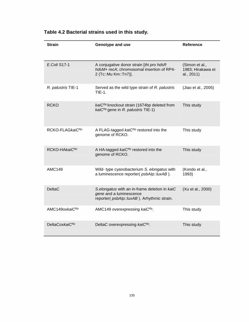
135
Table 4.2 Bacterial strains used in this study.
Strain Genotype and use Reference
E.Coli S17-1 A conjugative donor strain [thi pro hdsR hdsM+ recA; chromosomal insertion of RP4-2 (Tc::Mu Km::Tn7)].
(Simon et al., 1983; Hirakawa et al., 2011)
R. palustris TIE-1 Served as the wild type strain of R. palustris TIE-1.
(Jiao et al., 2005)
RCKO kaiCRp knockout strain (1674bp deleted from kaiCRp gene in R. palustris TIE-1)
This study
RCKO-FLAGkaiCRp A FLAG-tagged kaiCRp restored into the genome of RCKO.
This study
RCKO-HAkaiCRp A HA-tagged kaiCRp restored into the genome of RCKO.
This study
AMC149 Wild- type cyanobacterium S. elongatus with a luminescence reporter( psbAIp::luxAB ).
(Kondo et al., 1993)
DeltaC S.elongatus with an in-frame deletion in kaiC gene and a luminescence reporter( psbAIp::luxAB ). Arhythmic strain.
(Xu et al., 2000)
AMC149oxkaiCRp AMC149 overexpressing kaiCRp. This study
DeltaCoxkaiCRp DeltaC overexpressing kaiCRp. This study

136
Table 4.3 Plasmids used in this study.
Plasmid Description Reference
pJQ200KS Mobilizable suicide vector; sacB, Gmr
(Quandt and Hynes, 1993; Hirakawa et al., 2011)
pJQ200KS - RCKO Overlap extension PCR was used to amplify homologous regions of
1-kb upstream and 1-kb downstream of kaiCRp
locus, then the 2-kb fragment was ligated with Pjq200ks by BamHI and XbaI. This
plasmid was used to delete kaiCRp
gene in the wild-type R.
palustris. Gmr
This study
pJQ200KS -Insert Overlap extention PCR was used to amplify the 2 kb region surrounding the glmUSX-recG locus of R. palustris TIE-1, a NcoI site was incorporated into the middle of this PCR fragment which was ligated to pJQ- 200KS by SphI and SmaI, as described by Bose
and Newman. This plasmid was used to restore tagged kaiCRp
gene
into the RCKO strain. Gmr
(Bose and Newman, 2011) This study
pJQ200KS – Insert-
PkaiCRp
A 469-bp region upstream of kaiC
Rp
gene locus in R. palustris was
amplified as the promoter region of kaiCRp
gene. This PCR fragment was ligated to the NcoI site of pJQ200KS – Insert. A NdeI and a XbaI sites were incorporated to the downstream of the promoter
region. Gmr
This study
pJQ200KS – Insert -
PkaiCRp
- FLAGkaiCRp
kaiCRp
gene was amplified from the wild type R. palustris. A FLAG tag was incorporated into the N-terminus or the C-terminus. This PCR fragment was ligated to the downstream of promoter retion of
kaiCRp
gene in pJQ200KS – Insert-PkaiCRp
. This construct was
used to restore FLAG-tagged kaiCRp
gene to the RCKO strain. Gmr
This study
pJQ200KS – Insert -
PkaiCRp
- HAkaiCRp
kaiCRp
gene was amplified from the wild type R. palustris. A HA tag was incorporated into the N-terminus or the C-terminus. This PCR
fragment was ligated to the downstream of promoter retion of kaiCRp
gene in pJQ200KS – Insert-PkaiCRp
. This construct was used to
restore HA-tagged kaiCRp
gene to the RCKO strain. Gmr
This study
Ptrc NSII A vector containing homologous regions of the neutral site II of S.
elongatus and an IPTG- inducible Trc promoter. Kanr
(Xu et al., 2013)
Ptric NSII-kaiCRp
kaiCRp
gene was amplified from the wild-type R. palustris and ligated to Ptric NSII by NdeI under the control of IPTG- inducible Trc promoter. This plasmid was transformed into the cyanobacterial
strains AMC149 and DeltaC to overexpress kaiCRp
gene in S.
elongatus. Kanr
This study

137
Table 4.4 Primers used in this study.
Primer Sequence Description/ Reference
CupF ATAGGATCCACAGCTCGCCTCGCGGACCGGAA 1-kb upstream of kaiCRp gene
CupR56 TATGGCAATCGTGTGGATGGAGTCCGCCATCGCACCACAGG 1-kb upstream of kaiCRp gene
CdownF56 TCCATCCACACGATTGCCATAACGGACTAGGGGACGACGAT 1-kb downstream of kaiCRp gene
CdownR AAATCTAGACGCGCTGCGACAGATCGACCAGG 1-kb downstream of kaiCRp gene
TIE-1upfor GGCGCGCCGCATGCCACACCGGCAGGTTGTTGATGGCTG (Bose and Newman, 2011)
TIE-1fusionrev(new) CGGGTTAGTTACCACGCGTCATTACTAGTTCGCGACCATGGCTACCCGACCTTGTCCGGCGCCTTTTC
(Bose and Newman, 2011)
TIE-1dnrev ACTAGTCCCGGGCGAGATCGATTTTCTGGTCGGCAC (Bose and Newman, 2011)
TIE-1fusionfor(new) GAAAAGGCGCCGGACAAGGTCGGGTAGCCATGGTCGCGAACTAGTAATGACGCGTGGTAACTAACCCG
(Bose and Newman, 2011)
R10(PkaiF) GCGGCTAGCCCATGGCGTCACGTCGCGCTTTGC promoter region of kaiCRp gene
R30(PrkaicR2) CTGACCATGGTCTAGACATATGCGCACCACAGGTCGTTTTCTGA Promoter region of kaiCRp gene
RCup ATGCAACATATGGCGGACGGCATATC Forward primer of kaiCRp gene
RCdown ATGCAATCTAGACTAGTCCGTGTCATCGGC Reverse primer of kaiCRp gene
FlagCup ATGCAACATATGGACTATAAGGACGACGACGACAAGGCGGACGGCATATC Forward primer of kaiCRp gene
FlagCdown ATGCAATCTAGACTACTTGTCGTCGTCGTCCTTATAGTCGTCCGTGTCATCGGC
Reverse primer of kaiCRp gene
HACup ATGCAACATATGTATCCCTATGACGTGCCCGACTATGCGGCGGACGGCATATC
Forward primer of kaiCRp gene
HACdown ATGCAATCTAGACTACGCATAGTCGGGCACGTCATAGGGATAGTCCGTGTCATCGGC
Reverse primer of kaiCRp gene
RCNdeF ATACATATGagcATGGCGGACGGCATATC Forward primer of kaiCRp gene
RCNdeR ATACATATGagCTAGTCCGTGTCATCGGCCG Reverse primer of kaiCRp gene

138
Bose and Newman (Quandt and Hynes, 1993; Bose and Newman, 2011). To
include the native promoter region of the kaiCRp gene in the insertion plasmid, a
469-bp region upstream of the kaiCRp gene was cloned and ligated to the NcoI
site of pJQ-200KS-Insert, resulting in pJQ200KS-Insert-PkaiCRp. A NdeI-XbaI site
was incorporated downstream of the promoter region to allow the insertion of
genes of interest. The KaiCRp gene was cloned from genomic DNA of the R.
palustris TIE-1 strain, while a FLAG tag was fused to its N-terminus. This DNA
fragment was ligated with the plasmid pJQ200KS-Insert-PkaiCRp by NdeI and
XbaI where the FLAG- kaiCRp gene is under the control of the kaiCRp promoter.
The resultant plasmid, pJQ200KS-Insert-PkaiCRp-FLAGkaiCRp, was then
transformed into RCKO strain by conjugation with E.coli S17-1 (Simon et al.,
1983; Hirakawa et al., 2011; Bose and Newman, 2011). The following selection
procedures are similar to the construction of kaiCRp deletion strain. The
integration of FLAG-kaiCRp was confirmed by sequencing and western blot.
Construction of kaiCRp overexpression strains in cyanobacterium S.
elongatus
The kaiCRp gene was cloned from genomic DNA of R. palustris TIE-1
strain and ligated with the plasmid Ptrc-NSII by NdeI (Xu et al., 2013). The
resulting plasmid, Ptrc-NSII-kaiCRp, was transformed into cyanobacterial strains
including the wild-type AMC149 (Kondo et al., 1993) and an arhythmic strain
DeltaC (Xu et al., 2000). By transformation, the kaiCRp gene was incorporated
into the Neutral Site II of the genome of AMC149 and DeltaC under the control of

139
an IPTG-inducible promoter trc (Xu et al., 2013), resulting in two corresponding
kaiCRp overexpression strains AMC149oxkaiCRp and DeltaCoxkaiCRp. The kaiCRp
overexpression strains inherit the psbAIp::luxAB luminescence reporter located
on the Neutral Site I of AMC149 and DeltaC (Kondo et al., 1993; Xu et al., 2000).
Growth curve experiment
Batch liquid cultures of R. palustris strains including the wild-type R.
palustris TIE-1, the kaiCRp knockout strain RCKO, and the kaiCRp
complementation strain RCKO-FLAGkaiCRp were grown anaerobically in FW or
YP medium. For growth curve experiments and doubling time calculations,
cultures were grown at 30 °C or 23 °C with shaking under either LL conditions or
LD 12:12 conditions. Seed cultures of these strains were grown anaerobically in
FW medium under LL before inoculation. Growth was monitored by measuring
the optical density (OD) at 600nm. OD600 was then plotted against time for a
comparison of growth among strains. The growth constant k was generated by
fitting exponential curves to the growth curves at log scale, and doubling time
was calculated as this equation: doubling time (h) = (ln(2)/k) × 24.
Nitrogenase activity measurement
Nitrogenase activity of R. palustris was measured by acetylene reduction
assay as described elsewhere (Hardy et al., 1973). Assays were carried out in
sealed serum bottles containing the anaerobic cultures under LL or LD 12:12
conditions. 10% Acetylene (final concentration) was injected into the headspace

140
and incubated with cells under light (40-50 µE m−2 s−1). For time-course
experiments, samples were taken every 3 hours for 1 LD 12:12 cycle and/or 2
days (48 hours) in LL. 500µl gas in the headspace was analyzed by a gas
chromatograph (Shimadzu GC-2010 Plus) fitted with a flame ionization detector
and a Rt-Alumina BOND/MAPD PLOT column (Restech, PA). The temperatures
of the injector, detector, and oven were 200°C, 200°C, and 130°C, respectively.
Phylogenetic analysis
The amino acid sequences of KaiC proteins of selected cyanobacterial
species and purple non-sulfur bacterial species were retrieved from the NCBI
GenBank. The phylogenetic tree for KaiC was constructed using Phylogeny.fr
web service integrated MUSCLE alignment, Gblocks curation and PhyML method
(Dereeper et al., 2010; Dereeper et al., 2008; Edgar, 2004; Castresana, 2000;
Guindon and Gascuel, 2003; Anisimova and Gascuel, 2006; Chevenet et al.,
2006). The KaiC sequences were aligned using PRALINE multiple sequence
alignment program with Homology-extended alignment strategy (Simossis et al.,
2005; Heringa, 2000; Heringa, 2002; heringa, 1999; Simossis and Heringa, 2003;
Simossis and Heringa, 2005).
Measuring luminescence rhythms in cyanobacterial strains
Cyanobacterial kaiCRp overexpressing strains, AMC149oxkaiCRp and
DeltaCoxkaiCRp , were cultured on BG-11 solid medium supplemented with
appropriate antibiotics. Before measuring luminescence rhythms, toothpick

141
colonies of these strains were grown at 30 °C for two LD-cycles. After the cells
were released to LL, the trc promoter inducer IPTG was added to the final
concentrations of 0, 5, 10, 100, 250, 500 and 1000 µM to induce the expression
of kaiCRp. Then the agar plates containing these strains were placed on a home-
made luminescence monitoring machine to measure the luminescence rhythms
for 5-7 days at 30 °C and constant light conditions (40-50 µE m−2 s−1). The wild-
type cyanobacterium AMC149 and the kaiC-deletion strain DeltaC were also
included on the agar plates as controls.
Quantitative reverse-transcription PCR
RNA was isolated from 3-5 ml exponential growing cells with a NucleoSpin
RNA II kit (Clontech). 300 ng of RNA was then used to synthesize cDNA by using
the iScript cDNA synthesis kit (Bio-Rad). With 1ul of the synthesized cDNA as
template, quantitative reverse-transcription PRC was conducted on a CFX96
Touch™ Real-Time PCR Detection System (Bio-Rad) by using iTaq SYBR green
supermix (Bio-Rad). The program was running at 95 oC for 30s, followed by 40
cycles of 95 oC for 5s and 60 oC for 30s. A final melting curve was performed for
each reaction to ensure that only a single peak was amplified. The primers were
designed by using Primer3 web service
(http://biotools.umassmed.edu/bioapps/primer3_www.cgi).
Immunoblot assay for FLAG-KaiCRp
The FLAG-kaiCRp strain was cultured under LD 12:12 cycles. When cell
142
density (OD600) reached 0.4 in the exponential growth phase, 5 ml cells were
collected every 3 hours for 1 LD 12:12 cycle and then the culture was released to
LL where cell collection was continued for another 48 hours. After collection, cells
were centrifuged at 4 oC, and cell pellets were immediately frozen by liquid
nitrogen. Total protein was extracted from cell pellets resuspended in KaiC
extraction buffer (Xu et al., 2000) by sonication, and the amount of total protein
was quantified by the Lowry protein assay (Lowry et al., 1951). The extract was
then separated by SDS-polyacrylamide gel electrophoresis (PAGE) (10%) and
transferred onto nitrocellulose membranes. The immunoblot was treated with
FLAG antibody at 1:1000 dilutions and detected by using Pierce ECL Western
Blotting Substrate (Thermo Scientific). The signal was captured by an Alpha
Innotech gel image system (Alpha Innotech). Equal loadings were confirmed by
Coomassie Brilliant Blue (CBB) staining in the gel, Ponceau Red staining on the
membrane, and/or by the density of nonspecific bands on the immunoblots.
ATPase activity assay
The GST-tagged KaiCRp protein was expressed and purified in DH5a
E.coli cells following the protocol of expressing and purifying S. elongatus KaiC
(Mori et al., 2007; Egli et al., 2012). GST fusion proteins were purified by affinity
chromatography on glutathione-agarose resin (Pierce/Thermo Scientific) and
cleaved from GST using human rhinovirus 3C protease. The proteins were
further purified by ion-exchange chromatography on Q Sepharose with a gradient
of NaCl. 1mM ATP was incubated with the purified protein (3.5uM) at 0 oC, 4 oC,

143
23 oC, 30 oC and 37 oC for about 24 hours. The hydrolysis of ATP by KaiCRp was
quantified by using High-performance liquid chromatography (HPLC) as
described (Sudo et al., 2000) by using a … HPLC column.
Statistical analysis
The modified cosiner method (Kucho et la., 2005) was used to determine
if the data can be an oscillation/rhythm, or alternatively if they show non-cyclic
variation. The nitrogen fixation data was first detrended linearly as described by
Kucho et al.. And the detrended data was then fitted to a series of cosine curves
with different periods ranging from 12 hours to 24 hours. Along with curve fitting,
the peak time and amplitude were calculated as described (Kucho et al., 2005).
An oscillation or rhythm was determined by two criteria: first, a true circadian
rhythm should have an error factor (Ef) less than 0.2; second, the amplitude
should be greater than 1.2.

144
Chapter V
Conclusions and Future directions
Conclusions
It was believed for a long time that prokaryotic organisms were not able to
generate circadian rhythms due to the short doubling time and relatively simple
cell structures (Edmunds, 1983; Kippert, 1987; Johnson et al., 1996). However,
research from several groups have not only discovered that cyanobacteria (the
most abundant prokaryotes on the earth) have circadian clocks, but also
comprehensively demonstrated the molecular basis and the adaptive significance
of cyanobacterial circadian clocks (Grobbelaar et al., 1986; Mitsui, 1986; Kondo
et al., 1993; Kondo et al., 1994; Ishiura, 1998; Ouyang et al., 1998; Iwasaki et al.,
2002; Woelfle et al., 2004; Johnson et al., 2008; Kitayama et al., 2013). Following
these pioneer works, my dissertation mainly involves two studies: first, the
potential mechanism by which the cyanobacteria circadian clock enhances
fitness; second, the function of kaiCRp in the purple bacterium
Rhodopseudomonas palustris (R. palustris) (to test if circadian clocks exist in
another prokaryotic organism).

145
The mechanism by which the circadian clock system enhances fitness in
cyanobacterium Synechococcus elongatus PCC 7942 (S. elongatus) cannot
be explained by existing hypotheses
Circadian clocks are considered to be an adaptation to the daily cycles on
the earth, and this theory has been rigorously tested in the cyanobacterium S.
elongatus (Ouyang et al., 1998; Woelfle et al., 2004; Chapter II). Ouyang et al.
(1998) reported that cyanobacterial strains whose free running periods (FRPs)
were close to the environmental cycles were the most fit, as compared with
strains whose FRPs were significantly longer or shorter than the environmental
periods. Later, Woelfle et al. (2004) demonstrated the circadian clock-mediated
fitness enhancement by competing the wild-type strain with arrhythmic mutants.
In Chapter II, the clock-mediated fitness enhancement was tested in a variety of
conditions, including on solid agar, in continuous cultures, and in a codon-
optimized strain (OptkaiBC) (Xu et al., 2013). All of the results support the idea
that cyanobacterial circadian clock confers adaptive significance under light-dark
(LD) cycles within a physiological range of environmental conditions.
To explain the underlying mechanism of the clock-mediated fitness
enhancement, several models have been proposed, including the “limiting
resource model”, “diffusible inhibitor model” and “cell-cell communication model”
(Woelfle and Johnson, 2009; Ma et al., 2013; Chapter II). Although numerous
experiments have been done to test these models, none of them has been
conclusively established. For example, when the WT strain and CLAb were

146
cultured and separated by a membrane which allows the diffusion of small
molecules, no growth defect was observed in the CLAb culture under the
competition conditions, as would have been predicted by the “diffusible inhibitor
model.” In addition, no significant difference was observed in the initial growth
rates between WT and CLAb, leaving the “limiting resource model” unconfirmed.
Taken together, results in Chapter II suggested that the mechanism of the clock-
mediated fitness enhancement cannot be explained by the existing hypotheses.
Metabolic profiles of the clock mutants are affected under LD cycles
In cyanobacteria, circadian clocks and metabolism form an autoregulatory
feedback network. On one hand, circadian clocks regulate many metabolic
processes; on the other hand, some metabolic processes may provide entraining
information to the circadian clock (Rust et al., 2011; Kim et al., 2013). This
relationship indicates that the coupling between circadian clock and metabolism
may play an important role in cyanobacterial physiology. In Chapter III, I tested if
the disruption of the circadian clock could alter metabolism in S. elongatus, and
whether the metabolic status under the competition conditions can help us to
understand the clock-conferred fitness.
As predicted, metabolic profiles of two clock mutants, CLAb and CLAc,
displayed significant differences from the WT strain under LD cycles. Not only
was the number of cycling metabolites largely reduced in these two mutants,
but also abnormal production of metabolites were observed in some pathways,
suggesting that the metabolic profiles were affected when the circadian clock is

147
disrupted. Especially in CLAb, some free fatty acids were over-expressed during
the night, which might damage the cells and make this strain vulnerable.
Although more work needs to be done to confirm that the altered metabolism
contributes to the reduced fitness during the competition, these results definitely
open a window for us to uncover the underlying mechanism of the clock-
mediated fitness enhancement.
Metabolic profiles of the wild-type S. elongatus show dramatic difference in
light-dark cycles vs. constant light (LL) condition.
For photosynthetic cyanobacteria, light is an essential factor that regulates
their metabolism. Results in Chapter III demonstrated that ~40% of the
metabolites in the WT strain were produced cyclically under LD cycles. Another
question that I wanted to address was to test if constant light could over-ride the
circadian regulation of the metabolism. To answer this question, the metabolic
profiles of the WT strain were compared under different light conditions (LD vs.
LL). Results in Chapter III showed that under LL conditions, the number of
cycling metabolites was reduced, and that the global metabolic profiles was
significantly different from that in LD cycles, indicating that the regulation by
constant light exposure can over-ride the regulation by the circadian clock. Based
on the results in the WT strain, it is possible that light also plays a dominant role
in the regulation of metabolism in the arrhythmic mutants, which could be the
reason that the WT strain only outcompetes CLAb or CLAc under LD cycles.

148
A new kind of timing mechanism driven by kaiCRp was discovered in the
purple bacterium R. palustris
Although cyanobacteria are the only prokaryotic phylum in which circadian
clocks have been conclusively demonstrated, one of the central clock genes,
kaiC, has been identified in many other bacteria and archaea (Dvornyk et al.,
2003). Little is known about the function of kaiC in these organisms. Considering
the adaptive fitness conferred by the cyanobacterial circadian clock, it is
reasonable to predict that other bacteria and/or archaea that inhabit cyclic
environments, especially those performing photosynthesis, would also benefit
from a daily timing mechanism. Therefore, in Chapter IV, I tested the hypothesis
that the purple bacterium R. palustris, which possesses kaiC in its genome and
also performs photosynthesis, has a timing mechanism driven by kaiCRp.
To test this hypothesis, a kaiCRp deletion strain (RCKO) was generated
and nitrogen fixation activities were measured under LD cycles and in LL
conditions. Under LD cycles, the nitrogen fixation activities of the wild-type strain
(WT) were entrained to the environmental cycles and displayed a stable phase
angle, while the RCKO strain did not exhibit a stable phase angle under LD
cycles in its nitrogen fixation activity, indicating that a timing mechanism driven
by kaiCRp might be present in R. palustris. However, in LL conditions, although
some oscillations of nitrogen fixation activities were observed in both WT and
RCKO, the rhythm was not confirmed to be a circadian rhythm by statistical

149
analysis, suggesting that this timekeeping mechanism is likely to be an “hour
glass” timer or damped oscillator instead of a self-sustained circadian clock.
Moreover, consistent with the results in S. elongatus, data reported in
Chapter IV confirmed that this kaiCRp-driven timing mechanism of R. palustris
confers an adaptive value under LD cycles, indicating that the adaptive value of
timing systems may be a universal feature.
Future Directions
Uncover the mechanism of the clock-mediated fitness enhancement by
metabolic approaches
In Chapter III, I demonstrated that the metabolic profiles were affected
when the circadian clock is disrupted, suggesting that circadian clocks regulate
metabolism under LD conditions. While the altered metabolism may be the
reason that these clock mutants were defeated by the WT strain during
competition, more work needs to done to confirm this idea. First, targeted mass-
spectrometry should be applied to confirm the identity of the metabolites that
were produced abnormally. Second, once some pathways or specific metabolites
are confirmed, they can be genetically manipulated in the WT strain to test if the
competition could be affected. For example, some free fatty acids were over-
expressed in CLAb. A competition experiment can be conducted between CLAb
and an engineered WT strain that over-expresses the same free fatty acids. If the
engineered “WT” strain no longer outcompetes CLAb, this result could indicate

150
that the over-production of free fatty acids could be involved in the mechanism of
the competition experiment.
Besides focusing on these arrhythmic mutants (i.e., CLAb and CLAc), an
alternative approach could use a long period mutant such that the metabolism
can be manipulated by changing light conditions. Ouyang et al. (1998) reported
that when the long period mutant C28a whose free running period is about 30
hours was co-cultured with the WT strain under LD 12:12 conditions, it was
defeated by the WT strain. However, the WT strain was out-competed by C28a
when the two strains were co-cultured under LD 15:15 conditions (Ouyang et al.,
1998), indicating that both the WT and C28a have different metabolic profiles
under different LD cycles. Therefore, if the metabolic profiles of C28a and WT
can be characterized under LD 12:12 and LD 15:15 conditions, a clearer picture
might be obtained to help us to understand how the circadian regulation of
metabolism enhances the fitness of cyanobacteria.
The molecular basis of the timing system in R. palustris
The study in Chapter IV demonstrated that R. palustris has a timing
mechanism driven by KaiCRp. The molecular basis of this timing system,
however, remains elusive. In S. elongatus, both the phosphorylation status and
the ATPase activity play essential roles in KaiC function (Kitayama et al., 2013).
My preliminary result has shown that KaiCRp has ATPase activity, while the
phosphorylation status of KaiCRp is unclear. Therefore, to further characterize this

151
new type of timing mechanism in R. palustris, the phosphorylation status of
KaiCRp should be studied.
Besides kaiCRp, a homolog of kaiB is also present in R. palustris. Based
on the essential role of kaiB in S. elongatus, I would predict that kaiBRp is also
essential for this timing system. Because there is no kaiA present in R. palustris,
the way that KaiBRp interacts with KaiCRp might be different from that in S.
elongatus. In vivo and in vitro characterization of KaiBRp will be a critical step to
fully understand the timing mechanism of R. palustris.
A functional timing system should be composed of three components: an
input pathway, a pacemaker and an output pathway. In S. elongatus, each of
these parts contains several genes and these genes work together to generate
circadian rhythms (Ditty and Mackey, 2009). As the first step to study this new
kind of timing mechanism in R. palustris, my work revealed only a small piece of
information about the pacemaker. For future directions in the long term, more
genes involved in this timing mechanism should be identified, which will hopefully
help us to understand the evolution of microbial timing systems.
The diversity and evolution of microbial timing systems
After Dvornyk et al. (2003) reported that kaiC exists throughout eubacterial
and archaeal species, many questions arose. What are the functions of kaiC in
other bacteria? Is the kaiC-driven timing system ubiquitous among prokaryotic
organisms? Why do some species have multiple copies of kaiC? My study in R.

152
palustris suggests that kaiCRp drives a new type of timing mechanism in R.
palustris, but there are still numerous questions waiting to be answered.
Studies in the marine cyanobacterium Prochlorococcus indicates that this
species may have only an “hour glass” timer, possibly due to the lack of kaiA
(Holtzendorff et al., 2008; Axmann et al., 2009; Axmann et al., 2013). Together
with the results in R. palustris, it is possible that kaiA is essential for a circadian
clock but not for other kinds of timing systems. However, it is likely that the timing
systems of R. palustris and Prochlorococcus are also quite different from each
other. There is evidence showing that Prochlorococcus used to have a kaiA gene
but lost it recently in the process of genome reduction (Axmann et al., 2009). In
contrast, R. palustris possibly acquired kaiB and kaiC before the origin of kaiA in
cyanobacteria (Mullineaux and Stanewsky, 2009).
Another interesting observation is that some organisms possess multiple
copies of kaiC. In particular, the cyanobacterium Synechocystis sp. PCC 6803
(Synechocystis) possess three kaiC genes and one of them is phylogenetically
closer to KaiCRp than any other cyanobacterial KaiC (Chapter IV). Kucho et al.
(2005) reported that Synechocystis has a circadian clock that regulates 2-9% of
the genes in its genome. However, little is known about the individual role that
each KaiC plays in Synechocystis. Similar to R. palustris, Synechocystis is a
metabolically versatile organism and they can both directly utilize some organic
carbon sources from the environment (Anderson and McIntosh, 1991; Larimer et
al., 2004). Therefore, it is possible that the KaiCRp-like KaiC in Synechocystis

153
functions under metabolic conditions that are different from S. elongatus while
the KaiCSe-like KaiC in Synechocystis functions similarly to KaiC in S. elongatus.

154
REFERENCES
Altschul, S.F., Madden, T.L., Schaffer, A.A., Zhang, J., Zhang, Z., Miller, W., and Lipman, D.J. (1997). Gapped BLAST and PSI-BLAST: a new generation of protein database search programs. Nucleic acids research 25, 3389-3402.
Anisimova, M., and Gascuel, O. (2006). Approximate likelihood-ratio test for branches: A fast, accurate, and powerful alternative. Systematic biology 55, 539-552.
Aoki, S.K., Malinverni, J.C., Jacoby, K., Thomas, B., Pamma, R., Trinh, B.N., Remers, S., Webb, J., Braaten, B.A., Silhavy, T.J., et al. (2008). Contact-dependent growth inhibition requires the essential outer membrane protein BamA (YaeT) as the receptor and the inner membrane transport protein AcrB. Mol Microbiol 70, 323-340.
Aoki, S.K., Pamma, R., Hernday, A.D., Bickham, J.E., Braaten, B.A., and Low, D.A. (2005). Contact-dependent inhibition of growth in Escherichia coli. Science 309, 1245-1248.
Arita, K., Hashimoto, H., Igari, K., Akaboshi, M., Kutsuna, S., Sato, M., and Shimizu, T. (2007). Structural and biochemical characterization of a cyanobacterium circadian clock-modifier protein. J Biol Chem 282, 1128-1135.
Axmann, I.M., Duhring, U., Seeliger, L., Arnold, A., Vanselow, J.T., Kramer, A., and Wilde, A. (2009). Biochemical evidence for a timing mechanism in prochlorococcus. J Bacteriol 191, 5342-5347.
Axmann, I.M., Hertel, S., Wiegard, A., Dorrich, A.K., and Wilde, A. (2014). Diversity of KaiC-based timing systems in marine Cyanobacteria. Marine genomics 14C, 3-16.
Bailey, S.M., Udoh, U.S., and Young, M.E. (2014). Circadian regulation of metabolism. The Journal of endocrinology 222, R75-R96.
Beaver, L.M., Gvakharia, B.O., Vollintine, T.S., Hege, D.M., Stanewsky, R., and Giebultowicz, J.M. (2002). Loss of circadian clock function decreases reproductive fitness in males of Drosophila melanogaster. Proc Natl Acad Sci U S A 99, 2134-2139.
Bell-Pedersen, D., Cassone, V.M., Earnest, D.J., Golden, S.S., Hardin, P.E., Thomas, T.L., and Zoran, M.J. (2005). Circadian rhythms from multiple oscillators: lessons from diverse organisms. Nature reviews. Genetics 6, 544-556.
Bose, A., and Newman, D.K. (2011). Regulation of the phototrophic iron oxidation (pio) genes in Rhodopseudomonas palustris TIE-1 is mediated by the global regulator, FixK. Mol Microbiol 79, 63-75.
Castresana, J. (2000). Selection of conserved blocks from multiple alignments for their use in phylogenetic analysis. Molecular biology and evolution 17, 540-552.
Cerveny, J., Sinetova, M.A., Valledor, L., Sherman, L.A., and Nedbal, L. (2013). Ultradian metabolic rhythm in the diazotrophic cyanobacterium Cyanothece sp. ATCC

155
51142. Proc Natl Acad Sci U S A 110, 13210-13215.
Chabot, J.R., Pedraza, J.M., Luitel, P., and van Oudenaarden, A. (2007). Stochastic gene expression out-of-steady-state in the cyanobacterial circadian clock. Nature 450, 1249-1252.
Chevenet, F., Brun, C., Banuls, A.L., Jacq, B., and Christen, R. (2006). TreeDyn: towards dynamic graphics and annotations for analyses of trees. BMC bioinformatics 7, 439.
Darveau, R.P., and Hancock, R.E. (1983). Procedure for isolation of bacterial lipopolysaccharides from both smooth and rough Pseudomonas aeruginosa and Salmonella typhimurium strains. J Bacteriol 155, 831-838.
De Livera, A.M., and Bowne, J.B. A collection of functions for analysing metabolomics data. R package.
DeCoursey, P.J., Walker, J.K., and Smith, S.A. (2000). A circadian pacemaker in free-living chipmunks: essential for survival? J Comp Physiol A 186, 169-180.
Dereeper, A., Audic, S., Claverie, J.M., and Blanc, G. (2010). BLAST-EXPLORER helps you building datasets for phylogenetic analysis. BMC Evol Biol 10, 8.
Dereeper, A., Guignon, V., Blanc, G., Audic, S., Buffet, S., Chevenet, F., Dufayard, J.F., Guindon, S., Lefort, V., Lescot, M., et al. (2008). Phylogeny.fr: robust phylogenetic analysis for the non-specialist. Nucleic acids research 36, W465-469.
Dilworth, M.J. (1966). Acetylene reduction by nitrogen-fixing preparations from Clostridium pasteurianum. Biochimica et biophysica acta 127, 285-294.
Ditty, J.L., and Mackey, S.R. (2009). Classic circadian characteristics: historical perspective and properties relative to the Synechococcus elongatus PCC 7942 model. . In Bacterial circadian programs, J.L.M. Ditty, S. R.; Johnson, C. H. , ed. (Berlin Heidelberg: Springer-Verlag), pp. 1-18.
Dodd, A.N., Salathia, N., Hall, A., Kevei, E., Toth, R., Nagy, F., Hibberd, J.M., Millar, A.J., and Webb, A.A. (2005). Plant circadian clocks increase photosynthesis, growth, survival, and competitive advantage. Science 309, 630-633.
Dong, G., Yang, Q., Wang, Q., Kim, Y.I., Wood, T.L., Osteryoung, K.W., van Oudenaarden, A., and Golden, S.S. Elevated ATPase activity of KaiC applies a circadian checkpoint on cell division in Synechococcus elongatus. Cell 140, 529-539.
Dunlap, J.C. (2004). Chronobiology: Biological timekeeping (Sunderland, Mass.: Sinauer).
Dvornyk, V., Vinogradova, O., and Nevo, E. (2003). Origin and evolution of circadian clock genes in prokaryotes. Proc Natl Acad Sci U S A 100, 2495-2500.
Edgar, R.C. (2004). MUSCLE: a multiple sequence alignment method with reduced time and space complexity. BMC bioinformatics 5, 113.
156
Edmunds, L.N., Jr. (1983). Chronobiology at the cellular and molecular levels: models and mechanisms for circadian timekeeping. Am J Anat 168, 389-431.
Egli, M., Mori, T., Pattanayek, R., Xu, Y., Qin, X., and Johnson, C.H. (2012). Dephosphorylation of the core clock protein KaiC in the cyanobacterial KaiABC circadian oscillator proceeds via an ATP synthase mechanism. Biochemistry 51, 1547-1558.
Egli, M., Pattanayek, R., Sheehan, J.H., Xu, Y., Mori, T., Smith, J.A., and Johnson, C.H. (2013). Loop-loop interactions regulate KaiA-stimulated KaiC phosphorylation in the cyanobacterial KaiABC circadian clock. Biochemistry 52, 1208-1220.
Endler, J. (1986). Methods for the detection of natural selection wild. In Natural selection in the wild, J. Endler, ed. (Princeton, NJ: Princeton
University Press), pp. 52-97.
Futuyma, D.J. (1998). Evolutionary Biology (Sinauer Associates, Incorporated).
Gaudana, S.B., Krishnakumar, S., Alagesan, S., Digmurti, M.G., Viswanathan, G.A., Chetty, M., and Wangikar, P.P. (2013). Rhythmic and sustained oscillations in metabolism and gene expression of Cyanothece sp. ATCC 51142 under constant light. Frontiers in microbiology 4, 374.
Gleason, F.K., and Paulson, J.L. (1984). Site of action of the natural algicide, cyanobacterin, in the blue-green alga, Synechococcus sp. Archives of Microbiology 138, 273-277.
Golden, S.S. (1988). Mutagenesis of cyanobacteria by classical and gene-transfer-based methods. Methods Enzymol 167, 714-727.
Golden, S.S., Brusslan, J., and Haselkorn, R. (1987). Genetic engineering of the cyanobacterial chromosome. Methods Enzymol 153, 215-231.
Gonze, D., Roussel, M.R., and Goldbeter, A. (2002). A model for the enhancement of fitness in cyanobacteria based on resonance of a circadian oscillator with the external light-dark cycle. Journal of theoretical biology 214, 577-597.
Goodspeed, D., Chehab, E.W., Min-Venditti, A., Braam, J., and Covington, M.F. (2012). Arabidopsis synchronizes jasmonate-mediated defense with insect circadian behavior. Proc Natl Acad Sci U S A 109, 4674-4677.
Green, R.M., Tingay, S., Wang, Z.Y., and Tobin, E.M. (2002). Circadian rhythms confer a higher level of fitness to Arabidopsis plants. Plant Physiol 129, 576-584.
Grobbelaar, N.H., T.C.; Lin, H.Y.; Chow, T.J. (1986). Dinitrogen-fixing endogenous rhythm in Synechococcus RF-1. FEMS Microbiology Letters 37, 173-177.
Gruber, T.M., and Gross, C.A. (2003). Multiple sigma subunits and the partitioning of bacterial transcription space. Annu Rev Microbiol 57, 441-466.
Guindon, S., and Gascuel, O. (2003). A simple, fast, and accurate algorithm to estimate

157
large phylogenies by maximum likelihood. Systematic biology 52, 696-704.
Harder, W., and Kuenen, J.G. (1977). A review. Microbial selection in continuous culture. The Journal of applied bacteriology 43, 1-24.
Hardy, R.W., and Burns, R.C. (1968). Biological Nitrogen Fixation. Annual Review of Biochemistry 37, 331-358.
Hardy, R.W., Holsten, R.D., Jackson, E.K., and Burns, R.C. (1968). The acetylene-ethylene assay for n(2) fixation: laboratory and field evaluation. Plant Physiol 43, 1185-1207.
Hardy, R.W., and Knight, E., Jr. (1967). ATP-dependent reduction of azide and HCN by N2-fixing enzymes of Azotobacter vinelandii and Clostridium pasteurianum. Biochimica et biophysica acta 139, 69-90.
Hellweger, F.L. (2010). Resonating circadian clocks enhance fitness in cyanobacteria in silico. Ecological Modelling, 1620-1629.
Heringa, J. (2000). Computational methods for protein secondary structure prediction using multiple sequence alignments. Current protein & peptide science 1, 273-301.
Heringa, J. (2002). Local weighting schemes for protein multiple sequence alignment. Computers & chemistry 26, 459-477.
Hibbing, M.E., Fuqua, C., Parsek, M.R., and Peterson, S.B. Bacterial competition: surviving and thriving in the microbial jungle. Nat Rev Microbiol 8, 15-25.
Highkin, H.R., and Hanson, J.B. (1954). Possible Interaction between Light-dark Cycles and Endogenous Daily Rhythms on the Growth of Tomato Plants. Plant Physiol 29, 301-302.
Hirakawa, H., Oda, Y., Phattarasukol, S., Armour, C.D., Castle, J.C., Raymond, C.K., Lappala, C.R., Schaefer, A.L., Harwood, C.S., and Greenberg, E.P. (2011). Activity of the Rhodopseudomonas palustris p-coumaroyl-homoserine lactone-responsive transcription factor RpaR. J Bacteriol 193, 2598-2607.
Holtzendorff, J., Partensky, F., Mella, D., Lennon, J.F., Hess, W.R., and Garczarek, L. (2008). Genome streamlining results in loss of robustness of the circadian clock in the marine cyanobacterium Prochlorococcus marinus PCC 9511. J Biol Rhythms 23, 187-199.
Ikemura, T. (1981). Correlation between the abundance of Escherichia coli transfer RNAs and the occurrence of the respective codons in its protein genes: a proposal for a synonymous codon choice that is optimal for the E. coli translational system. Journal of molecular biology 151, 389-409.
Ishiura, M., Kutsuna, S., Aoki, S., Iwasaki, H., Andersson, C.R., Tanabe, A., Golden, S.S., Johnson, C.H., and Kondo, T. (1998). Expression of a gene cluster kaiABC as a circadian feedback process in cyanobacteria. Science 281, 1519-1523.

158
Ito, H., Mutsuda, M., Murayama, Y., Tomita, J., Hosokawa, N., Terauchi, K., Sugita, C., Sugita, M., Kondo, T., and Iwasaki, H. (2009). Cyanobacterial daily life with Kai-based circadian and diurnal genome-wide transcriptional control in Synechococcus elongatus. Proc Natl Acad Sci U S A 106, 14168-14173.
Ivleva, N.B., Bramlett, M.R., Lindahl, P.A., and Golden, S.S. (2005). LdpA: a component of the circadian clock senses redox state of the cell. Embo J 24, 1202-1210.
Ivleva, N.B., Gao, T., LiWang, A.C., and Golden, S.S. (2006). Quinone sensing by the circadian input kinase of the cyanobacterial circadian clock. Proc Natl Acad Sci U S A 103, 17468-17473.
Iwasaki, H., Nishiwaki, T., Kitayama, Y., Nakajima, M., and Kondo, T. (2002). KaiA-stimulated KaiC phosphorylation in circadian timing loops in cyanobacteria. Proc Natl Acad Sci U S A 99, 15788-15793.
Jackson, E.K., Parshall, G.W., and Hardy, R.W. (1968). Hydrogen reactions of nitrogenase. Formation of the molecule HD by nitrogenase and by an inorganic model. J Biol Chem 243, 4952-4958.
Jiao, Y., Kappler, A., Croal, L.R., and Newman, D.K. (2005). Isolation and characterization of a genetically tractable photoautotrophic Fe(II)-oxidizing bacterium, Rhodopseudomonas palustris strain TIE-1. Applied and environmental microbiology 71, 4487-4496.
Jing, S., Fu, Y., Shang, J., and Ouyang, Y. (1998). [Rapid determination of essential fatty acids of edible oils by conversion to their hydroxamic acids]. Se pu = Chinese journal of chromatography / Zhongguo hua xue hui 16, 53-55.
Johnson, C.H. (2004). Circadian rhythms: as time glows by in bacteria. Nature 430, 23-24.
Johnson, C.H. (2005). Testing the adaptive value of circadian systems. Methods Enzymol 393, 818-837.
Johnson, C.H., Egli, M., and Stewart, P.L. (2008). Structural insights into a circadian oscillator. Science 322, 697-701.
Johnson, C.H., and Golden, S.S. (1999). Circadian programs in cyanobacteria: adaptiveness and mechanism. Annu Rev Microbiol 53, 389-409.
Johnson, C.H., Golden, S.S., Ishiura, M., and Kondo, T. (1996). Circadian clocks in prokaryotes. Mol Microbiol 21, 5-11.
Johnson, C.H., Stewart, P.L., and Egli, M. (2011). The cyanobacterial circadian system: from biophysics to bioevolution. Annual review of biophysics 40, 143-167.
Kageyama, H., Nishiwaki, T., Nakajima, M., Iwasaki, H., Oyama, T., and Kondo, T. (2006). Cyanobacterial circadian pacemaker: Kai protein complex dynamics in the KaiC phosphorylation cycle in vitro. Molecular cell 23, 161-171.

159
Kim, Y.I., Vinyard, D.J., Ananyev, G.M., Dismukes, G.C., and Golden, S.S. (2012). Oxidized quinones signal onset of darkness directly to the cyanobacterial circadian oscillator. Proc Natl Acad Sci U S A 109, 17765-17769.
Kippert, F. (1987). Endocytobiotic coordination, intracellular calcium signaling, and the origin of endogenous rhythms. Ann N Y Acad Sci 503, 476-495.
Kitayama, Y., Iwasaki, H., Nishiwaki, T., and Kondo, T. (2003). KaiB functions as an attenuator of KaiC phosphorylation in the cyanobacterial circadian clock system. Embo J 22, 2127-2134.
Kitayama, Y., Nishiwaki-Ohkawa, T., Sugisawa, Y., and Kondo, T. (2013). KaiC intersubunit communication facilitates robustness of circadian rhythms in cyanobacteria. Nature communications 4, 2897.
Klarsfeld, A., and Rouyer, F. (1998). Effects of circadian mutations and LD periodicity on the life span of Drosophila melanogaster. J Biol Rhythms 13, 471-478.
Koksharova, O.A., Klint, J., and Rasmussen, U. (2005). The protein map of Synechococcus sp. PCC 7942 - the first overlook. Quantitative Biology.
Kondo, T., Strayer, C.A., Kulkarni, R.D., Taylor, W., Ishiura, M., Golden, S.S., and Johnson, C.H. (1993). Circadian rhythms in prokaryotes: luciferase as a reporter of circadian gene expression in cyanobacteria. Proc Natl Acad Sci U S A 90, 5672-5676.
Kondo, T., Tsinoremas, N.F., Golden, S.S., Johnson, C.H., Kutsuna, S., and Ishiura, M. (1994). Circadian clock mutants of cyanobacteria. Science 266, 1233-1236.
Kucho, K., Okamoto, K., Tsuchiya, Y., Nomura, S., Nango, M., Kanehisa, M., and Ishiura, M. (2005). Global analysis of circadian expression in the cyanobacterium Synechocystis sp. strain PCC 6803. J Bacteriol 187, 2190-2199.
Kushige, H., Kugenuma, H., Matsuoka, M., Ehira, S., Ohmori, M., and Iwasaki, H. (2013). Genome-wide and heterocyst-specific circadian gene expression in the filamentous Cyanobacterium Anabaena sp. strain PCC 7120. J Bacteriol 195, 1276-1284.
Larimer, F.W., Chain, P., Hauser, L., Lamerdin, J., Malfatti, S., Do, L., Land, M.L., Pelletier, D.A., Beatty, J.T., Lang, A.S., et al. (2004). Complete genome sequence of the metabolically versatile photosynthetic bacterium Rhodopseudomonas palustris. Nature biotechnology 22, 55-61.
Larson, A., and Losos, J.B. (1996). Phylogenetic systematics of adaptation. In Adaptation, M. Rose, and G.V. Lauder, eds. (San Diego: Academic Press), pp. 187-220.
Lee, J., Moulik, M., Fang, Z., Saha, P., Zou, F., Xu, Y., Nelson, D.L., Ma, K., Moore, D.D., and Yechoor, V.K. (2013). Bmal1 and beta-cell clock are required for adaptation to circadian disruption, and their loss of function leads to oxidative stress-induced beta-cell failure in mice. Molecular and cellular biology 33, 2327-2338.
Lee, J.E., and Edery, I. (2008). Circadian regulation in the ability of Drosophila to combat

160
pathogenic infections. Curr Biol 18, 195-199.
Liu, Y. (2003). Molecular mechanisms of entrainment in the Neurospora circadian clock. J Biol Rhythms 18, 195-205.
Liu, Y., Tsinoremas, N.F., Johnson, C.H., Lebedeva, N.V., Golden, S.S., Ishiura, M., and Kondo, T. (1995). Circadian orchestration of gene expression in cyanobacteria. Genes Dev 9, 1469-1478.
Lowry, O.H., Rosebrough, N.J., Farr, A.L., and Randall, R.J. (1951). Protein measurement with the Folin phenol reagent. J Biol Chem 193, 265-275.
Lu, Y.F., Jin, T., Xu, Y., Zhang, D., Wu, Q., Zhang, Y.K., and Liu, J. (2013). Sex differences in the circadian variation of cytochrome p450 genes and corresponding nuclear receptors in mouse liver. Chronobiology international 30, 1135-1143.
Ma, P., Woelfle, M.A., and Johnson, C.H. (2013). An Evolutionary Fitness Enhancement Conferred by the Circadian System in Cyanobacteria. Chaos, solitons, and fractals 50, 65-74.
Markson, J.S., Piechura, J.R., Puszynska, A.M., and O'Shea, E.K. (2013). Circadian control of global gene expression by the cyanobacterial master regulator RpaA. Cell 155, 1396-1408.
McKinlay, J.B., and Harwood, C.S. (2010). Carbon dioxide fixation as a central redox cofactor recycling mechanism in bacteria. Proc Natl Acad Sci U S A 107, 11669-11675.
Millar, A.J. (2003). A suite of photoreceptors entrains the plant circadian clock. J Biol Rhythms 18, 217-226.
Min, H., Guo, H., and Xiong, J. (2005). Rhythmic gene expression in a purple photosynthetic bacterium, Rhodobacter sphaeroides. FEBS letters 579, 808-812.
MITSUI, A., KUMAZAWA, S., Takahashi, A., IKEMOTO, H., CAO, S., and ARAI, T. (1986). Strategy by which nitrogen-fixing unicellular cyanobacteria grow photoautotrophically. Nature, 720-722.
Miyagishima, S.Y., Wolk, C.P., and Osteryoung, K.W. (2005). Identification of cyanobacterial cell division genes by comparative and mutational analyses. Mol Microbiol 56, 126-143.
Molloy, M.P. (2008). Isolation of bacterial cell membranes proteins using carbonate extraction. Methods Mol Biol 424, 397-401.
Mori, T., Williams, D.R., Byrne, M.O., Qin, X., Egli, M., McHaourab, H.S., Stewart, P.L., and Johnson, C.H. (2007). Elucidating the ticking of an in vitro circadian clockwork. PLoS biology 5, e93.
Mukherjee, A., Walker, J., Weyant, K.B., and Schroeder, C.M. (2013). Characterization of flavin-based fluorescent proteins: an emerging class of fluorescent reporters. PloS one 8, e64753.

161
Mullineaux, C.W., and Stanewsky, R. (2009). The rolex and the hourglass: a simplified circadian clock in prochlorococcus? J Bacteriol 191, 5333-5335.
Nakajima, M., Imai, K., Ito, H., Nishiwaki, T., Murayama, Y., Iwasaki, H., Oyama, T., and Kondo, T. (2005). Reconstitution of circadian oscillation of cyanobacterial KaiC phosphorylation in vitro. Science 308, 414-415.
Ng, W.L., and Bassler, B.L. (2009). Bacterial quorum-sensing network architectures. Annual review of genetics 43, 197-222.
Ng, W.L., Perez, L.J., Wei, Y., Kraml, C., Semmelhack, M.F., and Bassler, B.L. (2011). Signal production and detection specificity in Vibrio CqsA/CqsS quorum-sensing systems. Mol Microbiol 79, 1407-1417.
Nishiwaki, T., Iwasaki, H., Ishiura, M., and Kondo, T. (2000). Nucleotide binding and autophosphorylation of the clock protein KaiC as a circadian timing process of cyanobacteria. Proc Natl Acad Sci U S A 97, 495-499.
Njus, D., McMurry, L., and Hastings, J.W. (1977). Conditionality of circadian rhythmicity: synergistic action of light and temperature. J Comp Physiol A 117, 335-344.
Ouyang, Y., Andersson, C.R., Kondo, T., Golden, S.S., and Johnson, C.H. (1998). Resonating circadian clocks enhance fitness in cyanobacteria. Proc Natl Acad Sci U S A 95, 8660-8664.
Parales, R.E., Parales, J.V., Pelletier, D.A., and Ditty, J.L. (2008). Diversity of microbial toluene degradation pathways. Adv Appl Microbiol 64, 1-73, 72 p following 264.
Partensky, F., Hess, W.R., and Vaulot, D. (1999). Prochlorococcus, a marine photosynthetic prokaryote of global significance. Microbiology and molecular biology reviews : MMBR 63, 106-127.
Pattanayak, G., and Rust, M.J. (2014). The cyanobacterial clock and metabolism. Current opinion in microbiology 18, 90-95.
Pattanayek, R., Williams, D.R., Pattanayek, S., Xu, Y., Mori, T., Johnson, C.H., Stewart, P.L., and Egli, M. (2006). Analysis of KaiA-KaiC protein interactions in the cyano-bacterial circadian clock using hybrid structural methods. EMBO J 25, 2017-2028.
Pittendrigh, C.S. (1981). Circadian system: general perspective and entrainment. . In Handbook of behavioral neurobiology, A. J, ed. (New York: Plenum), pp. 57-77.
Proctor, L.M. (1997). Nitrogen-fixing, photosynthetic, anaerobic bacteria associated with pelagic copepods. Aquat. Microb. Ecol. 12, 105-113.
Quandt, J., and Hynes, M.F. (1993). Versatile suicide vectors which allow direct selection for gene replacement in gram-negative bacteria. Gene 127, 15-21.
Rensing, L., Meyer-Grahle, U., and Ruoff, P. (2001). Biological timing and the clock metaphor: oscillatory and hourglass mechanisms. Chronobiology international 18, 329-369.

162
Ringnér, M. (2008). What is principal component analysis. Nature biotechnology 26, 303-304.
Rougier, F., Claude, D., Maurin, M., Sedoglavic, A., Ducher, M., Corvaisier, S., Jelliffe, R., and Maire, P. (2003). Aminoglycoside nephrotoxicity: modeling, simulation, and control. Antimicrob Agents Chemother 47, 1010-1016.
Roussel, M.R., Gonze, D., and Goldbeter, A. (2000). Modeling the differential fitness of cyanobacterial strains whose circadian oscillators have different free-running periods: comparing the mutual inhibition and substrate depletion hypotheses. Journal of theoretical biology 205, 321-340.
Ruffing, A.M. (2013). RNA-Seq analysis and targeted mutagenesis for improved free fatty acid production in an engineered cyanobacterium. Biotechnology for biofuels 6, 113.
Ruffing, A.M., and Jones, H.D. (2012). Physiological effects of free fatty acid production in genetically engineered Synechococcus elongatus PCC 7942. Biotechnology and bioengineering 109, 2190-2199.
Rust, M.J., Golden, S.S., and O'Shea, E.K. (2011). Light-driven changes in energy metabolism directly entrain the cyanobacterial circadian oscillator. Science 331, 220-223.
Rutherford, S.T., van Kessel, J.C., Shao, Y., and Bassler, B.L. (2011). AphA and LuxR/HapR reciprocally control quorum sensing in vibrios. Genes Dev 25, 397-408.
Schaefer, A.L., Greenberg, E.P., Oliver, C.M., Oda, Y., Huang, J.J., Bittan-Banin, G., Peres, C.M., Schmidt, S., Juhaszova, K., Sufrin, J.R., et al. (2008). A new class of homoserine lactone quorum-sensing signals. Nature 454, 595-599.
Schmitz, O., Katayama, M., Williams, S.B., Kondo, T., and Golden, S.S. (2000). CikA, a bacteriophytochrome that resets the cyanobacterial circadian clock. Science 289, 765-768.
Sharif, D.I., Gallon, J., Smith, C.J., and Dudley, E. (2008). Quorum sensing in Cyanobacteria: N-octanoyl-homoserine lactone release and response, by the epilithic colonial cyanobacterium Gloeothece PCC6909. Isme J 2, 1171-1182.
Shi, G., Xing, L., Liu, Z., Qu, Z., Wu, X., Dong, Z., Wang, X., Gao, X., Huang, M., Yan, J., et al. (2013). Dual roles of FBXL3 in the mammalian circadian feedback loops are important for period determination and robustness of the clock. Proc Natl Acad Sci U S A 110, 4750-4755.
Simon, L.D., Randolph, B., Irwin, N., and Binkowski, G. (1983). Stabilization of proteins by a bacteriophage T4 gene cloned in Escherichia coli. Proc Natl Acad Sci U S A 80, 2059-2062.
Simossis, V.A., and Heringa, J. (2005). PRALINE: a multiple sequence alignment toolbox that integrates homology-extended and secondary structure information. Nucleic acids research 33, W289-294.

163
Simossis, V.A., Kleinjung, J., and Heringa, J. (2005). Homology-extended sequence alignment. Nucleic acids research 33, 816-824.
Smith, R.M., and Williams, S.B. (2006). Circadian rhythms in gene transcription imparted by chromosome compaction in the cyanobacterium Synechococcus elongatus. Proc Natl Acad Sci U S A 103, 8564-8569.
Stal, L.J. (1995). Physiological ecology of cyanobacteria in microbial mats and other communities. New Phytol., 1-32.
Stokkan, K.A., Yamazaki, S., Tei, H., Sakaki, Y., and Menaker, M. (2001). Entrainment of the circadian clock in the liver by feeding. Science 291, 490-493.
Sun, J., Daniel, R., Wagner-Dobler, I., and Zeng, A.P. (2004). Is autoinducer-2 a universal signal for interspecies communication: a comparative genomic and phylogenetic analysis of the synthesis and signal transduction pathways. BMC Evol Biol 4, 36.
Takai, N., Nakajima, M., Oyama, T., Kito, R., Sugita, C., Sugita, M., Kondo, T., and Iwasaki, H. (2006). A KaiC-associating SasA-RpaA two-component regulatory system as a major circadian timing mediator in cyanobacteria. Proc Natl Acad Sci U S A 103, 12109-12114.
Taniguchi, Y., Katayama, M., Ito, R., Takai, N., Kondo, T., and Oyama, T. (2007). labA: a novel gene required for negative feedback regulation of the cyanobacterial circadian clock protein KaiC. Genes Dev 21, 60-70.
Terauchi, K., Kitayama, Y., Nishiwaki, T., Miwa, K., Murayama, Y., Oyama, T., and Kondo, T. (2007). ATPase activity of KaiC determines the basic timing for circadian clock of cyanobacteria. Proc Natl Acad Sci U S A 104, 16377-16381.
Tomita, J., Nakajima, M., Kondo, T., and Iwasaki, H. (2005). No transcription-translation feedback in circadian rhythm of KaiC phosphorylation. Science 307, 251-254.
Vakonakis, I., and LiWang, A.C. (2004). Structure of the C-terminal domain of the clock protein KaiA in complex with a KaiC-derived peptide: implications for KaiC regulation. Proc Natl Acad Sci U S A 101, 10925-10930.
van Heerden, K.M., de Jong, G., Fox, M.F., Kotze, G.M., Brusnicky, J., Dietzsch, E., Grobbelaar, J.J., and Retief, A.E. (1986). Balanced chromosome translocations and abnormal phenotypes. A report of 5 cases. S Afr Med J 69, 825-827.
Van Mooy, B.A., Hmelo, L.R., Sofen, L.E., Campagna, S.R., May, A.L., Dyhrman, S.T., Heithoff, A., Webb, E.A., Momper, L., and Mincer, T.J. (2012). Quorum sensing control of phosphorus acquisition in Trichodesmium consortia. ISME J 6, 422-429.
Van Praag, E., Degli Agosti, R., and Bachofen, R. (2000). Rhythmic activity of uptake hydrogenase in the prokaryote Rhodospirillum rubrum. J Biol Rhythms 15, 218-224.
Vaze, K.M., and Sharma, V.K. (2013). On the adaptive significance of circadian clocks for their owners. Chronobiology international 30, 413-433.
164
Wagner, S.C. (2011). Biological Nitrogen Fixation. Nature Education Knowledge 3.
Warnes, G.R., and Bolker, B. (2014). Various R programming tools for plotting data. R package.
Went, F.W. (1960). Photo- and thermoperiodic effects in plant growth. Cold Spring Harbor symposia on quantitative biology 25, 221-230.
Wiegard, A., Dorrich, A.K., Deinzer, H.T., Beck, C., Wilde, A., Holtzendorff, J., and Axmann, I.M. (2013). Biochemical analysis of three putative KaiC clock proteins from Synechocystis sp. PCC 6803 suggests their functional divergence. Microbiology 159, 948-958.
Woelfle, M.A., and Johnson, C.H. (2009). The adaptive value of the circadian clock system in cyanobacteria. In Bacterial circadian programs, J.L.M. Ditty, S. R.; Johnson, C. H. , ed. (Berlin Heidelberg: Springer-Verlag), pp. 205-222.
Woelfle, M.A., Ouyang, Y., Phanvijhitsiri, K., and Johnson, C.H. (2004). The adaptive value of circadian clocks: an experimental assessment in cyanobacteria. Curr Biol 14, 1481-1486.
Xu, Y., Ma, P., Shah, P., Rokas, A., Liu, Y., and Johnson, C.H. (2013). Non-optimal codon usage is a mechanism to achieve circadian clock conditionality. Nature 495, 116-120.
Xu, Y., Mori, T., and Johnson, C.H. (2000). Circadian clock-protein expression in cyanobacteria: rhythms and phase setting. EMBO J 19, 3349-3357.
Xu, Y., Mori, T., and Johnson, C.H. (2003). Cyanobacterial circadian clockwork: roles of KaiA, KaiB and the kaiBC promoter in regulating KaiC. Embo J 22, 2117-2126.
Xu, Y., Mori, T., Pattanayek, R., Pattanayek, S., Egli, M., and Johnson, C.H. (2004). Identification of key phosphorylation sites in the circadian clock protein KaiC by crystallographic and mutagenetic analyses. Proc Natl Acad Sci U S A 101, 13933-13938.
Xu, Y., Weyman, P.D., Umetani, M., Xiong, J., Qin, X., Xu, Q., Iwasaki, H., and Johnson, C.H. (2013). Circadian yin-yang regulation and its manipulation to globally reprogram gene expression. Curr Biol 23, 2365-2374.
Zaccaro, M.C., Kato, A., and Zulpa, G. (2006). Bioactivity of Scytonema hofmanni (Cyanobacteria) in Lilium alexandrae in vitro propagation. Electronic Journal of Biotechnology.