Embed Size (px)
Citation preview

Fit-for-purpose considerations for cell counting using image cytometry
April 10, 2017Cell Counting Workshop
Jean Qiu, PhDCTO, Founder
Nexcelom Bioscience

Does cell counting produce better results?
0.0
2.0
4.0
6.0
8.0
10.0
12.0
0 50 100 150 200
ECA
R (
mp
H/m
in/u
g)
Time (min)
DCIS GST HCT116 GSt
0.0
1.0
2.0
3.0
4.0
5.0
6.0
7.0
8.0
9.0
10.0
0 50 100 150 200
ECA
R (
mp
H/m
in/*
10
-4 C
ells
)Time (min)
DCIS GST HCT116 GSt
Glycolytic function measured using Seahorse
Normalization using protein quantity
Normalization using cell count
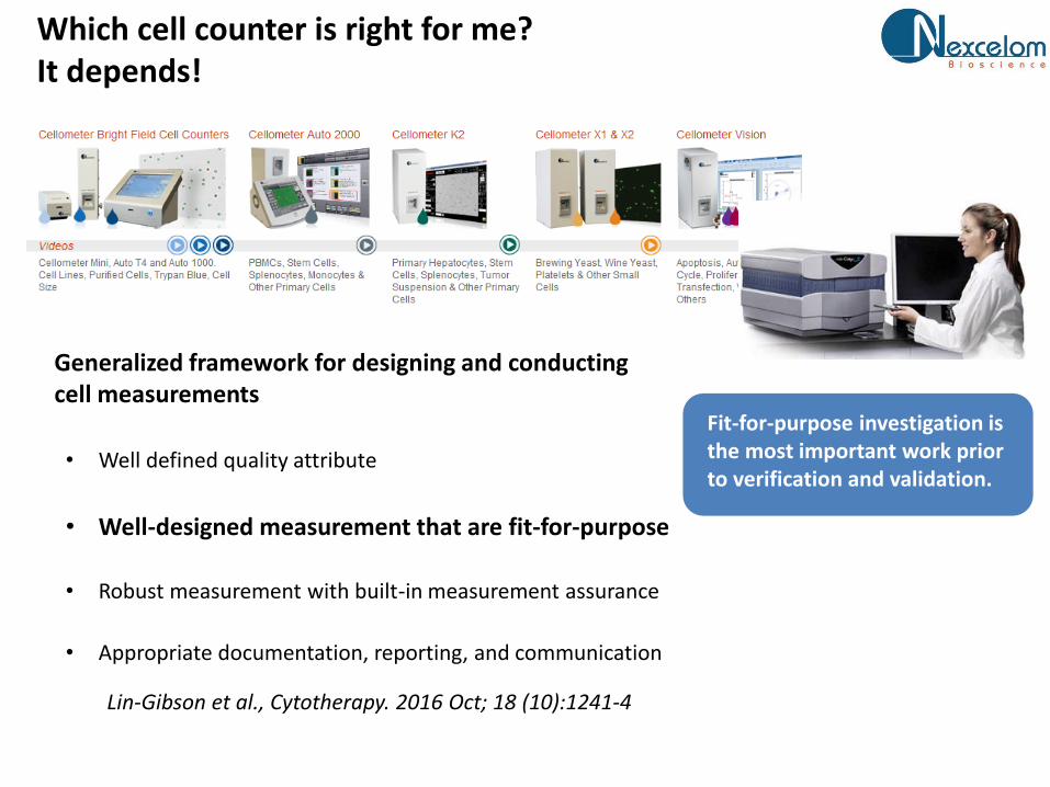
Which cell counter is right for me?It depends!
Generalized framework for designing and conducting cell measurements
• Well defined quality attribute
• Well-designed measurement that are fit-for-purpose
• Robust measurement with built-in measurement assurance
• Appropriate documentation, reporting, and communication
Lin-Gibson et al., Cytotherapy. 2016 Oct; 18 (10):1241-4
Fit-for-purpose investigation is the most important work prior to verification and validation.
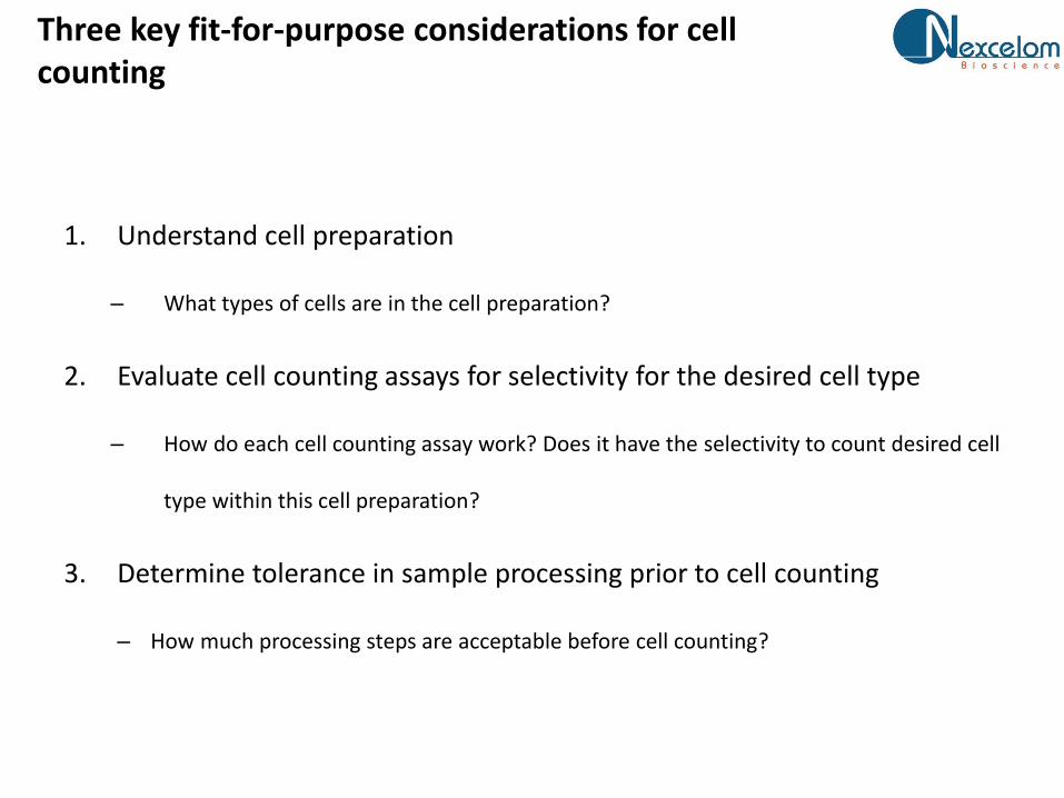
Three key fit-for-purpose considerations for cell counting
1. Understand cell preparation
– What types of cells are in the cell preparation?
2. Evaluate cell counting assays for selectivity for the desired cell type
– How do each cell counting assay work? Does it have the selectivity to count desired cell
type within this cell preparation?
3. Determine tolerance in sample processing prior to cell counting
– How much processing steps are acceptable before cell counting?
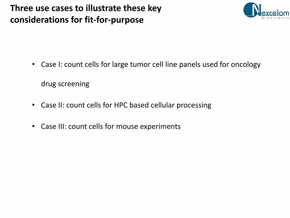
Three use cases to illustrate these key considerations for fit-for-purpose
• Case I: count cells for large tumor cell line panels used for oncology
drug screening
• Case II: count cells for HPC based cellular processing
• Case III: count cells for mouse experiments

A few definitions
• Biological operation
– A series of experiments to produce results
• Cell preparation
– Cell sample to be analyzed for cell count
• Cell counting assay
– The specific measurement to obtain cell counts
• Cell counting system
– Hardware, software, reagents to produce cell counting results
A laboratory conducts multiple biological operations with varied cell counting needs
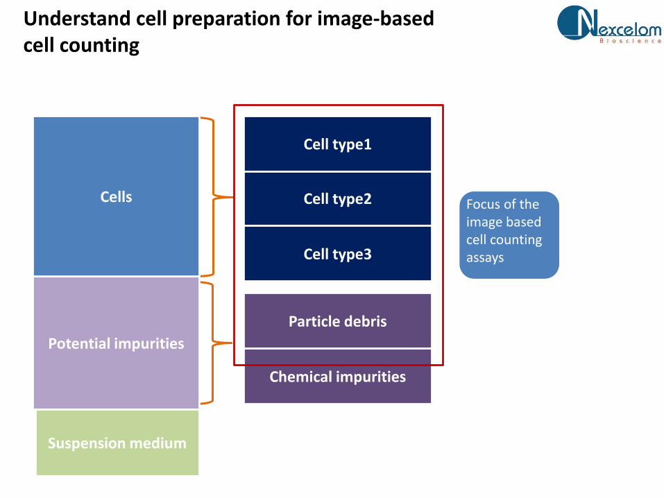
Understand cell preparation for image-based cell counting
Potential impurities
Suspension medium
Cells Cell type2
Cell type1
Cell type3
Particle debris
Chemical impurities
Focus of the image based cell counting assays
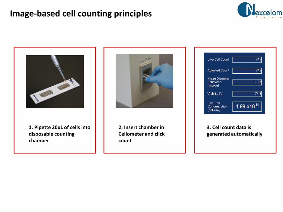
Image-based cell counting principles
1. Pipette 20uL of cells into disposable counting chamber
2. Insert chamber in Cellometer and click count
3. Cell count data is generated automatically

Cell counting assay using bright-field image
Most of the time you see this on a product literature
Real life can be messy

FL staining can be more specific for some cell types - cadiomyocytes
AO stains all cells
PI stains dead cells
BF is hard to quantify
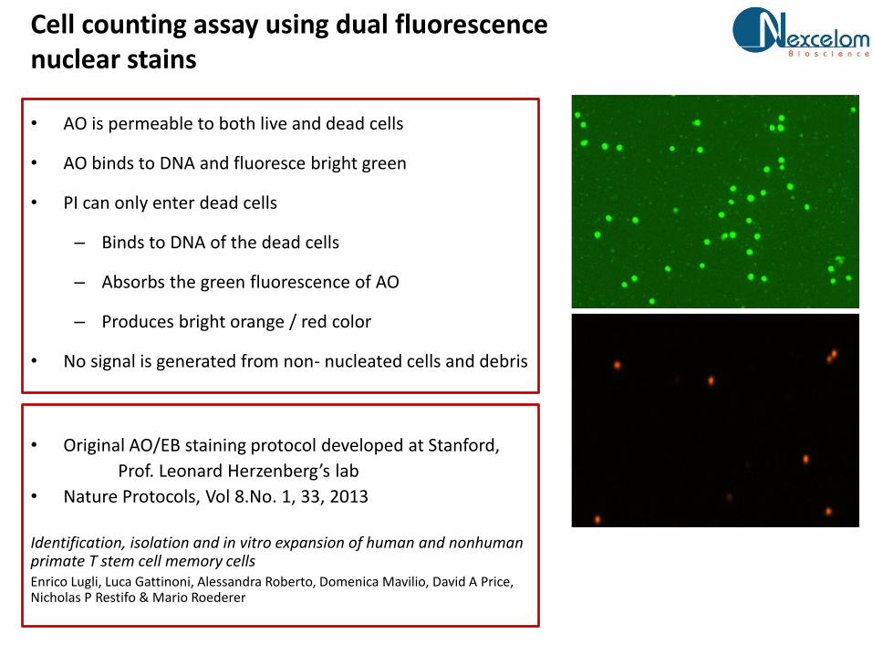
Cell counting assay using dual fluorescence nuclear stains
• AO is permeable to both live and dead cells
• AO binds to DNA and fluoresce bright green
• PI can only enter dead cells
– Binds to DNA of the dead cells
– Absorbs the green fluorescence of AO
– Produces bright orange / red color
• No signal is generated from non- nucleated cells and debris
• Original AO/EB staining protocol developed at Stanford,
Prof. Leonard Herzenberg’s lab
• Nature Protocols, Vol 8.No. 1, 33, 2013
Identification, isolation and in vitro expansion of human and nonhuman primate T stem cell memory cellsEnrico Lugli, Luca Gattinoni, Alessandra Roberto, Domenica Mavilio, David A Price, Nicholas P Restifo & Mario Roederer

Cell counting fit-for-purpose case I: oncology screening projects using
human cell line panels
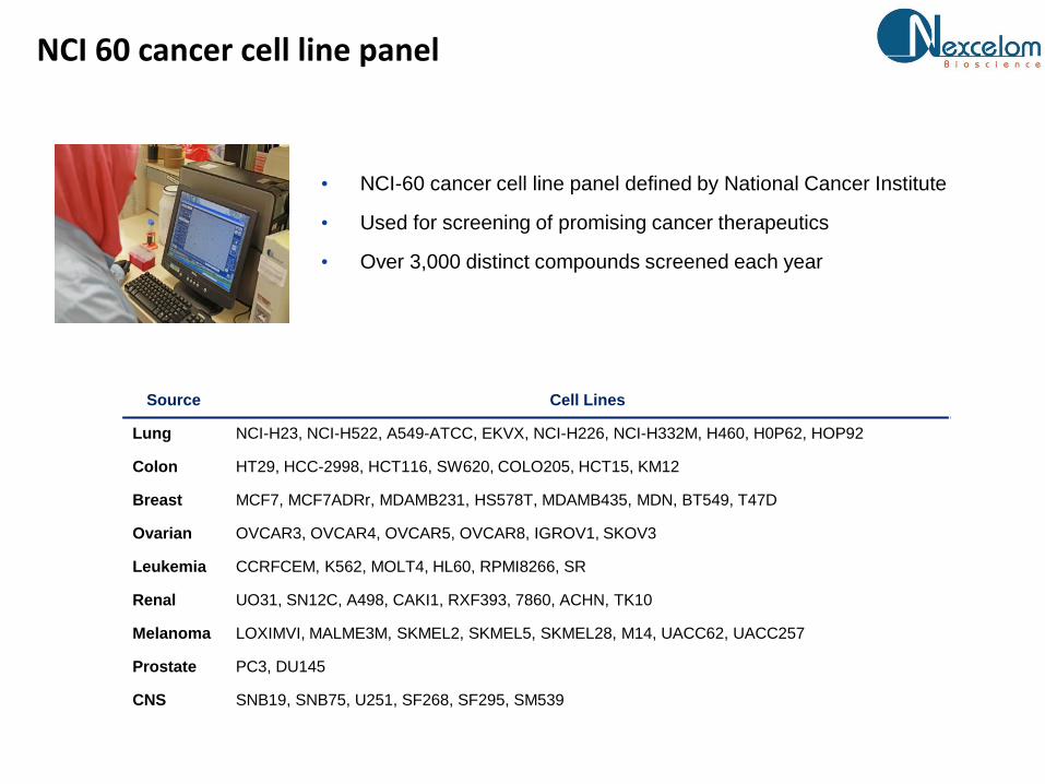
NCI 60 cancer cell line panel
13
• NCI-60 cancer cell line panel defined by National Cancer Institute
• Used for screening of promising cancer therapeutics
• Over 3,000 distinct compounds screened each year
Source Cell Lines
Lung NCI-H23, NCI-H522, A549-ATCC, EKVX, NCI-H226, NCI-H332M, H460, H0P62, HOP92
Colon HT29, HCC-2998, HCT116, SW620, COLO205, HCT15, KM12
Breast MCF7, MCF7ADRr, MDAMB231, HS578T, MDAMB435, MDN, BT549, T47D
Ovarian OVCAR3, OVCAR4, OVCAR5, OVCAR8, IGROV1, SKOV3
Leukemia CCRFCEM, K562, MOLT4, HL60, RPMI8266, SR
Renal UO31, SN12C, A498, CAKI1, RXF393, 7860, ACHN, TK10
Melanoma LOXIMVI, MALME3M, SKMEL2, SKMEL5, SKMEL28, M14, UACC62, UACC257
Prostate PC3, DU145
CNS SNB19, SNB75, U251, SF268, SF295, SM539

Much larger cell line panels
• Novartis /Broad (CCEL) - 1,000 cell types• MGH/Sanger – more than 1,600 cell types

Purpose of the cell counting
• Count cells for cell line expansion
• Count cells to seed assay plates (1536w, 384w, 96w)

What do cell preparations look like for NCI-60 cell lines?
With very large variations
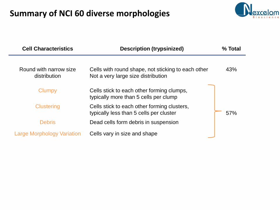
Summary of NCI 60 diverse morphologies
Cell Characteristics Description (trypsinized) % Total
Round with narrow size
distribution
Cells with round shape, not sticking to each other
Not a very large size distribution
43%
Clumpy Cells stick to each other forming clumps,
typically more than 5 cells per clump
57%
Clustering Cells stick to each other forming clusters,
typically less than 5 cells per cluster
Debris Dead cells form debris in suspension
Large Morphology Variation Cells vary in size and shape
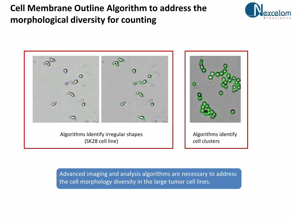
Cell Membrane Outline Algorithm to address the morphological diversity for counting
Advanced imaging and analysis algorithms are necessary to address the cell morphology diversity in the large tumor cell lines.
Algorithms Identify irregular shapes(SK28 cell line)
Algorithms identify cell clusters
Case I summary
• Each cell preparation contains only one cell type
• Cell preparation is generally in heathy conditions with viability higher than
90%
• Bright-field based cell counting assay can be used
• Special image analysis capability is required for devise morphologies
Cell counting fit-for-purpose case II: cell therapy bio-processing
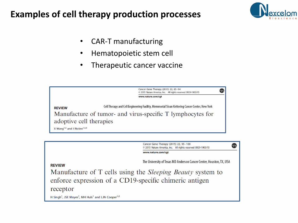
Examples of cell therapy production processes
• CAR-T manufacturing
• Hematopoietic stem cell
• Therapeutic cancer vaccine

Purpose of the cell counting
• Count cells from starting material, in-processing material and the final
products

Source of the starting material
PBMC by Ficoll separation Leukopak from leukophoresis
Other source materials: bone marrow, cord blood
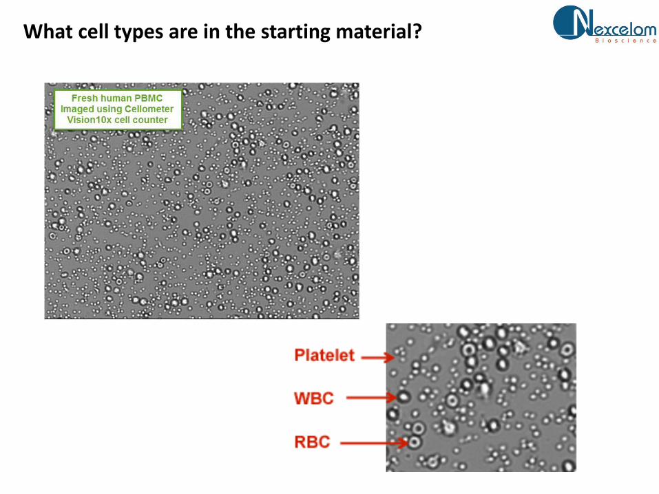
What cell types are in the starting material?
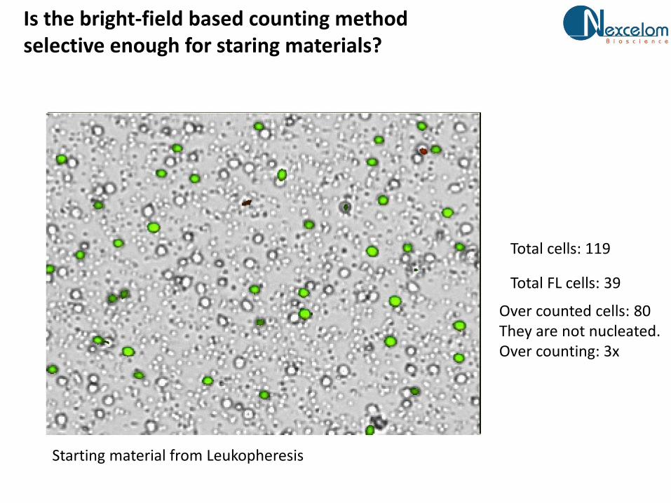
Is the bright-field based counting method selective enough for staring materials?
Total cells: 119
Total FL cells: 39
Over counted cells: 80They are not nucleated.Over counting: 3x
Starting material from Leukopheresis

Multiple Leuokopheresis samples shown varied amount of RBC
• Manual counting using Hemacytometer and trypan blue
• Automatic cell counting using Cellometer Vision and AO/PI stains
• Experiment using Leukopheresis samples
Sample ID A B C D E F
Ratio(Manual counting to Cellometer AO/PI) 7 5 12 3 7 5
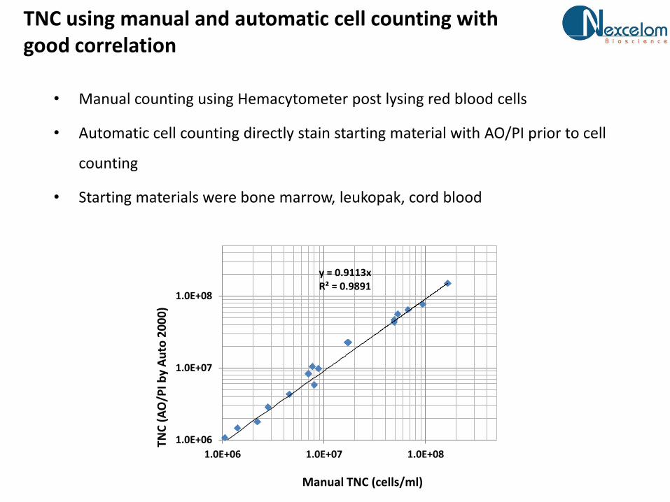
TNC using manual and automatic cell counting with good correlation
y = 0.9113xR² = 0.9891
1.0E+06
1.0E+07
1.0E+08
1.0E+06 1.0E+07 1.0E+08
TNC
(A
O/P
I by
Au
to 2
00
0)
Manual TNC (cells/ml)
• Manual counting using Hemacytometer post lysing red blood cells
• Automatic cell counting directly stain starting material with AO/PI prior to cell
counting
• Starting materials were bone marrow, leukopak, cord blood

Select a cell counting system with multiple cell counting assays
Source materials:
PB, BM, CB
In processing:
MNC
Finished product:
CD34+ cells
Bright-field image
Fluorescentimage
AO/PIAO/PI Trypan blue
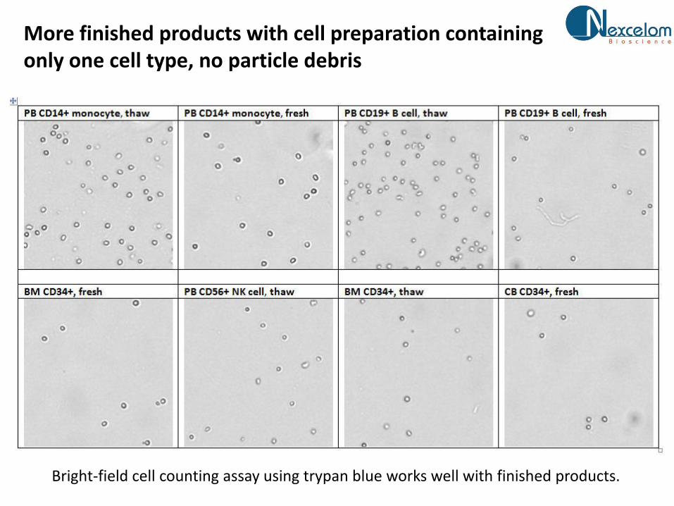
More finished products with cell preparation containing only one cell type, no particle debris
Bright-field cell counting assay using trypan blue works well with finished products.
Case II summary
• Cell preparation changes though cell process
• Without lysing, fluorescence staining method is more selective
• Bright-field based cell counting assay can be used for finished product
when cell preparation is
• Cell counting system provides multiple assays are essential to satisfy
varied cell preparation conditions

Cell counting fit-for-purpose case III: primary cells from mouse models

Example cell types and assays from mouse models
• Tissues
– Blood, Spleen, BAL, Lung
– Axillary LN, Mesenteric LN, Inguinal LN, Obturator LN, Iliac (Internal) LN,
Cervical LN, Mesenteric LN , Bronchial LN
– Primary bronchi, Tonsils, Nares Mucosa, Trachea, Rectum, Jejunum, Cervix,
Vagina, Uterus, Ileac
• Cells are used for:
– FACs Surface and Intracellular Staining
– Elispots
– qPCR

Cell preparation evaluation
• The operation contains multiple cell preparations
• All cell preparation contains multiple cells types
• Most cell preparation contains particular debris from tissue digestion
process
• The amount of cells vary from one animal to another animal
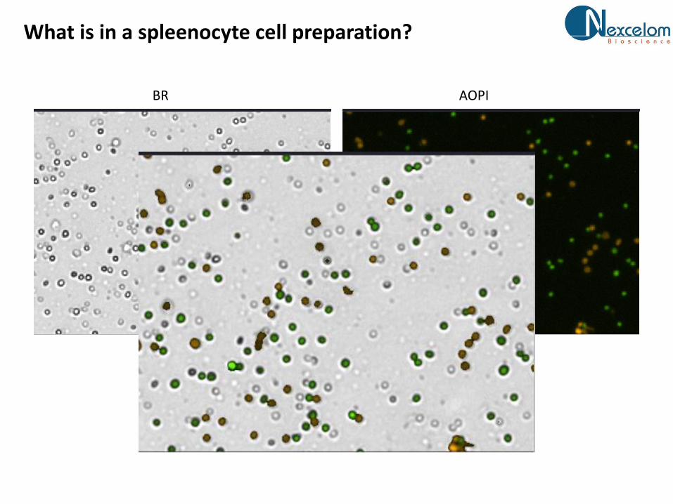
What is in a spleenocyte cell preparation?
BR AOPI
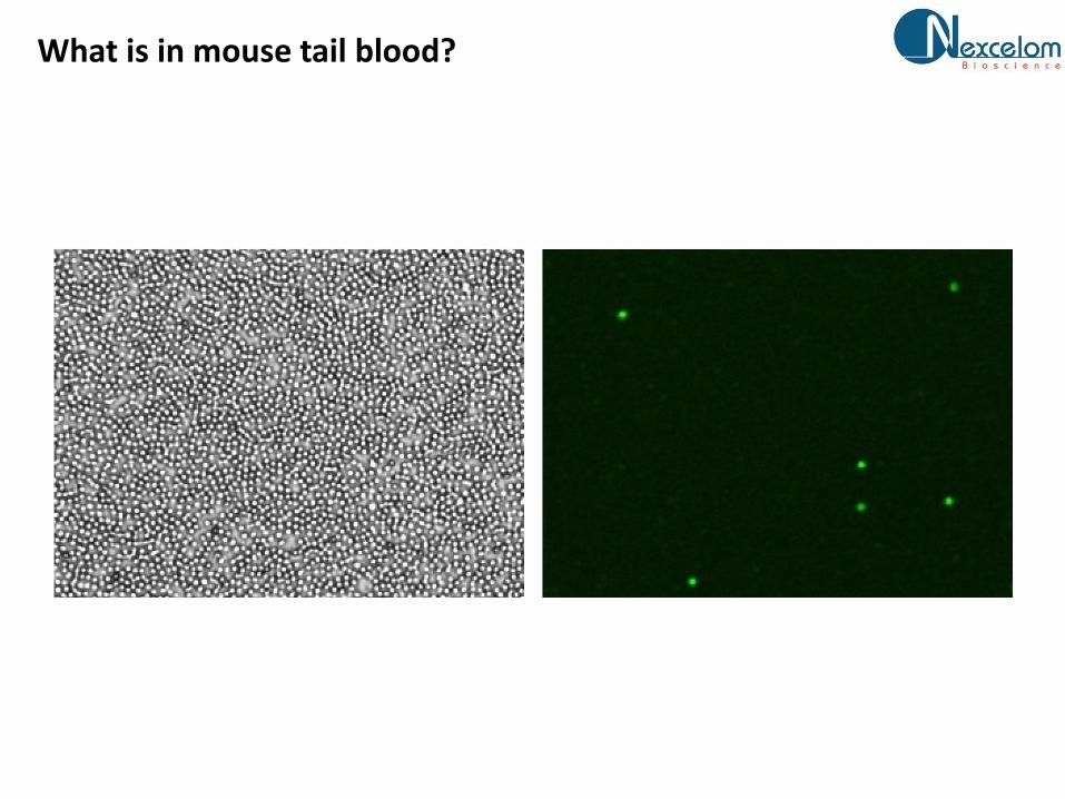
What is in mouse tail blood?
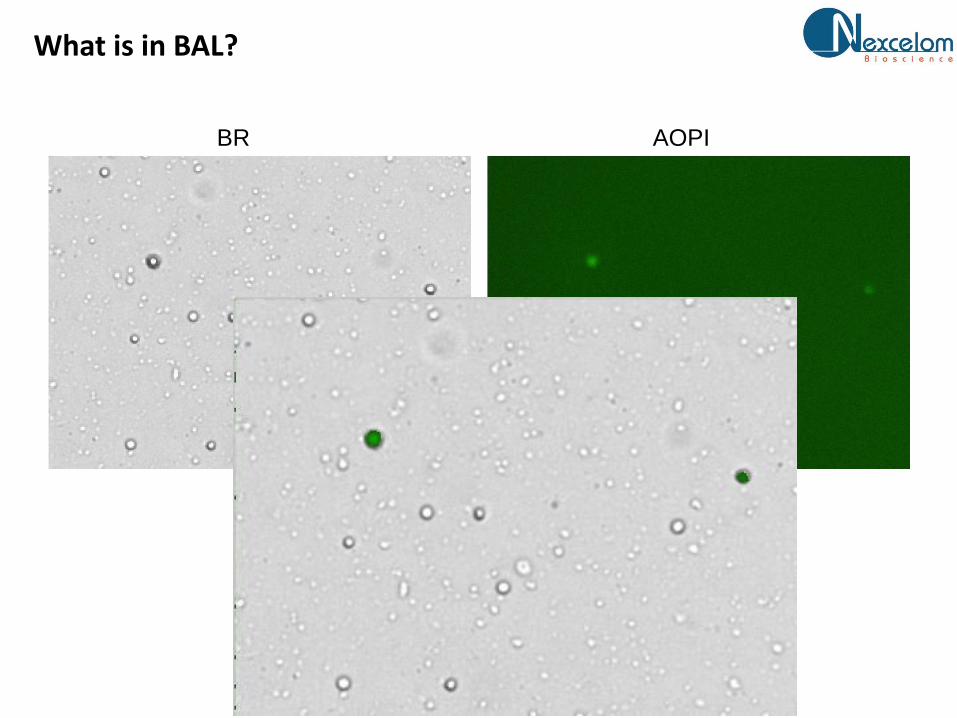
What is in BAL?
BR AOPI
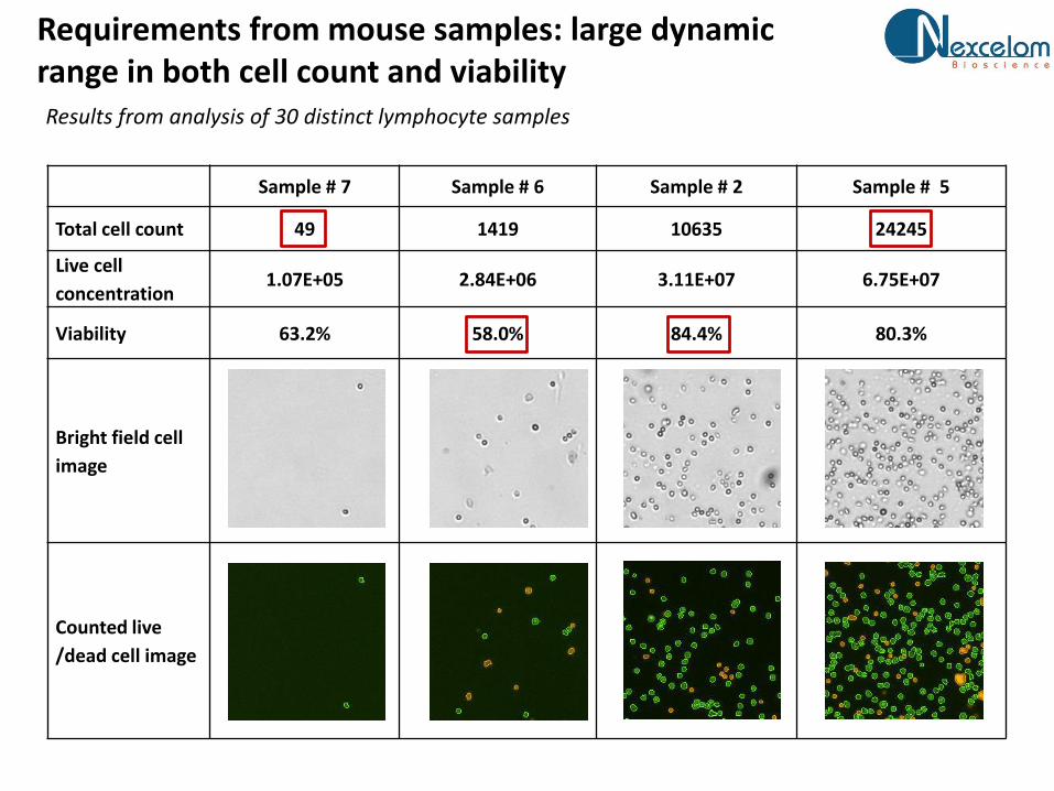
Requirements from mouse samples: large dynamic range in both cell count and viabilityResults from analysis of 30 distinct lymphocyte samples
Sample # 7 Sample # 6 Sample # 2 Sample # 5
Total cell count 49 1419 10635 24245
Live cell
concentration1.07E+05 2.84E+06 3.11E+07 6.75E+07
Viability 63.2% 58.0% 84.4% 80.3%
Bright field cell
image
Counted live
/dead cell image

Case III summary
• No tolerance in lysing and other cell processing steps prior to counting –
large number of samples per run
• Large variation in the amount and type of particle debris due to tissue
source and dissociation processes
• Cell concentration and viability vary in several order of magnitude
• Bright-field based cell counting assay cannot be used
• Dual FL stain provided good cell counting assay

Q&A
Jean [email protected]
Three key fit-for-purpose considerations for cell counting using imaging cytometry
1. Understand cell preparation
2. Evaluate cell counting assays for selectivity
3. Determine tolerance in sample processing