Embed Size (px)
Citation preview
Pak. J. Bot., 51(1): 341-360, 2019. DOI: 10.30848/PJB2019-1(15)
FIRST REPORT OF DYE YIELDING POTENTIAL AND COMPOUNDS OF LICHENS; A
CULTURAL HERITAGE OF HIMALAYAN COMMUNITIES, PAKISTAN
SUMMAIRA SHAHEEN, ZAFAR IQBAL AND MANZOOR HUSSAIN
Department of Botany, Hazara University Mansehra-21300, Khyber Pakhtunkhwa, Pakistan. *Corresponding author’s email: [email protected]
Abstract
Lichens are well-known dye yielding organisms since ancient times. The present study investigates the dye yielding
potential of nineteen lichen species belonging to eleven genera (Flavopunctelia, Flavoparmelia, Cladonia, Parmelia,
Umbilicaria, Xanthoria, Ochrolechia, Hyperphyscia, Hypogymnia, Dermatocarpon and Parmotrema) of Himalayan region
(Abbottabad) Pakistan. Wool and silk were dyed using the 3 different methods i.e. dimethyl sulphoxide (DEM), ammonia
fermentation (AFM) and boiling water (BWM). Over 57 different dye tests were made on silk. Predominant color was cerise
but yellow, brown, purple, green, pink and olive were produced. COSMIN software was used to detect HEX Colour codes with
RBG and HSL values. These dye colors were further altered by modifying: exposure to light, temperature and subsequent
additional extractions using the different method or the same one. After dying samples were tested for stability in sunlight and
the action of soap, some samples were faded to some degree and some of them changed color. Most dyes obtained through the
AFM and DEM method were stable while dyes from boiling water method were light stable. A correlation of dye color with
lichen secondary metabolites was also attempted. Spot test results showed the presence of different lichen substances
(gyrophoric, lecanoric acid, umbilicaric acids, usnic acid, atranorin, chloroatranorin, salazinic acid and parietin).
Key words: Lichens, Extraction, Secondary metabolites, Dye, Cultural heritage, Himalayas.
Introduction
Lichens are beautiful organisms; once one starts
looking for them he may be surprised at the diversity and quantity. These are symbiotic organisms composed up of a phycobiont and a photobiont (usually an alga) together form an independent and unique physiological unit. They are growing in terrestrial habitat on wood, leaves, rocks, soil and other fixture (Shah, 2011). They show slow growth in harsh living conditions that’s why they are able to produce a variety of secondary metabolites (Abdullahi, 2009; Santiago et al., 2010; Marijana & Rankovic, 2010; Krystle et al., 2010), which are believed to serve as anti-herbivore, antimicrobial and antigrowth agents (Gupta et al., 2007; Manojlovic et al., 2005). Besides these properties, these unique entities have an inherent ability to produce beautiful dye colors. A dye can be defined as a coloring substance that has an affinity to the applied substrate. The dye generally requires a mordant to improve the fastness of the fiber dye. The secondary metabolites known as “lichen acids” are the main source in lichen dyes (Veranja et al., 2005; Richardson, 1988a).
Natural dyes are usually derived from plants, invertebrates and minerals source, most of them are from plant and other organic sources such as fungi and lichens. Lichen dyes have a particular affinity for natural fibers (silk and cotton), other products can also be dyed with lichens such as leather, marble, wood, wine and food materials as well. Lichens are well-known organisms and have a long dye yielding history (Diadick, 2001). First documented dye produced by Roccella spp. was Orchil dye (purple color) through ammonia fermentation method (Margareta, 1981). Many dye colors are converted through the extraction process, not visible in the fresh lichen thallus. Indigenous knowledge, particularly associated with extraction of dyes from lichens is ancient (Shukla & Upreti 2015). Cow urine method (CUM) mostly used in ancient times, then ammonia fermentation method (AFM), dimethyl sulphoxide extraction method (DEM) and boiling water method (BWM) are most
familiar extraction methods for lichen dyes. Mostly purple, pink, yellow, brown, orange, and green dyes can be extracted from lichens. A list of more than hundred dye yielding lichen species was given by Casselman (2001).
After the discovery of first synthetic dye in 1856 natural dyes colors were completely replaced by synthetic dyes due to easy extraction and cost-effectiveness (Margareta, 1981). Synthetic dyes have immense detrimental environmental impacts due to their non-biodegradable nature and noxious effects. The demand of dyes for textile, food and cosmetics industries from natural sources has increased in the recent years due to the high rate of pollution level (Bolton, 1960). Recently several attempts are made for the development of environment and users friendly pigments mostly from the natural sources.
Lichens contain characteristic compounds known as depsides and depsidones that are made up of two/three phenolic units derived from the acetate-polymelonate pathway by a fungal partner (Asahina & Shubata 1954). Depsides were first discovered in the early part of the 20th century; are small molecules consisting of a series of linked phenol carboxylic acids esters. They are derived from Ordellinic acid (Asahina & Shibata 1954). Both compounds are the main source of dye which can color natural fibers (Richardson 1988b).
Pakistan is a country with different vegetation zones (Collinson, 1977), with high mountains in the north (Himalayas, Hindukush, Karakorum), subtropical Indus valley (dominated by arable land), deserts in Baluchistan and Thar Desert in the east. Consequently, the lichen biota is probably very rich and varied. However, unfortunately the lichen biota of Pakistan is so far largely unknown, especially with regard to different properties of lichens e.g. antimicrobial activity, ethno lichenology, dye yielding potential etc. There are 20,000 lichens species described all over the world, and Pakistan represents 367 (0.5%) of known lichens (Aptroot & Iqbal, 2012). The Himalayan region of Pakistan has rich lichens biota, including of large number of parmelioid lichens species that provide
SUMMAIRA SHAHEEN ET AL., 342
excellent source of lichens dyes (Upreti et al., 2010). The dye yielding properties of Pakistani lichens are not known until now. Thus in the present study an attempt has been made to screen out the most common and abundantly growing Himalayan lichens for their potential of dye yielding properties and as well as their substances.
Materials and Methods
Collection and identification of lichen sample
Collection lichen sample: In the present study, the
lichens samples collection was performed in (34°16"88 N,
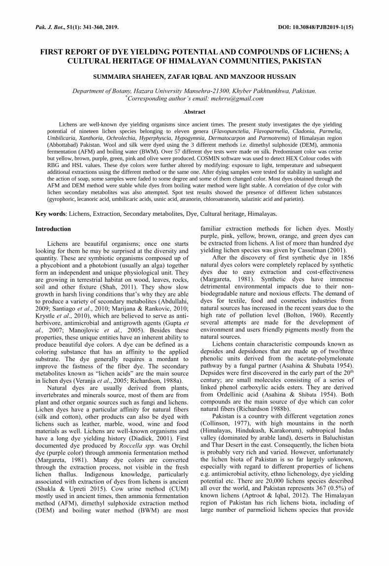
73°22"15 E), Abbottabad District, Pakistan (Fig. 1a).
Abbottabad, with a geographical area of about 1,969km2
is situated between 34.1304° North latitudes and 73.1822°
E. East longitudes and 1,260 meters (4,134 ft.) altitude.
Few species of Lichens were reported from this district by
Aptroot & Iqbal (2012), however most areas of the
Abbottabad remained unexplored especially with respect
to dye yielding potential of lichens. To bridge this gap, the
studies on lichens of Abbottabad initiated in the year
2015. The lichens material was processed immediately in
the Botany Department, Hazara University Mansehra
Pakistan, to reduce the chance of contamination. Samples
were carefully collected; dust, soil, and rock debris were
removed, shade dried to a constant weight (dry weight)
and were kept at room temperature until extraction in the
sterile Petri plate.
Fig. 1a. Map of district Abbottabad.
http://103.240.220.71/btt/repos/files/2015/12/District-Abbottabad-A4.jpg
PAKISTANI HIMALAYAN LICHEN'S DYE 343
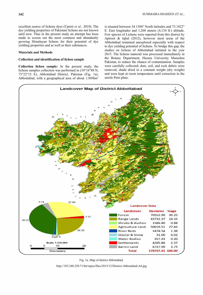
Identification of lichen samples: Lichen specimens were
collected from Mushakpuri, Nathia Gali, Namli Maira,
Chairsajikot, Bagnotar, Chamnaka, Muslimabad, Harnoi,
Banda Sapan, Salhad, Dhamtour, Taqia, Bodla, Chamhad
and Havelian village areas (Fig. 1b & Table 5) of District
Abbottabad during 2015. The samples collected were
Flavoparmelia caperata, Dermatocarpon miniatum,
Flavopunctelia soredica, Hyperphyscia adglutinata,
Hypogymnia physodes, Parmelia neodiscordans,
Menegazzia terebrata, Parmelia saxatilis, Parmelia
sulcate, Parmotrema reticulatum, Parmotrema tinctorum,
Cladonia arbuscular, Cladonia furcata, Umbilicaria
mammulata, Umbilicaria polyphylla, Umbilicaria vellea,
Xanthoria elegans, Xanthoria parietina and Ochrolechia
turneri. The identification was done by morphology using
relevant keys and monographs (Smith, 1918; Chopra, 1934;
Culberson & Kristinsson, 1970; Culberson, 1972; Shyam et
al., 2010; Aptroot & Iqbal, 2012; Tucker, 2014; Awasthi,
1988). Microchemical colour tests were also performed for
identification of their secondary metabolites.
Fig 1b. Map of district Abbottabad showing lichens collection areas.
Spot tests: Colour tests were performed by chemical
reagents; applying on the thallus, resulting change in
colour. No change in colour was denoted by a negative (-)
symbol and positive change is denoted by a positive (+)
symbol followed by the colour produced. The chemical
reagents used are as follows.
K test (Potassium): 10-25% aqueous solution of
potassium hydroxide was applied to the thallus.
C test (Calcium hypochlorite): A freshly prepared
aqueous solution of calcium hypochlorite or bleaching
powder, it was prepared by dissolving calcium
hypochlorite in distilled water in 2% ratio.
KC test (Potassium and Calcium hypochlorite): At a
particular spot of the thallus potassium hydroxide was
applied first and immediately followed by calcium
hypochlorite.
Pd test (Paraphenylenediamine): Solution of paraphenylenediamine was prepared in ethanol in a small quantity for the use of a single day because it was unstable and could not be used for the next day. A more stable solution called Steiner’s PD solution was prepared by dissolving 1 gm of paraphenylenediamine and 10 gm of sodium sulfite in 100 ml of distilled water with 1 ml of a liquid detergent.
I test (Iodide): Three gm of iodine was dissolved in water with 0.5 gm of potassium iodide.
These chemicals were applied to cortex and medulla of lichen thallus (Table 2).
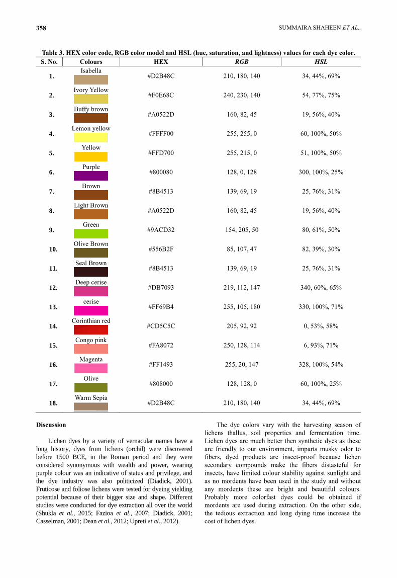
Extraction of dyes from lichen samples: In history traditionally cow urine method (CUM) was employed for extraction of dyes then replaced with ammonia fermentation method (AFM) and later boiling water method (BWM) was introduced. In addition to the traditional methods, Dimethylsulphoxide Extraction Method (DEM) was used for extraction of lichen dyes. In this study the dyes were extracted with ammonia fermentation (AFM), boiling water (BWM) and Di-methyl sulphoxide extraction method (DEM).Lichen samples were segregated, cleaned off substratum, thoroughly washed and dried. Dried samples of lichens were crushed, powdered then weighted and used for extraction of dyes and dying experiments were carried out. Tussar silk fibers were obtained from local market. Fibers samples were weighted and thoroughly washed with distilled water before they were used for dying so that the dye penetrate and fix well into the fibers. Equal weights of dry lichens and silk fibers were used. Both dye extractions and dying were done in 250 ml flask at room temperature except BWM. No mordant was used because lichen dyes were unique in that they did not require any mordant. Three dying methods used were:
Ammonia fermentation method (AFM): Four grams of lichens were added to diluted ammonium hydroxide solution (1:10) thoroughly mixed and left for a month in a flask. Then the extract was filtered by using Whatman filter paper. Four grams of silk were added. After one month threads were removed, dried and the colour was noted.
Boiling water method (BWM): Four grams of powdered lichen was added to water and heated till boiling and filtered into a flask. The fibers were then immersed in a dye bath containing filtrate and were heated at 90°C for two hours. After cooling dye bath threads were removed, rinsed in water, dried and color was noted.
Di-methylsulphoxide Extraction Method (DEM): Four grams of crushed lichen was added to 50ml of Dimethyl sulphoxide solution in a flask. The content was stirred vigorously and maintained at room temperature for one month. After one month extract was filtered and threads were added for dying. After one-month threads were removed, washed in cold water, dried and colors were noted.
Stability of dyes against the light was tested following Sharma & Grover (2011). Dyed fibers were exposed to sunlight for 8 hours/day for a week. The fibers were also washed with detergent to observe the fastness of color. The colors were named with those matching Ridgway colors undyed colored fibers were used as control (Ridgway, 1912). COSMIN software was used to detect HEX Colour codes with RBG and HSL values.
SUMMAIRA SHAHEEN ET AL., 344
Results
Although The Himalayan lichen biota of Pakistan is
not fully explored no doubt it has a rich diversity of foliose
lichens that are a potential source of natural dyes which can
provide brilliant colors in different solvents. These can be
used as a source for making dyes due to their unique
chemistry and abundant biomass. A variety of colour dyes
like yellow, brown, purple, green, pink and olive were
obtained from lichens Fig. 2(a-s) in this study. All the three
methods employed in this study have given different colors
to the fibers. All the lichens were tested for dying tussar
silk fiber. A bright and attractive color by at least one of the
methods employed was obtained. Tussar silk fiber is used
for dying tests, as it had buff color, so after dying the fiber
appeared different from white silk and cotton fibers. Out of
three methods employed dye colors extracted from
Ammonia fermentation method (AFM) were much brighter
as compared to boiling water method (BWM) and
Dimethylsulphoxide Extraction Method (DEM) (Fig. 3).
COSMIN software was used to detect HEX Colour codes
with RBG and HSL values (Table 3).
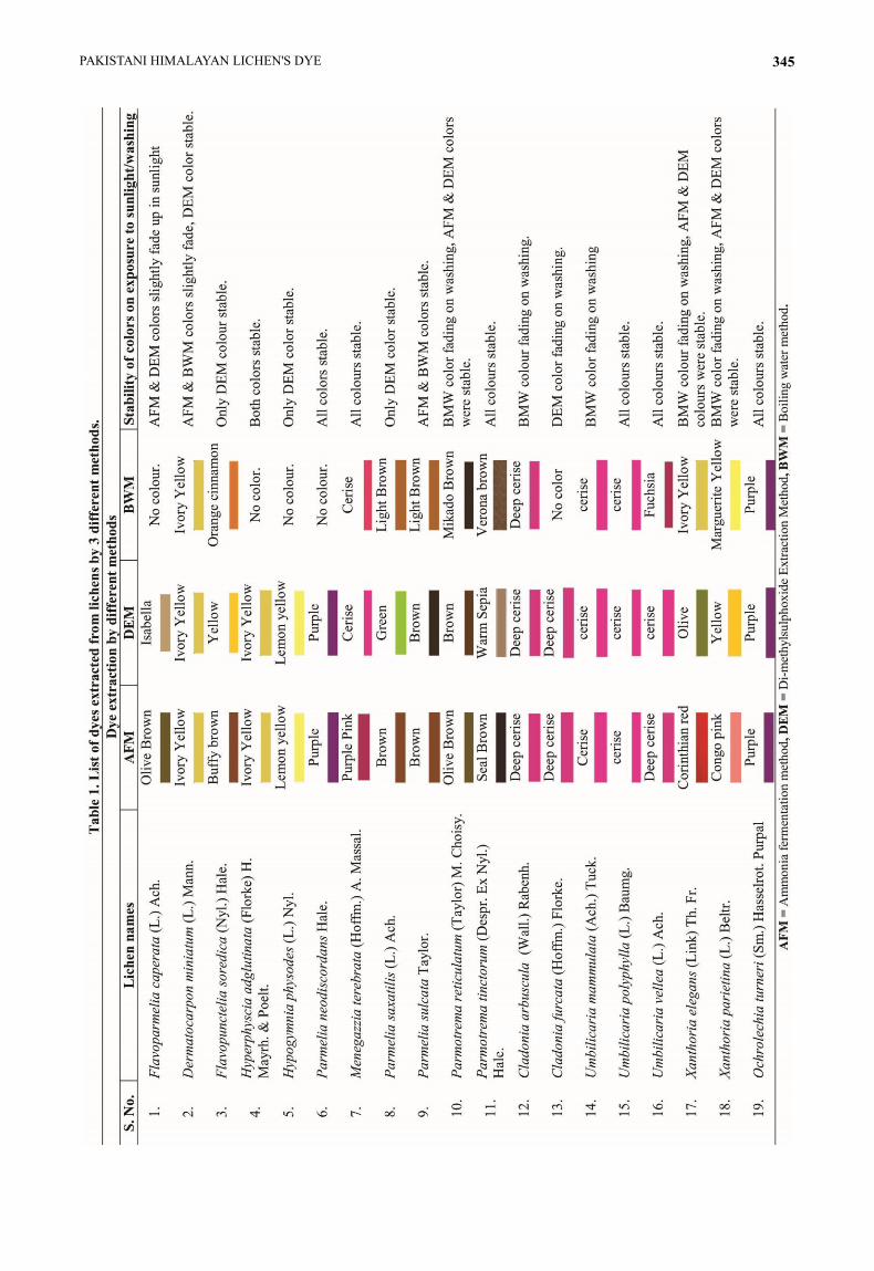
Colour dyes obtained from AFM ranged from Olive
Brown, Yellow, Buffy brown, Ivory Yellow, Lemon
yellow, Purple, Purple Pink, Brown, Seal Brown, Deep
cerise, cerise, red, Pink and Magenta, whereas DEM
produced Isabella, Ivory Yellow, Purple, Cerise, Green,
Brown, Deep cerise, Olive and Magenta. Yellow, Orange,
Cerise, Brown, Deep cerise, Cerise and Fuchsia obtained
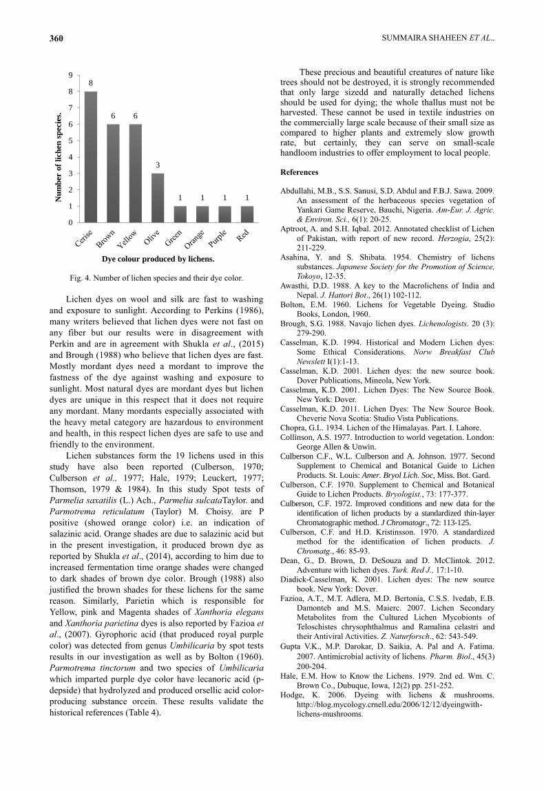
through BWM (Table 1). Out of nineteen lichens selected
for dye extraction 6 lichen species produced Brown
shades, 6 Yellow, 2 Purple, 1 red, 1 Green, 3 Olive, 1
Orange and 8 cerise color shades (Fig. 4).
Colour fastness: On exposure to sunlight/washing dyes
differed in the stability of colours. Parmelia neodiscordans
Hale., Menegazzia terebrata (Hoffm.) A. Massal.,
Parmotrema tinctorum (Despr. ex Nyl.) Hale., Umbilicaria
polyphylla (L.) Baumg., Umbilicaria vellea (L.) Ach. and
Ochrolechia turneri (Sm.) Hasselrot.showed more stable
colours as compared to others. 15 AFM results gave stable
colours while in case of DEM there were 14 stable colour
results. BWM showed less effective results as compared to
the other two methods. The change in colour is due to photo-
oxidation chromophores (colour producing structure).
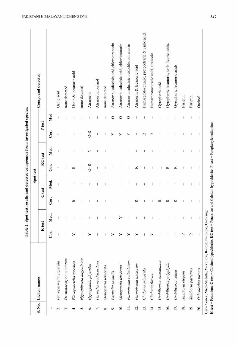
Correlation of dye colour with lichen secondary
metabolites: We tried to find a correlation between the
dye colours with the secondary metabolites of lichen
species. Spot tests results were not only used for
identification of lichens but also used for detection of
major secondary metabolites (Table 2). Spot tests of
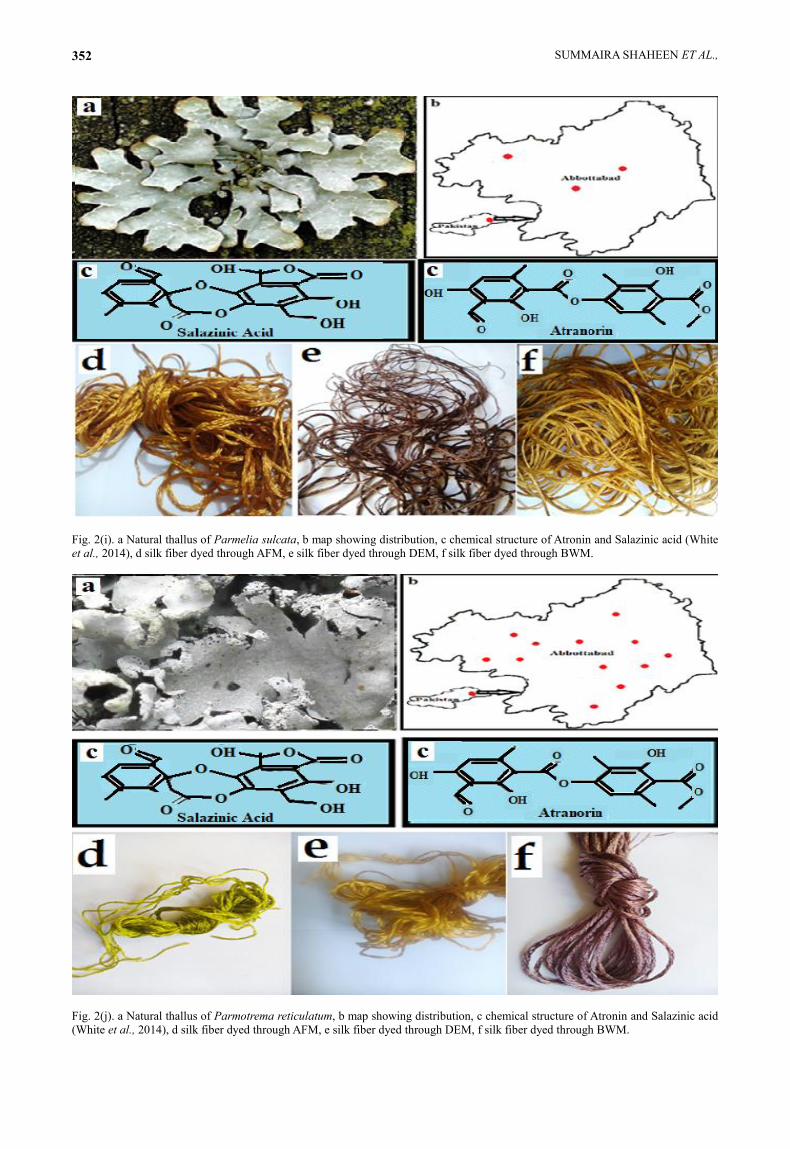
Parmelia saxatilis (L.) Ach., Parmelia sulcataTaylor and
Parmotrema reticulatum (Taylor) M. Choisy indicated the
presence of salazinic acid due which these lichens
produced shades of brown colour. Lichens containing
only salazinic acid were responsible for orange and brown
dyes while both salazinic acid and atranorin produced
yellow colour. Parietin is responsible for Yellow, pink and
Magenta shades of Xanthoria elegans and Xanthoria
parietina dyes while atranorin produced yellow colour in
Hypogymnia physodes (L.) Nyl. Orange colour dye of
Flavoparmelia caperata (L.) Ach. is due to usnic acid
while gray colour might be due to ceparatic acid. Usnic
acid and lecanoric acid caused orange shades in
Flavopunctelia soredica (Nyl.) Hale dye. Parmotrema
tinctorum (Despr. ex Nyl.) Hale. imparted orange dye to
fibers because it had both atranorin which caused yellow
and lecanoric acid i.e. converted to orcein (red). Orange
shades were also reported from Flavopunctelia soredica
(Nyl.) Hale (Shukla et al., 2014). Gyrophoric acid was
detected from genus Umbilicaria that produced royal
purple colour.
Spot test results (Table 2) revealed that most of the
lichen species had lecanoric acid. Lecanoric acid is
actually p-depside that hydrolysis to orsellic acid and
undergoes a series of reactions to form colour producing
chemical i.e. orcein having chemical formula C28H24N2O7.
Orcein is not approved as a food dye. Most of the
secondary metabolites detected in this study had ortho-
hydroxy aldehyde group, which reacted with free amino
acid group and formed stable Schiff base which ultimately
imparted colours to fibers.
Chemistry of Dye producing Lichen’s secondary
metabolites: Lichens produced a variety of secondary
metabolites. Eighty percent of these metabolites are
specifically produced by lichens while 20% are commonly
produced by plants or in higher fungi (Casselman, 2001).
These secondary products of lichens undergo a series of
chemical reactions in the presence of air, water and
solvents to produce colour compounds used for dyeing
purpose. These secondary products in lichens commonly
called lichen acids are of fungal origin. More than thousand
secondary metabolites are known worldwide with lichens
reference (Dean et al., 2012). Majority of these compounds
are phenolic in nature dibenzofuranes (usnic acid), (b-
orcinol derivatives and orcinol), depsidones (salazinic
acid), depsides (barbatic acid), lactones (protolichesterinic
& nephrosterinic acid), depsones (picrolichenic acid),
quinones (parietin) and pulvinic acid derivatives (Upreti et
al., 2010). The depsidones and depsides are aromatic in
nature formed by two or three phenolic units. Lichens have
diverse biosynthetic pathways (shikimic acid, polymalonate
and mevalonic acid pathways) to produce these different
compounds depsides, depsidones and esters are precursors
of orcein (coloured compound) in lichen dyes (Upreti et al.,
2012). These chemicals hydrolyze and are converted into
orsellic acid which further undergoes decarboxylation
reaction to produce orcein. Then after condensation
reactions orcein give rise to various derivatives. Orcein
derivatives in different concentration actually impart dye
colour (Shukla & Upreti 2015). The coloring of any
substrate is due to the chemical reaction between chemical
constituents and Orcein derivatives of dye substrate (Upreti
et al., 2010). The ortho-hydroxyl aldehyde group of lichen
dyes reacts with free amino group of natural protein fibers
and converted into Schiff base (compounds having C=N
function) (White et al., 2014). Orcein as large colorless
crystals can be extracted by ethanol extraction. Orcein
contains a variety of phenazones. Orcein is a mixture of
hydroxy-orecins, amino-orceins and amino-orceinimines
(Veranja et al., 2005).
PAKISTANI HIMALAYAN LICHEN'S DYE 345

SUMMAIRA SHAHEEN ET AL., 346
PAKISTANI HIMALAYAN LICHEN'S DYE 347
SUMMAIRA SHAHEEN ET AL., 348
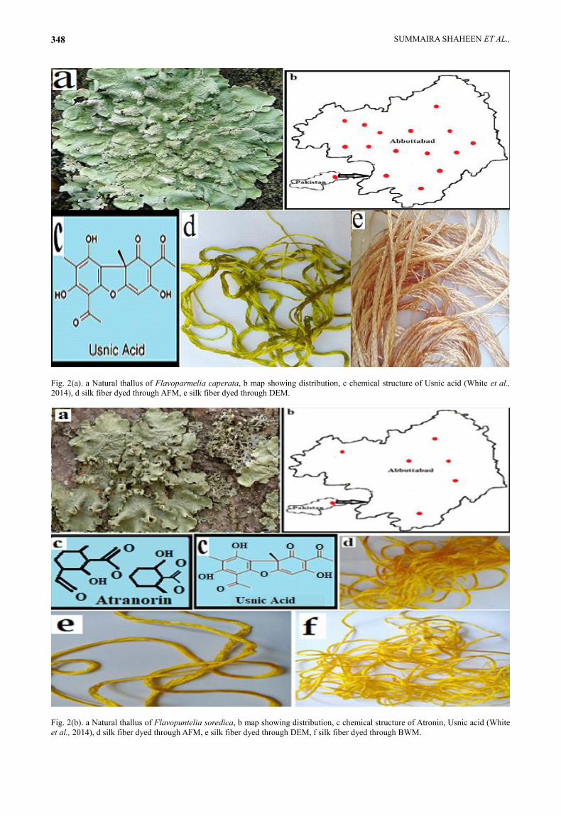
Fig. 2(a). a Natural thallus of Flavoparmelia caperata, b map showing distribution, c chemical structure of Usnic acid (White et al.,
2014), d silk fiber dyed through AFM, e silk fiber dyed through DEM.
Fig. 2(b). a Natural thallus of Flavopuntelia soredica, b map showing distribution, c chemical structure of Atronin, Usnic acid (White
et al., 2014), d silk fiber dyed through AFM, e silk fiber dyed through DEM, f silk fiber dyed through BWM.
PAKISTANI HIMALAYAN LICHEN'S DYE 349
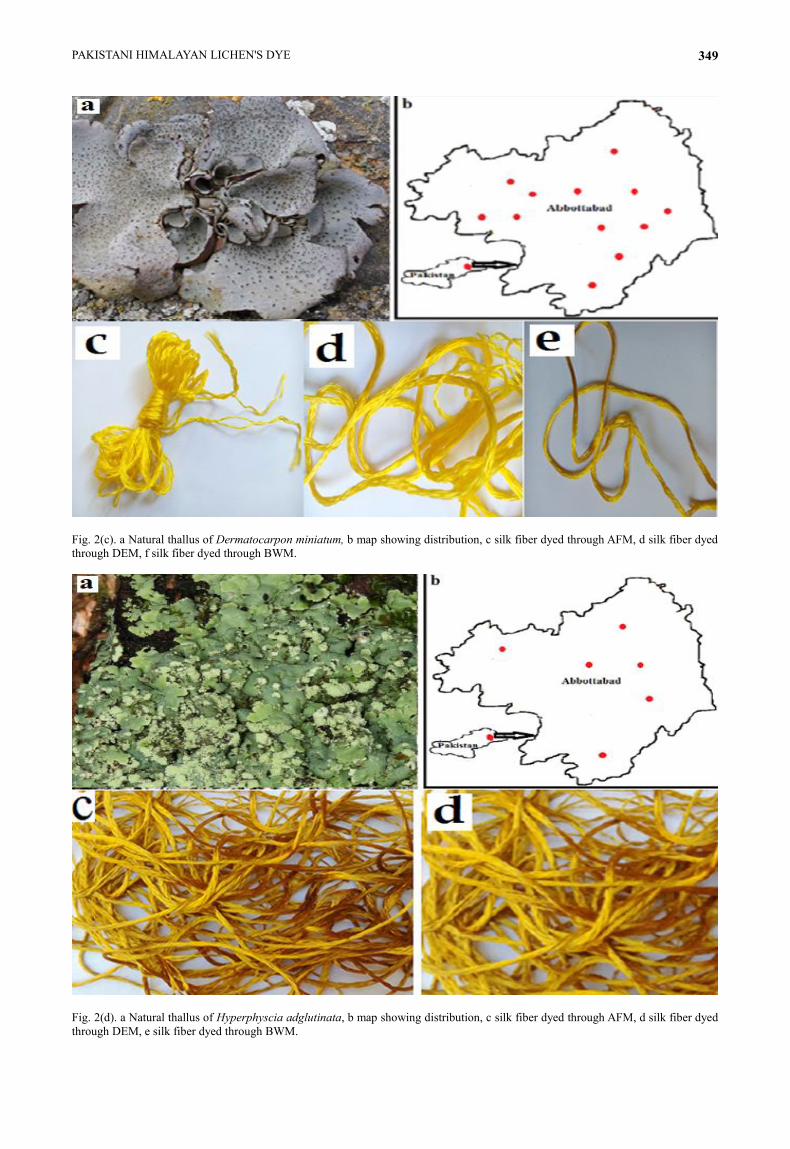
Fig. 2(c). a Natural thallus of Dermatocarpon miniatum, b map showing distribution, c silk fiber dyed through AFM, d silk fiber dyed
through DEM, f silk fiber dyed through BWM.
Fig. 2(d). a Natural thallus of Hyperphyscia adglutinata, b map showing distribution, c silk fiber dyed through AFM, d silk fiber dyed
through DEM, e silk fiber dyed through BWM.
SUMMAIRA SHAHEEN ET AL., 350
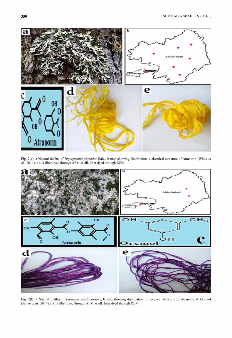
Fig. 2(e). a Natural thallus of Hypogymnia physodes Hale., b map showing distribution, c chemical structure of Atranorin (White et
al., 2014), d silk fiber dyed through AFM, e silk fiber dyed through DEM.
Fig. 2(f). a Natural thallus of Parmelia neodiscordans, b map showing distribution, c chemical structure of Atranorin & Orcinol
(White et al., 2014), d silk fiber dyed through AFM, e silk fiber dyed through DEM.
PAKISTANI HIMALAYAN LICHEN'S DYE 351
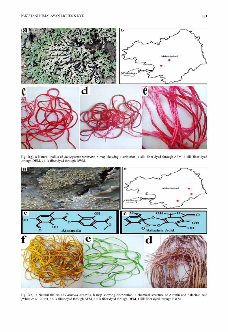
Fig. 2(g). a Natural thallus of Menegazzia terebrata, b map showing distribution, c silk fiber dyed through AFM, d silk fiber dyed
through DEM, e silk fiber dyed through BWM.
Fig. 2(h). a Natural thallus of Parmelia saxatilis, b map showing distribution, c chemical structure of Atronin and Salazinic acid
(White et al., 2014), d silk fiber dyed through AFM, e silk fiber dyed through DEM, f silk fiber dyed through BWM.
SUMMAIRA SHAHEEN ET AL., 352
Fig. 2(i). a Natural thallus of Parmelia sulcata, b map showing distribution, c chemical structure of Atronin and Salazinic acid (White
et al., 2014), d silk fiber dyed through AFM, e silk fiber dyed through DEM, f silk fiber dyed through BWM.
Fig. 2(j). a Natural thallus of Parmotrema reticulatum, b map showing distribution, c chemical structure of Atronin and Salazinic acid
(White et al., 2014), d silk fiber dyed through AFM, e silk fiber dyed through DEM, f silk fiber dyed through BWM.
PAKISTANI HIMALAYAN LICHEN'S DYE 353
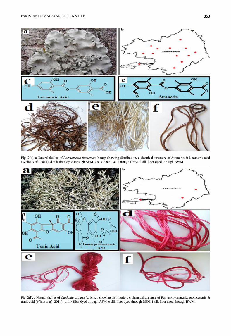
Fig. 2(k). a Natural thallus of Parmotrema tinctorum, b map showing distribution, c chemical structure of Atranorin & Lecanoric acid
(White et al., 2014), d silk fiber dyed through AFM, e silk fiber dyed through DEM, f silk fiber dyed through BWM.
Fig. 2(l). a Natural thallus of Cladonia arbuscula, b map showing distribution, c chemical structure of Fumarprotocetraric, protocetraric &
usnic acid (White et al., 2014), d silk fiber dyed through AFM, e silk fiber dyed through DEM, f silk fiber dyed through BWM.
SUMMAIRA SHAHEEN ET AL., 354
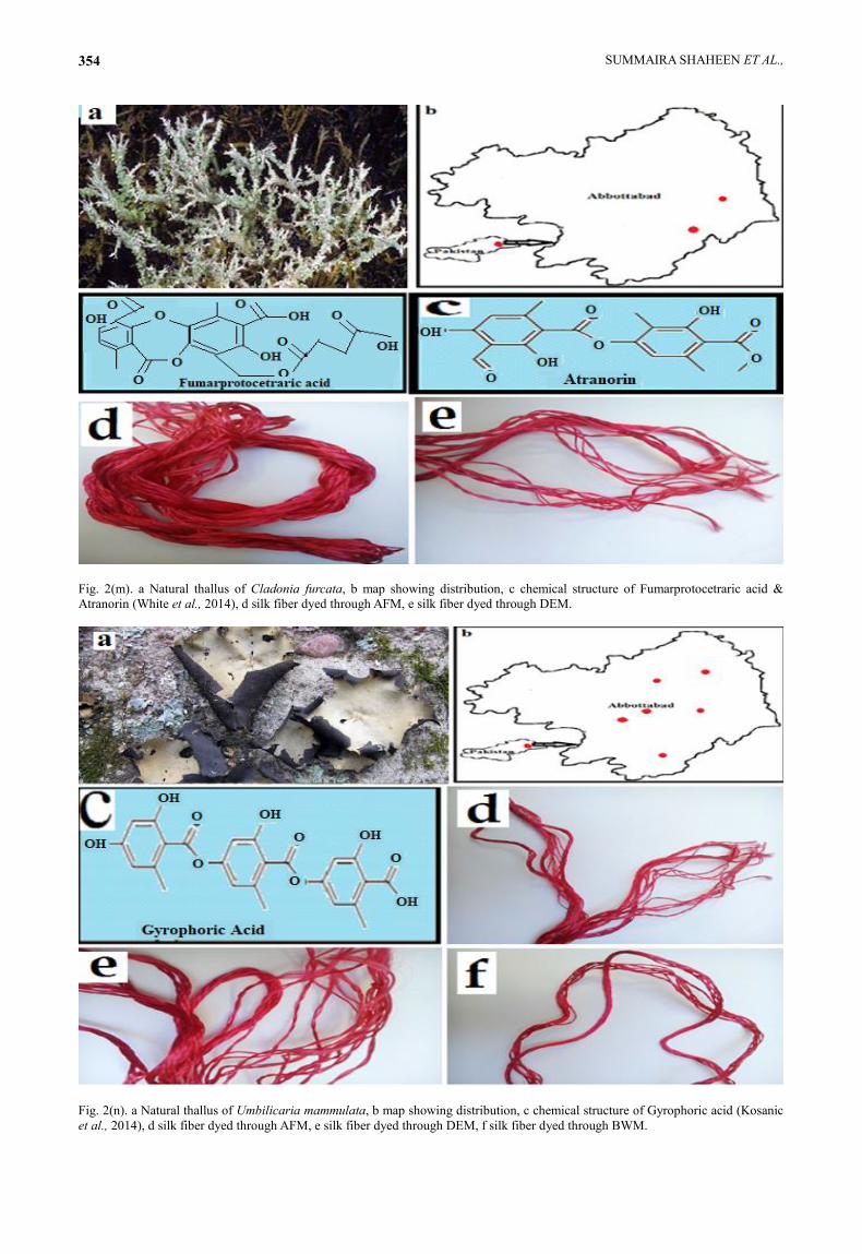
Fig. 2(m). a Natural thallus of Cladonia furcata, b map showing distribution, c chemical structure of Fumarprotocetraric acid &
Atranorin (White et al., 2014), d silk fiber dyed through AFM, e silk fiber dyed through DEM.
Fig. 2(n). a Natural thallus of Umbilicaria mammulata, b map showing distribution, c chemical structure of Gyrophoric acid (Kosanic
et al., 2014), d silk fiber dyed through AFM, e silk fiber dyed through DEM, f silk fiber dyed through BWM.
PAKISTANI HIMALAYAN LICHEN'S DYE 355
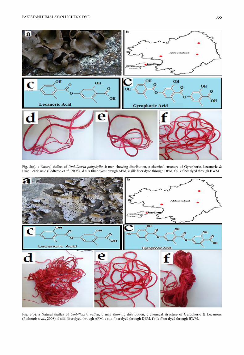
Fig. 2(o). a Natural thallus of Umbilicaria polyphylla, b map showing distribution, c chemical structure of Gyrophoric, Lecanoric &
Umbilicaric acid (Podterob et al., 2008) , d silk fiber dyed through AFM, e silk fiber dyed through DEM, f silk fiber dyed through BWM.
Fig. 2(p). a Natural thallus of Umbilicaria vellea, b map showing distribution, c chemical structure of Gyrophoric & Lecanoric
(Podterob et al., 2008), d silk fiber dyed through AFM, e silk fiber dyed through DEM, f silk fiber dyed through BWM.
SUMMAIRA SHAHEEN ET AL., 356
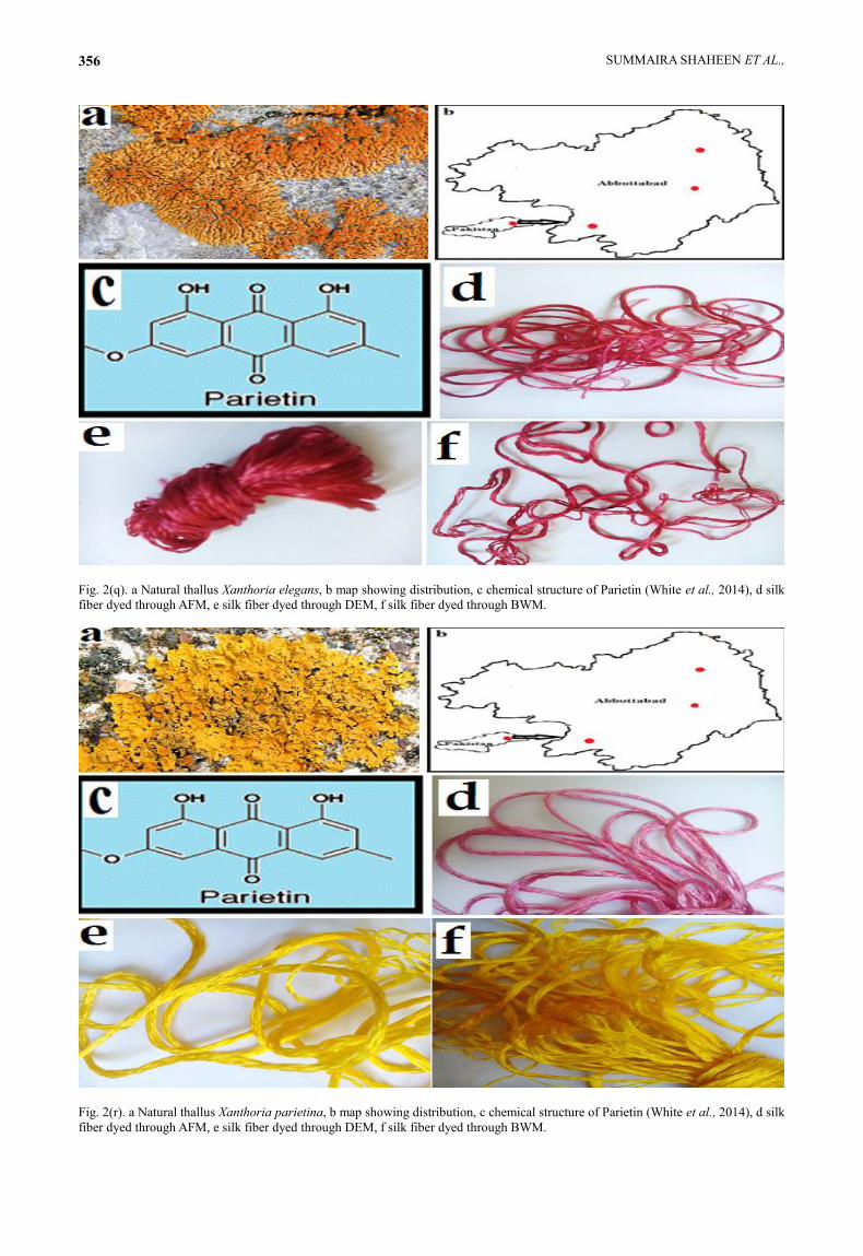
Fig. 2(q). a Natural thallus Xanthoria elegans, b map showing distribution, c chemical structure of Parietin (White et al., 2014), d silk
fiber dyed through AFM, e silk fiber dyed through DEM, f silk fiber dyed through BWM.
Fig. 2(r). a Natural thallus Xanthoria parietina, b map showing distribution, c chemical structure of Parietin (White et al., 2014), d silk
fiber dyed through AFM, e silk fiber dyed through DEM, f silk fiber dyed through BWM.
PAKISTANI HIMALAYAN LICHEN'S DYE 357
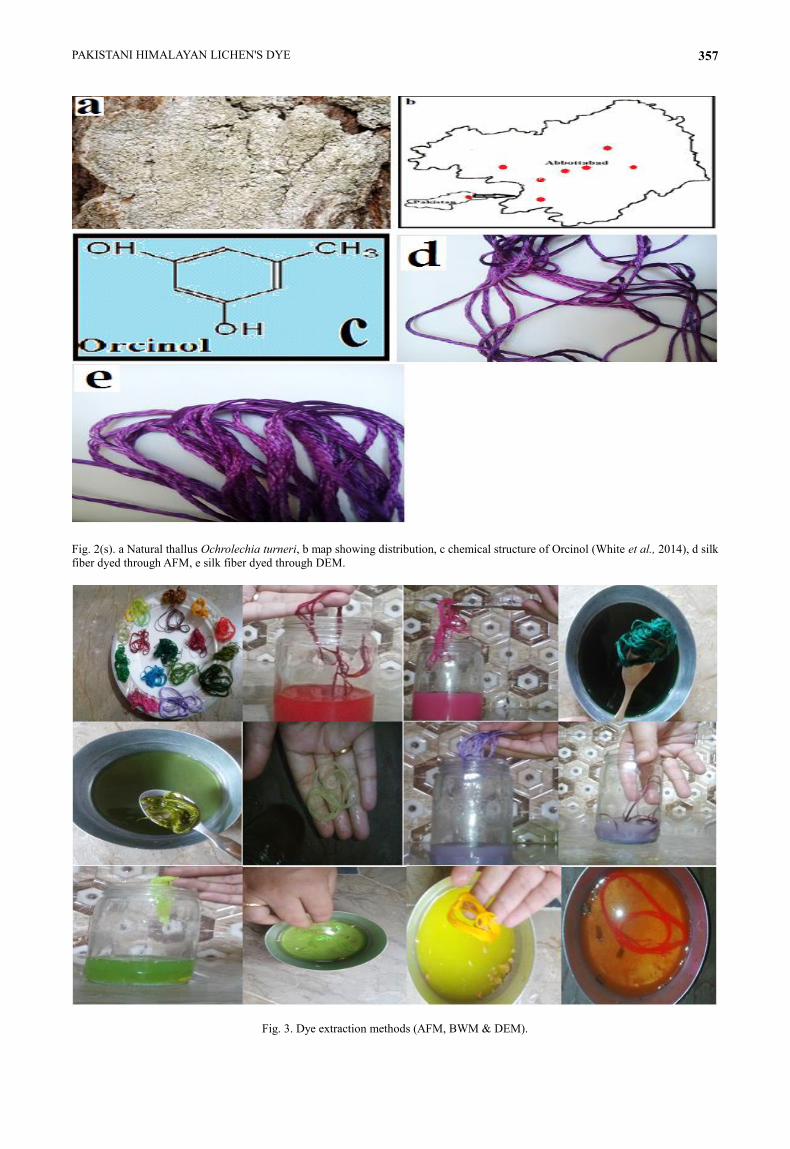
Fig. 2(s). a Natural thallus Ochrolechia turneri, b map showing distribution, c chemical structure of Orcinol (White et al., 2014), d silk
fiber dyed through AFM, e silk fiber dyed through DEM.
Fig. 3. Dye extraction methods (AFM, BWM & DEM).
SUMMAIRA SHAHEEN ET AL., 358
Table 3. HEX color code, RGB color model and HSL (hue, saturation, and lightness) values for each dye color.
S. No. Colours HEX RGB HSL
1. Isabella
#D2B48C 210, 180, 140 34, 44%, 69%
2. Ivory Yellow
#F0E68C 240, 230, 140 54, 77%, 75%
3. Buffy brown
#A0522D 160, 82, 45 19, 56%, 40%
4. Lemon yellow
#FFFF00 255, 255, 0 60, 100%, 50%
5. Yellow
#FFD700 255, 215, 0 51, 100%, 50%
6. Purple
#800080 128, 0, 128 300, 100%, 25%
7. Brown
#8B4513 139, 69, 19 25, 76%, 31%
8. Light Brown
#A0522D 160, 82, 45 19, 56%, 40%
9. Green
#9ACD32 154, 205, 50 80, 61%, 50%
10. Olive Brown
#556B2F 85, 107, 47 82, 39%, 30%
11. Seal Brown
#8B4513 139, 69, 19 25, 76%, 31%
12. Deep cerise
#DB7093 219, 112, 147 340, 60%, 65%
13. cerise
#FF69B4 255, 105, 180 330, 100%, 71%
14. Corinthian red
#CD5C5C 205, 92, 92 0, 53%, 58%
15. Congo pink
#FA8072 250, 128, 114 6, 93%, 71%
16. Magenta
#FF1493 255, 20, 147 328, 100%, 54%
17. Olive
#808000 128, 128, 0 60, 100%, 25%
18. Warm Sepia
#D2B48C 210, 180, 140 34, 44%, 69%
Discussion
Lichen dyes by a variety of vernacular names have a
long history, dyes from lichens (orchil) were discovered
before 1500 BCE, in the Roman period and they were
considered synonymous with wealth and power, wearing
purple colour was an indicative of status and privilege, and
the dye industry was also politicized (Diadick, 2001).
Fruticose and foliose lichens were tested for dyeing yielding
potential because of their bigger size and shape. Different
studies were conducted for dye extraction all over the world
(Shukla et al., 2015; Fazioa et al., 2007; Diadick, 2001;
Casselman, 2001; Dean et al., 2012; Upreti et al., 2012).
The dye colors vary with the harvesting season of
lichens thallus, soil properties and fermentation time.
Lichen dyes are much better then synthetic dyes as these
are friendly to our environment, imparts musky odor to
fibers, dyed products are insect-proof because lichen
secondary compounds make the fibers distasteful for
insects, have limited colour stability against sunlight and
as no mordents have been used in the study and without
any mordents these are bright and beautiful colours.
Probably more colorfast dyes could be obtained if
mordents are used during extraction. On the other side,
the tedious extraction and long dying time increase the
cost of lichen dyes.
PAKISTANI HIMALAYAN LICHEN'S DYE 359
SUMMAIRA SHAHEEN ET AL., 360
Fig. 4. Number of lichen species and their dye color.
Lichen dyes on wool and silk are fast to washing
and exposure to sunlight. According to Perkins (1986),
many writers believed that lichen dyes were not fast on
any fiber but our results were in disagreement with
Perkin and are in agreement with Shukla et al., (2015)
and Brough (1988) who believe that lichen dyes are fast.
Mostly mordant dyes need a mordant to improve the
fastness of the dye against washing and exposure to
sunlight. Most natural dyes are mordant dyes but lichen
dyes are unique in this respect that it does not require
any mordant. Many mordants especially associated with
the heavy metal category are hazardous to environment
and health, in this respect lichen dyes are safe to use and
friendly to the environment.
Lichen substances form the 19 lichens used in this
study have also been reported (Culberson, 1970;
Culberson et al., 1977; Hale, 1979; Leuckert, 1977;
Thomson, 1979 & 1984). In this study Spot tests of
Parmelia saxatilis (L.) Ach., Parmelia sulcataTaylor. and
Parmotrema reticulatum (Taylor) M. Choisy. are P
positive (showed orange color) i.e. an indication of
salazinic acid. Orange shades are due to salazinic acid but
in the present investigation, it produced brown dye as
reported by Shukla et al., (2014), according to him due to
increased fermentation time orange shades were changed
to dark shades of brown dye color. Brough (1988) also
justified the brown shades for these lichens for the same
reason. Similarly, Parietin which is responsible for
Yellow, pink and Magenta shades of Xanthoria elegans
and Xanthoria parietina dyes is also reported by Fazioa et
al., (2007). Gyrophoric acid (that produced royal purple
color) was detected from genus Umbilicaria by spot tests
results in our investigation as well as by Bolton (1960).
Parmotrema tinctorum and two species of Umbilicaria
which imparted purple dye color have lecanoric acid (p-
depside) that hydrolyzed and produced orsellic acid color-
producing substance orcein. These results validate the
historical references (Table 4).
These precious and beautiful creatures of nature like trees should not be destroyed, it is strongly recommended that only large sizedd and naturally detached lichens should be used for dying; the whole thallus must not be harvested. These cannot be used in textile industries on the commercially large scale because of their small size as compared to higher plants and extremely slow growth rate, but certainly, they can serve on small-scale handloom industries to offer employment to local people.
References
Abdullahi, M.B., S.S. Sanusi, S.D. Abdul and F.B.J. Sawa. 2009.
An assessment of the herbaceous species vegetation of
Yankari Game Reserve, Bauchi, Nigeria. Am-Eur. J. Agric.
& Environ. Sci., 6(1): 20-25.
Aptroot, A. and S.H. Iqbal. 2012. Annotated checklist of Lichen
of Pakistan, with report of new record. Herzogia, 25(2):
211-229.
Asahina, Y. and S. Shibata. 1954. Chemistry of lichens
substances. Japanese Society for the Promotion of Science,
Tokoyo, 12-35.
Awasthi, D.D. 1988. A key to the Macrolichens of India and
Nepal. J. Hattori Bot., 26(1) 102-112.
Bolton, E.M. 1960. Lichens for Vegetable Dyeing. Studio
Books, London, 1960.
Brough, S.G. 1988. Navajo lichen dyes. Lichenologists. 20 (3):
279-290.
Casselman, K.D. 1994. Historical and Modern Lichen dyes:
Some Ethical Considerations. Norw Breakfast Club
Newslett I(1):1-13.
Casselman, K.D. 2001. Lichen dyes: the new source book.
Dover Publications, Mineola, New York.
Casselman, K.D. 2001. Lichen Dyes: The New Source Book.
New York: Dover.
Casselman, K.D. 2011. Lichen Dyes: The New Source Book.
Cheverie Nova Scotia: Studio Vista Publications.
Chopra, G.L. 1934. Lichen of the Himalayas. Part. I. Lahore.
Collinson, A.S. 1977. Introduction to world vegetation. London:
George Allen & Unwin.
Culberson C.F., W.L. Culberson and A. Johnson. 1977. Second
Supplement to Chemical and Botanical Guide to Lichen
Products. St. Louis: Amer. Bryol Lich. Soc, Miss. Bot. Gard.
Culberson, C.F. 1970. Supplement to Chemical and Botanical
Guide to Lichen Products. Bryologist., 73: 177-377.
Culberson, C.F. 1972. Improved conditions and new data for the
identification of lichen products by a standardized thin-layer
Chromatographic method. J Chromatogr., 72: 113-125.
Culberson, C.F. and H.D. Kristinsson. 1970. A standardized
method for the identification of lichen products. J.
Chromatg., 46: 85-93.
Dean, G., D. Brown, D. DeSouza and D. McClintok. 2012.
Adventure with lichen dyes. Turk. Red J., 17:1-10.
Diadick-Casselman, K. 2001. Lichen dyes: The new source
book. New York: Dover.
Fazioa, A.T., M.T. Adlera, M.D. Bertonia, C.S.S. lvedab, E.B.
Damonteb and M.S. Maierc. 2007. Lichen Secondary
Metabolites from the Cultured Lichen Mycobionts of
Teloschistes chrysophthalmus and Ramalina celastri and
their Antiviral Activities. Z. Naturforsch., 62: 543-549.
Gupta V.K., M.P. Darokar, D. Saikia, A. Pal and A. Fatima.
2007. Antimicrobial activity of lichens. Pharm. Biol., 45(3)
200-204.
Hale, E.M. How to Know the Lichens. 1979. 2nd ed. Wm. C.
Brown Co., Dubuque, Iowa, 12(2) pp. 251-252.
Hodge, K. 2006. Dyeing with lichens & mushrooms.
http://blog.mycology.crnell.edu/2006/12/12/dyeingwith-
lichens-mushrooms.
8
6 6
3
1 1 1 1
0
1
2
3
4
5
6
7
8
9
Nu
mb
er o
f li
chen
sp
ecie
s.
Dye colour produced by lichens.
PAKISTANI HIMALAYAN LICHEN'S DYE 361
Khan S.A., M. Hamayun, M.H.J. Yoon, H.Y. Kim, S.J. Suh and
S.K. Hwang. 2008. Plant growth promotion and
Penicillium citrinum. BMC Micr., 8: 231-237.
Kok, A. 1966. A short history of the orchil dyes. Lichenologist,
3: 248-272.
Kosanic, M., B. Rankovic, T. Stanojkovic, P.A. Vasiljevic and N.
Manojlovic. 2014. Biological activities and chemical
composition of lichens from Serbia. EXCLI J., 13: 1226-
1238.
Krystle, A., A. Santiago, N.C. Borricano, J.N. Jayne, M.A.
Denisse, C.P. Myleen and E.C. Thomas. 2010. Antibacterial
activities of fruticose lichens collected from selected sites
in Luzon Island, Philippines. Phil. Sci. Lett., 3(2): 18-29.
Leuckert, C., J. Poelt and G. Hahnel. 1977. Zur Chemotaxonomie
der eurasischen Arten der Flechtengattung Rhizoplaca. Nova
Hedwigia, 28: 71-129.
Manojlovic, T.N., M. Novakovi, V. Stevovi and S. Soluji. 2005.
Antimicrobial activity of three Serbian Caloplaca. Pharm
Biol., 43(8): 718-722.
Margareta, S.F. 1981. The chemistry of plants animal dyes. J.
Chem Educ., 58(4): 301-305.
Marijana, K. and B. Rankovic. 2010. Screening of antimicrobial
activity of Lichen species in vitro. Kragujevac J. Sci., 32:
65-72.
Perkins, P. 1986. Ecology, beauty, profits: trade in lichen
dyestuffs. J. Soc. Dyers Colour, 102: 221-227.
Podterob, A.P. 2008. Chemical composition of lichens and their
medical applications. Pharm Chem. J., 65: 207-302.
Richardson, D.H.S. 1988a. Medicinal and other economic
aspects of lichens. In: (Ed.): Galun, M. Handbook of
lichenology, vol 3. pp. 93-108.
Richardson, D.H.S. 1988b. Handbook of lichenology. Vol. 3,
(CRC Press, Boca Raton), 93-108.
Ridgway, R. 1912. Colours standard and colours nomenclature
Washington, D.C. Columbia University Libraries
Electronic Books. pp. 143-149.
Santiago, K.A.A., E. Sangvichien, K. Boonpragob and C. Tee.
2010. Secondary metabolic profiling and antibacterial
activities of different species of Usnea collected in
Northern Philippines. Mycosphere, 110-124.
Shah, N.C. 2011. Lichen of commercial Importance in India.
Herb. Tech. Ind., 10(01): Jan-Feb.
Sharma, A. and E. Grover. 2011. Colour fastness of walnut dye
on cotton. Indian J. Nat. Prod. Resour., 2(2): 164-169.
Shukla, P. and D.K. Upreti. 2014a. Natural dyes from
Himalayan lichens. Ind. J. Tradi Knowl., 13(1): 195-201.
Shukla, P. and D.K. Upreti. 2015. Recent Advances in
Lichenology : Modern Methods and Approaches in Lichen
Systematics and Culture Techniques, Volume 1. Springers,
pp. 209-229.
Shukla, P., and D.K. Upreti,. 2014b. Assessment of dye yielding
potential of Indian lichens. Ind. J. Plant Sci., 3(1): 57-63.
Shukla, P., D.K. Upreti, S. Nayaka and P. Tiwari. 2014. Natural
dyes from Himalayan lichens. Ind. J. Tradit Know., 13(1),
pp 195-201.
Shukla, V., R. Kumari, D.K. Patel and D.K. Upreti. 2015.
Characterization of the diversity of mycosporine-like amino
acids in lichens from high altitude region of Himalaya.
Amino Acids, 48(1): 129-36.
Shyam, K., N. Thajuddin and D.K. Upreti. 2010. Diversity of
Lichen in Kollihills of Tamil Nadu, India. Int. J. Biodiv.
Conserv., 3(2): 36-39.
Smith, A.L. 1918. A Monograph of the British Lichens, Vol. 1, 2
edn. London: British Museum (Natural History).
Thomson, J.W. 1979. Lichens of Anaktuvuk Pass, Alaska, with
emphasis on the impact of caribou grazing. Bryologist., 82:
393-408.
Thomson, J.W. 1984. American Arctic Lichens. The Macrolichens.
Columbia University Press, New York. 504 pp.
Tucker, C.T. 2014. Catalog of lichens, lichenicoles and allied
fungi in California (second revision). Constancea, 85.
Upreti, D.K., P. Tiwari, P. Shukla and A. Dwivedi. 2012. Natural
thalli and cultured mycobiont of U. ghattensis G. Awasthi: a
potential source of purple dye yielding lichen from India.
Ind. J. Nat. Prod. Res., 3(4): 489-492.
Upreti, D.K., S. Joshi and S. Nayaka. 2010. Chemistry of
common dye yielding lichens of India. ENVIS Forestr
Bull., 10 (1): 122-133.
Veranja, K., K. Bombuwela, S. Kathirgamanathar and V.M.
Thadhani. 2005. Lichen: A chemically important Biota. J.
Natl. Sci. Found., 33(3): 169-186.
White, P.A.S., R.C.M. Oliveira, A.P. Oliveira, M.R. Serafini,
A.A. S. Araujo, D.P. Gelain, J.C.F. Moreira, J.R.G.S.
Almeida, J.S.S. Quintans, L.J.Q. Junior and M.R.V. Santos.
2014. Antioxidant Activity and Mechanisms of Action of
Natural Compounds Isolated from Lichens: A Systematic
Review. Molecules, 19: 14496-14527.
(Received for publication 10 January 2018)