Embed Size (px)
Citation preview
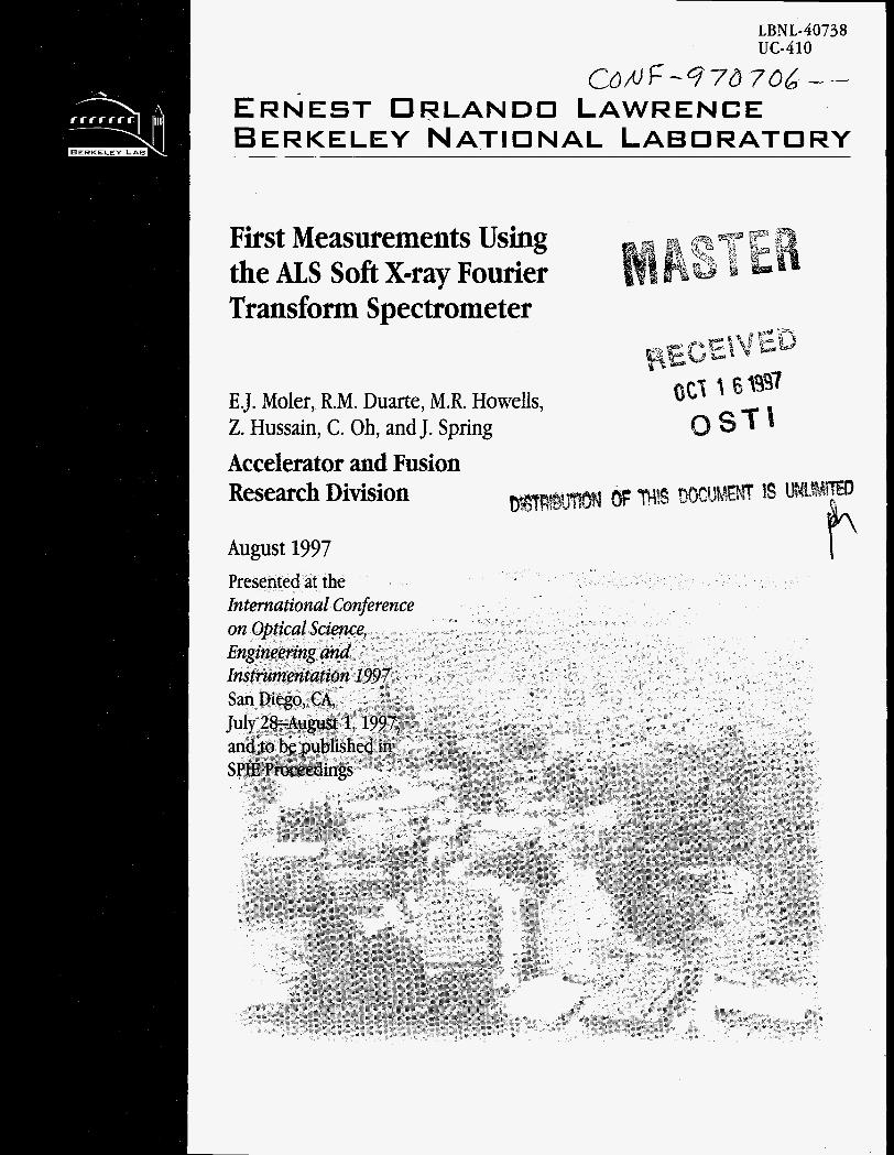
LBNL-40738 UC-410
C~/uF-%'d706 -- ERNEST ORLANDO LAWRENCE BERKELEY NATIONAL LABORATORY
First Measurements Using the ALS Soft X-ray Fourier Transform Spectrometer
E.J. Moler, R.M. Duarte, M.R. Howelk, 2. Hussain, C. Oh, and J. Spring Accelerator and Fusion Research Division
DISCLAIMER
This document was prepared as an account of work sponsored by the United Stales Government. While this document is believed to contain correct information, neither the United States Government nor any agency thereof, nor The Regents of the University of California, nor any of their employees, makes any warranty, express or implied, or assumes any legal responsibility for the accuracy, completeness, or usefulness of any information, apparatus, product, or process disclosed, or represents that its use would not infringe privately owned rights. Reference herein to any specific commercial product, process, or service by its trade name. trademark, manufacturer, or otherwise, does not necessarily constitute or imply its endorsement, recommendation, or favoring by the United States Government or any agency thereof, or The Regents of the University of California. The views and opinions of authors expressed herein do not necessarily state or reflect those of the United States Government or any agency thereof, or The Regents of the University of California.
Ernest Orlando Lawrence Berkeley National Laboratory is an equal opportunity employer.
LBNL-4073 LSBL-40 uc-411
First Measurements Using the ALS Soft X-ray Fourier Transform Spectrometer
E.J. Molera, R.M. Duartea, M.R. Howellsa, Z. Hussaina, C. Ohb, J. Springa
aAdvanced Light Source Ernest Orlando Lawrence Berkeley National Laboratory
University of California, Berkeley, California 94720
Korea bKyungpook National University
Light Source Note:
hthor(s) Initials C ~ A 9 Group Leader's initials @?-yf -. 1 1 w -
First measurements using the ALS soft x-ray Fourier transform spectrometer
E.J. Molera* , R.M. Duartea, M. R. Howellsa, Z. Hussaina, C. Oh', J. Springa aAdvanced Light Source, Lawrence Berkeley National Laboratory
bKyungpook National University, Korea
ABSTRACT
Commissioning of a Fourier Transform Soft X-ray spectrometer (FT-SX) is under way at the Advanced Light Source (ALS), Lawrence Berkeley National Laboratory, as a branch of beamline 9.3.2. The spectrometer is a novel soft x-ray interferometer designed fo: ultra-high resolution spectroscopy in the photon energy region of 60-120 eV with a theoretical resolving power E/AE-10 . This instrument is expected to provide experimental results which sensitively test models of correlated electron processes in atomic and molecular physics. The design criteria and consequent technical challenges posed by the short wavelengths of x-rays and desired resolving power are discussed. The fundamental and practical aspects of soft x-ray interferometry are also explored.
Keywords: Fourier transform, interferometry, soft x-ray, synchrotron radiation, spectroscopy, helium double-excitations
1. INTRODUCTION AND SCIENTIFIC MOTIVATION
Correlated electron motion is at the center of any chemical process and thus constitutes a very important arena of basic scientific research. The many-body nature of the problem precludes analytical solutions leaving only approximations to the quantum problem potentially tractable. The central problem in theoretical electron correlation studies is to fmd the appropriate approximations that describe all of the main characteristics of the experimentally observed features of such systems. It is ~tural to start with excitations of the helium atom since this is the simplest correlated electron system and, with the availability of synchrotron radiation sources, increasingly detailed experimental observations have become available. The FT-SX spectrometer has been designed to probe this prototypical system with unprecedented resolving power. Of particular interest are the regions of overlapping double excitation series where classically chaotic behavior is mixed with quantization effects 1.
2. SYNCHROTRON RADIATION AND RESOLUTION LLMITS OF STANDARD SOFT X-RAY MONOCHROMATORS
Synchrotron radiation has proved to be an extremely useful tools in basic atomic and molecular physics studies. The high photon flux and tunable photon energy enables detailed studies of excitation processes which are considerably challenging to the theoretical physics community. Third generation light sources, such as the Advanced Light Source (ALS) at Lawrence Berkeley National Laboratory, coupled with high resolution beam lines are providing extremely detailed observations of scientifically important systems such as the helium double excitation series, a correlated electron system, in the soft x-ray range. However, there is more to be gained with increased resolving power in x-ray absorption experiments.
The highest resolving power soft x-ray beam lines available consist of a low emittance synchrotron source and a highly optimised, grating based monochromator, usually a Spherical Grating Monochromator (SGM). The low emittance source allows for relatively high photon flux while using the small aperatures necessary to operate the SGM at maximum resolving power. While the claim of experimentally achieved resolving power can vary depending on the interpretation of the data and caution of the investigators, it is apparent that the theoretically maximum resolving power of a spherical grating monochromator, (E/AE=50,000) has been closely approached by Kaindle, et al. in the region of the helium double excitations (- 65 eV)*.
Email: [email protected]; LBNL mail stop 7-100; phone: 5 10-486-7637
DRIVE SHAFT TO MOV/NG
F/X€D FRAME FLEXURE HING& (76) FLEXURE HING& h f -
- ._ - - - . . . - . . . . -
(4) MIRROR PR/SM-\ (ON MO VING STAGE)
t- 36.83 Cm.-/
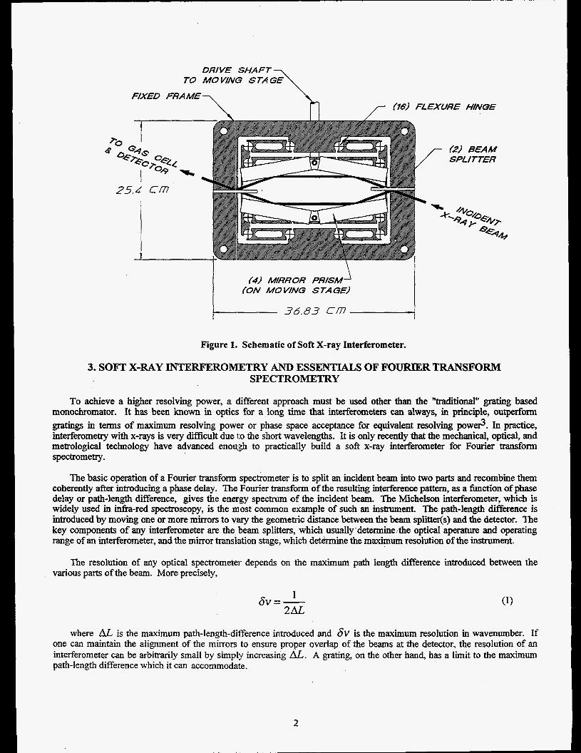
Figure 1. Schematic of Soft X-ray Interferometer.
3. SOB'" X-RAY INTERFEROMETRY AND ESSENTIALS OF FOURIER TRANSFORM SPECTROMETRY
To achieve a higher resolving power, a different approach must be used other than the "traditional" grating based monochromator. It has been known in optics, for a long time that interferometers can always, in principle, outperform gratings in terms of maximum resolving power or phase space acceptance for equivalent resolving powe?. In practice, interferometry with x-rays is very difficult due to the short wavelengths. It is only recently that the mechanical, optical, and metrological technology have advanced enough to practically build a soft x-ray interferometer for Fourier transform spectrometry.
The basic operation of a Fourier transform spectrometer is to split an incident beam into two parts and recombine them coherently after introducing a phase delay. The Fourier transform of the resulting interference pattern, as a function of phase delay or path-length difference, gives the energy spectrum of the incident bean The Michelson interferometer, which is widely used in infia-red spectroscopy, is the most common example of such an instrument. The path-length difference is introduced by moving one or more mirrors to v~vy the geometric distance between the beam s p l i i s ) and the detector. The key components of any interferometer are the beam splitters, which usually.detennine-the optical aperature and operating range of an interferometer, and the mirror translation stage, which determine the maximum resolution of the instrument.
The resolution of any optical spectrometer depends on the maximum path length difference introduced between the various parts of the beam. More precisely,
1 6V=- 2AL
where AL is the maximum path-length-dif'ference introduced and 6 V is the maximum resolution in wavenumber. If one can maintain the alignment of the mirrors to ensure proper overlap of the beams at the detector, the resolution of an interferometer can be arbitrarily small by simpl!, increasing Al,. A grating, on the other hand, has a limit to the maximum path-length difference which it can accommodate.
2
i I I 1
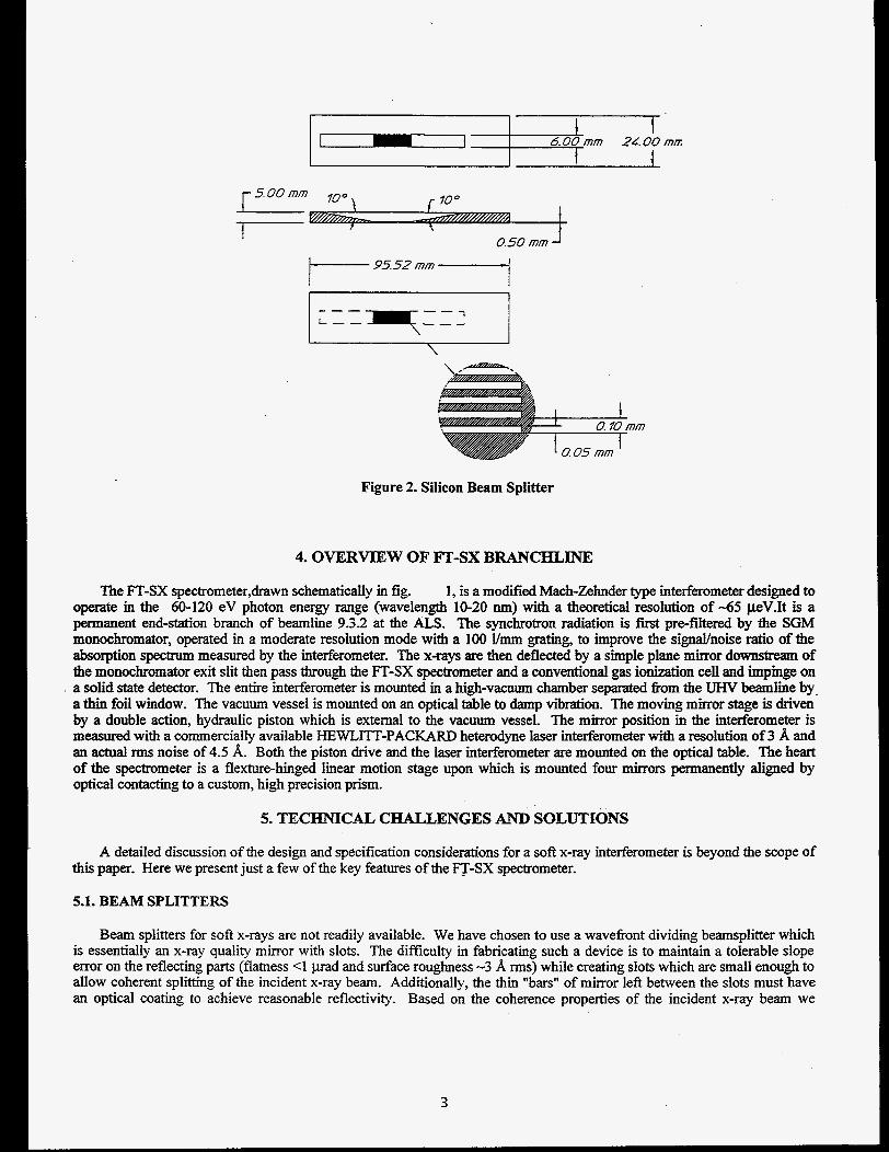
Figure 2. Silicon Beam Splitter
4. OVERVIEW OF FT-SX BRANCHLINE
The FT-SX spectrometer,drawn schematically in fig. 1, is a modified Mach-Zehnder type interferometer designed to operate in the 60-120 eV photon energy range (wavelength 10-20 nm) with a theoretical resolution of -65 pV.It is a permanent end-station branch of beamline 9.32 at the ALS. The synchrotron radiation is first pre-filtered by the SGM monochromator, operated in a moderate resolution mode with a 100 Vmm grating, to improve the signallnoise ratio of the absorption spectrum measured by the interferometer. The x-rays are then deflected by a simple plane m h r downstream of the monochromator exit slit then pass through the FT-SX spectrometer and a conventional gas ionization cell and impinge on a solid state detector. The entire interferometer is mounted in a high-vacuum chamber separated from the UHV beamlie by a thin foil window. The vacuum vessel is mounted on an optical table to damp vibration. The moving mirror stage is driven by a double action, hydraulic piston which is external to the vacuum vessel. The mirror position in the interferometer is measured with a commercially available HEWLITT-PACKARD heterodyne laser interferometer with a resolution of 3 A and an actual xms noise of 4.5 A. Both the piston drive and the laser interferometer are mounted on the optical table. The heart of the spectrometer is a flexture-hinged linear motion stage upon which is mounted four mirrors permanently aligned by optical contacting to a custom, high precision prism.
5. TECHNICAL CHALLENGES AND SOLUTIONS
A detailed discussion of the design and specification considerations for a sofe x-ray interferometer is beyond the scope of this paper. Here we present just a few of the key features of the p - S X spectrometer.
5.1. BEAM SPLITTERS
Beam splitters for soft x-rays are not readily available. We have chosen to use a wavefront dividing beamsplitter which is essentially an x-ray quality mirror with slots. The difficulty in fabricating such a device is to maintain a tolerable slope error on the reflecting parts (flatness <I prad and surface roughness -3 A rms) while creating slots which are small enough to allow coherent splitting of the incident x-ray beam. Additionally, the thin "bars" of mirror left between the slots must have an optical coating to achieve reasonable reflectivity. Based on the coherence properties of the incident x-ray beam we
3

determined a slot width of 50 pm and a period of 100 pm was sufficient to split the x-ray beam coherently. Our design is shown in fig. 2. The scheme developed to fabricate the beam splitters is as follows:
a) a single crystal Si wafer is polished tal an x-ray quality finish with a specific orientation relative to the (1 10) crystal plane.
b) the back side of the wafer is ultrasoniically machined to thin the center of the wafer where the slots will be and to cutout chamfers to allow the grazing incident x-rays to pass through the slots.
c) the wafer is masked and the slots ane chemically etched with an etchant that preferentially cuts along the [I101 direction.
d) the optical coating is applied in a manner to maintain a stress free thin film. For the ALS spectrometer we have selected molybdenum.
The beam splitters have been successfully fabricated by Rockwell Power Systems Division of Rockwell International.
5.2. THE MECHANICAL MIRROR STAGE
The mirror stage must allow enough motion to introduce the desired path-length-difference between the two halves of the x-ray beams and must maintain an anguk alignment which ensures coherent recombination of the two beams at the detector. For the FT-SX spectrometer the total range of motion is - 1.5 cm and the angular tolerance is 20.5 p a d across the entire range. We have achieved this by using "cartwheel" type flexural hinges (see fig. 1) which were wire-EDM cut from a monolithic maraging steel slab.
6. OPTICAL ALIGNMENT
Alignment of the beam splitters to the mirrors and to each other is currently under way. This has been initially accomplished by using light from a gas discharge lamp,using the visible emission lines of neon with the goal of moving up to the W V emission lines as the alignment improves. We have also found that using the fringes generated by a HeNe laser are useful for getting the beamsplitters parallel to each other. However, the large transverse coherence of the laser light renders it
insensitive to displacement of the recombined beams, which leads to lower contrast in the interferogram and thus lower signalhoke ratio in the recovered spectrum. 13gure 3 shows an interferogram of the visible neon emission Iies. Note that
-0.08
-0.10 t-
-0.16
6915 6920 6925 Mirror Stage Position (pm)
Figure 3. Diagnostic Interferograrm from the Visible Emission Lines of a Neon Discharge Lamp.
4
I I 1 1 1 6930 6935 6940 6945
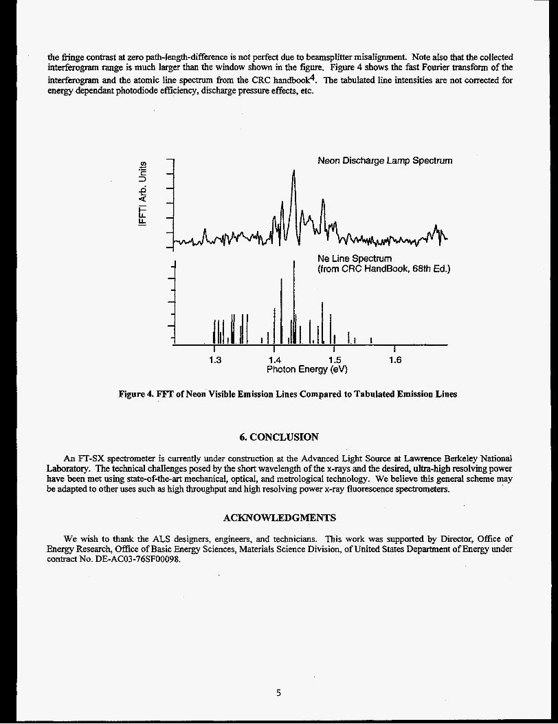
the f i g e contrast at zero path-Iength-difference is not perfect due to beamspIitter misalignment. Note aIso that the coilected intderogram range is much larger than the window shown in the figure. Figure 4 shows the fast Fourier transform of the interferogram and the atomic line spectrum from the CRC handbood. The tabulated line intensities are not corrected for energy dependant photodiode efficiency, discharge pressure effects, etc.
Neon Discharge Lamp Spectrum
Ne Line Spectrum (from CRC HandBook, 68th Ed.)
1.5 1.6 1.3 1.4 ..-
Photon Energy (ev>
Figure 4. F m of Neon Visible Emission Lines Compared to Tabulated Emission Lines
6. CONCLUSION
An FT-SX spectrometer is currently under construction at the Advanced Light Source at Lawrence Berkeley National Laboratory. The technical challenges posed by the short wavelength of the x-rays and the desired, ultra-high resolving power have been met using state-of-the-art mechanical, optical, and metrological technology. We believe this general scheme may be adapted to other uses such as high throughput and high resolving power x-ray fluorescence spectrometers.
ACKNOWLEDGMENTS
We wish to thank the ALS designers, engineers, and technicians. This work was supported by Director, Office of Energy Research, Office of Basic Energy Sciences, Materials Science Division, of United States Department of Energy under contract No. DE-AC03-76SF00098.
5
REFERENCES
1.
2.
3.
4.
M. Domke, C. Xue, A. Puschman, T. Mandel, E. Hudson, D. A. Shirley, G. Kaindl, C. H. Greene, G. R Sadeghpour, “Extensive Double-Excitation States in Atomic Helium”, Physical Reveiw Letters 66,344 1-3443 ,1991.
G. Kahdl, “Ultra-High Resolution Spectroscopy of the He Double Excited States”, Oji International Seminar on Atomic and Molecular Photoionization, Tsukuba, Japan1 995.
J. Chamberlain, The Principles of hterferometric Spectroscopy John Wiley and Sons, Chichester, 1979.
R. C. Weast, Ed., CRC Handbook of’Chemisrry and Physics Chemical Rubber Company Publishing, Boca Raton, Florida, 1987.
6