Embed Size (px)
Citation preview
ORIGINAL ARTICLE
First Branchial Cleft Anomalies: Avoiding the Misdiagnosis
Rajeev Kumar • Kapil Sikka • Prem Sagar •
Aanchal Kakkar • Alok Thakar
Received: 23 November 2012 / Accepted: 9 March 2013 / Published online: 19 March 2013
� Association of Otolaryngologists of India 2013
Abstract First branchial cleft anomalies are a very rare
entities accounting for less than 1 % of all branchial cleft
malformations. They are often misdiagnosed for other
cystic lesions occurring in parotid gland and inadequately
treated (incision and drainage or incomplete excision)
leading to multiple recurrences. We report a series of four
patients who were previously operated (incision and
drainage) for misdiagnosed first branchial cleft anomalies
with subsequent recurrences. All patients underwent
superficial parotidectomy with complete tract excision
using facial nerve monitoring to prevent iatrogenic injury
because of extensive fibrosis. We discuss the literature
pertaining to first branchial cleft anomalies, their varied
presentations and their relationship to facial nerve in par-
otid gland and importance of facial nerve monitoring in
revision surgery.
Keywords First branchial cleft anamolies � Facial nerve �Superficial parotidectomy � Facial nerve monitoring
Introduction
First branchial cleft anomalies are rare and account for 1 %
of all branchial cleft malformations [1, 2]. Presentation can
be either in the form of cyst, sinus or fistula in close
relation to external auditory canal (EAC), parotid and
anterior neck. Acierno has further classified first cleft
anomalies as type I and type II [1]. Type I anomalies run
lateral to facial nerve, composed of ectoderm histologically
and present as swellings near the ear. Type II anomalies, on
the other, run medial to facial nerve, composed of both
ectoderm and mesoderm and present between EAC and
hyoid bone [1]. Correct diagnosis is often delayed because
of their rarity resulting in inadequate treatment and sub-
sequent recurrences. We report a series of four such
patients of first branchial cleft anomalies presenting as
recurrent parotid swelling along with other clinical pre-
sentations of first branchial cleft anomalies, their relation-
ship to facial nerve and management issues.
Materials and Methods
A retrospective chart analysis was performed for first
branchial cleft anomalies. Four cases were retrieved.
Demographic data in terms of age, sex, duration of
symptoms, mode of presentation, pre-operative facial nerve
status, number of previous surgeries, type of surgical pro-
cedures, intraoperative findings including relationship with
facial nerve are listed in Table 1. Male to female ratio was
1:1 with almost all cases presenting in first and second
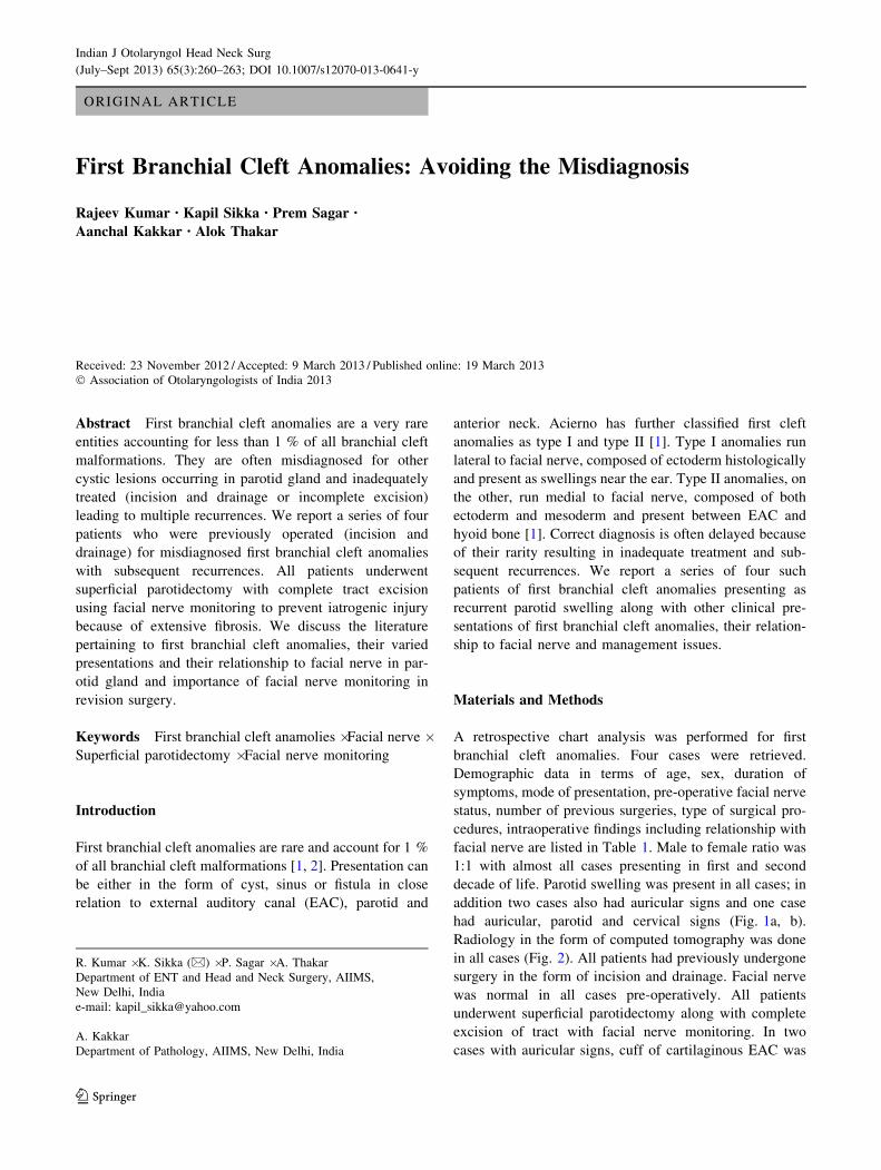
decade of life. Parotid swelling was present in all cases; in
addition two cases also had auricular signs and one case
had auricular, parotid and cervical signs (Fig. 1a, b).
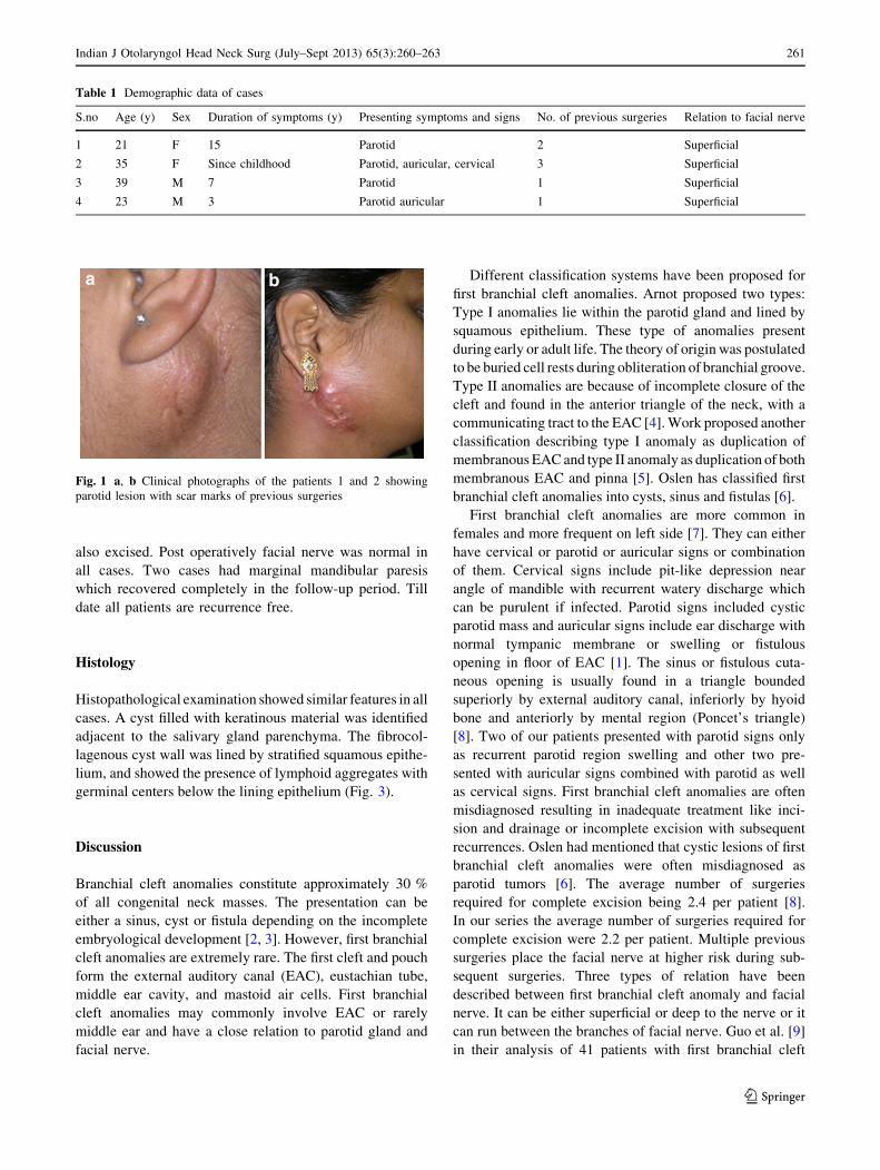
Radiology in the form of computed tomography was done
in all cases (Fig. 2). All patients had previously undergone
surgery in the form of incision and drainage. Facial nerve
was normal in all cases pre-operatively. All patients
underwent superficial parotidectomy along with complete
excision of tract with facial nerve monitoring. In two
cases with auricular signs, cuff of cartilaginous EAC was
R. Kumar � K. Sikka (&) � P. Sagar � A. Thakar
Department of ENT and Head and Neck Surgery, AIIMS,
New Delhi, India
e-mail: [email protected]
A. Kakkar
Department of Pathology, AIIMS, New Delhi, India
123
Indian J Otolaryngol Head Neck Surg
(July–Sept 2013) 65(3):260–263; DOI 10.1007/s12070-013-0641-y
also excised. Post operatively facial nerve was normal in
all cases. Two cases had marginal mandibular paresis
which recovered completely in the follow-up period. Till
date all patients are recurrence free.
Histology
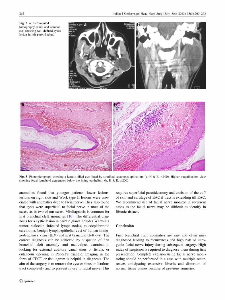
Histopathological examination showed similar features in all
cases. A cyst filled with keratinous material was identified
adjacent to the salivary gland parenchyma. The fibrocol-
lagenous cyst wall was lined by stratified squamous epithe-
lium, and showed the presence of lymphoid aggregates with
germinal centers below the lining epithelium (Fig. 3).
Discussion
Branchial cleft anomalies constitute approximately 30 %
of all congenital neck masses. The presentation can be
either a sinus, cyst or fistula depending on the incomplete
embryological development [2, 3]. However, first branchial
cleft anomalies are extremely rare. The first cleft and pouch
form the external auditory canal (EAC), eustachian tube,
middle ear cavity, and mastoid air cells. First branchial
cleft anomalies may commonly involve EAC or rarely
middle ear and have a close relation to parotid gland and
facial nerve.
Different classification systems have been proposed for
first branchial cleft anomalies. Arnot proposed two types:
Type I anomalies lie within the parotid gland and lined by
squamous epithelium. These type of anomalies present
during early or adult life. The theory of origin was postulated
to be buried cell rests during obliteration of branchial groove.
Type II anomalies are because of incomplete closure of the
cleft and found in the anterior triangle of the neck, with a
communicating tract to the EAC [4]. Work proposed another
classification describing type I anomaly as duplication of
membranous EAC and type II anomaly as duplication of both
membranous EAC and pinna [5]. Oslen has classified first
branchial cleft anomalies into cysts, sinus and fistulas [6].
First branchial cleft anomalies are more common in
females and more frequent on left side [7]. They can either
have cervical or parotid or auricular signs or combination
of them. Cervical signs include pit-like depression near
angle of mandible with recurrent watery discharge which
can be purulent if infected. Parotid signs included cystic
parotid mass and auricular signs include ear discharge with
normal tympanic membrane or swelling or fistulous
opening in floor of EAC [1]. The sinus or fistulous cuta-
neous opening is usually found in a triangle bounded
superiorly by external auditory canal, inferiorly by hyoid
bone and anteriorly by mental region (Poncet’s triangle)
[8]. Two of our patients presented with parotid signs only
as recurrent parotid region swelling and other two pre-
sented with auricular signs combined with parotid as well
as cervical signs. First branchial cleft anomalies are often
misdiagnosed resulting in inadequate treatment like inci-
sion and drainage or incomplete excision with subsequent
recurrences. Oslen had mentioned that cystic lesions of first
branchial cleft anomalies were often misdiagnosed as
parotid tumors [6]. The average number of surgeries
required for complete excision being 2.4 per patient [8].
In our series the average number of surgeries required for
complete excision were 2.2 per patient. Multiple previous
surgeries place the facial nerve at higher risk during sub-
sequent surgeries. Three types of relation have been
described between first branchial cleft anomaly and facial
nerve. It can be either superficial or deep to the nerve or it
can run between the branches of facial nerve. Guo et al. [9]
in their analysis of 41 patients with first branchial cleft
Table 1 Demographic data of cases
S.no Age (y) Sex Duration of symptoms (y) Presenting symptoms and signs No. of previous surgeries Relation to facial nerve
1 21 F 15 Parotid 2 Superficial
2 35 F Since childhood Parotid, auricular, cervical 3 Superficial
3 39 M 7 Parotid 1 Superficial
4 23 M 3 Parotid auricular 1 Superficial
Fig. 1 a, b Clinical photographs of the patients 1 and 2 showing
parotid lesion with scar marks of previous surgeries
Indian J Otolaryngol Head Neck Surg (July–Sept 2013) 65(3):260–263 261
123
anomalies found that younger patients, lower lesions,
lesions on right side and Work type II lesions were asso-
ciated with anomalies deep to facial nerve. They also found
that cysts were superficial to facial nerve in most of the
cases, as in two of our cases. Misdiagnosis is common for
first branchial cleft anomalies [10]. The differential diag-
nosis for a cystic lesion in parotid gland includes Warthin’s
tumor, sialocele, infected lymph nodes, mucoepidermoid
carcinoma, benign lymphoepithelial cyst of human immu-
nodeficiency virus (HIV) and first branchial cleft cyst. The
correct diagnosis can be achieved by suspicion of first
branchial cleft anomaly and meticulous examination
looking for external auditory canal sinus or fistula, or
cutaneous opening in Poncet’s triangle. Imaging in the
form of CECT or fistulogram is helpful in diagnosis. The
aim of the surgery is to remove the cyst or sinus or fistulous
tract completely and to prevent injury to facial nerve. This
requires superficial parotidectomy and excision of the cuff
of skin and cartilage of EAC if tract is extending till EAC.
We recommend use of facial nerve monitor in recurrent
cases as the facial nerve may be difficult to identify in
fibrotic tissues.
Conclusion
First branchial cleft anomalies are rare and often mis-
diagnosed leading to recurrences and high risk of iatro-
genic facial nerve injury during subsequent surgery. High
index of suspicion is required to diagnose them during first
presentation. Complete excision using facial nerve moni-
toring should be performed in a case with multiple recur-
rences anticipating extensive fibrosis and distortion of
normal tissue planes because of previous surgeries.
Fig. 2 a, b Computed
tomography (axial and coronal
cut) showing well defined cystic
lesion in left parotid gland
Fig. 3 Photomicrograph showing a keratin filled cyst lined by stratified squamous epithelium (a, H & E, 9100). Higher magnification view
showing focal lymphoid aggregates below the lining epithelium (b, H & E, 9200)
262 Indian J Otolaryngol Head Neck Surg (July–Sept 2013) 65(3):260–263
123
References
1. Acierno PS, Waldhausen JHT (2007) Congenital cervical cysts,
sinuses and fistulae. Otolaryngol Clin N Am 40:161–176
2. Waldhausen JHT (2006) Branchial cleft and arch anamolies in
children. Semin Pediatr Surg 15:64–69
3. Enepekides DJ (2001) Management of congenital anamolies of
the neck. Facial Plast Surg Clin N Am 9:131–145
4. Arnot RS (1971) Defects of the first branchial cleft. S Afr J Surg
9:93–98
5. Work WP (1972) Newer concepts of first branchial cleft.
Laryngoscope 82:1581–1593
6. Oslen KD, Maragos NE, Weiland LH (1980) First branchial cleft
anamolies. Laryngoscope 90:423–436
7. D’Souza AR, Uppal HS, De R, Zeitoun H (2002) Updating
concepts of first branchial cleft defects : a literature review. Int J
Pediatr Otorhinolaryngol 62:103–109
8. Ford GR, Balakrishman A, Evans JN et al (1992) Branchial cleft
and pouch anamolies. J Laryngol Otol 37:685–690
9. Guo Y-X, Guo C-B (2012) Relation between a first branchial cleft
anomaly and the facial nerve. Br J Oral Maxillofac Surg 50:259–263
10. Tham YS, Low WK (2005) First branchial cleft anomalies have
relevance in otology and more. Ann Acad Med Singapore
34:335–338
Indian J Otolaryngol Head Neck Surg (July–Sept 2013) 65(3):260–263 263
123