Embed Size (px)
Citation preview
Chapter 1
© 2012 Borcic and Braut, licensee InTech. This is an open access chapter distributed under the terms of the Creative Commons Attribution License (http://creativecommons.org/licenses/by/3.0), which permits unrestricted use, distribution, and reproduction in any medium, provided the original work is properly cited.
Finite Element Analysis in Dental Medicine
Josipa Borcic and Alen Braut
Additional information is available at the end of the chapter
http://dx.doi.org/10.5772/50038
1. Introduction
Studying dental structures and surrounding tissues in the oral cavity presents the basis for understanding the occurrence of pathological process and enables the correct approach and treatment. Oral rehabilitation is inherently difficult, due to the functional and parafunctional forces within the mouth that result in extremely complex structural responses by the oral tissue [1]. The success of restorative materials depends on their properties to withstand and resist occlusal forces and successfully support the remaining oral structure [2]. Studies examining the biomechanical behavior of oral structures require sophisticated simulations of the fundaments of the stomatognathic system [3].
There were numerous ways and attempts of experimental research, but due to the complexity of dental structures, composed of various tissue materials mechanically and chemically interconnected, and due to complex tooth morphology and surrounding structures, these attempts failed to obtain precise and reliable results. Researches have used photoelastic methods, computer simulation methods and finite element analysis to conduct stress analyses of sound and restored teeth in order to predict their fracture resistance. Conventional methods such as photoelasticity and the strain-gauge methods are inadequate to predict reliable stress distribution in the tooth [4]. The use of traditional load-to-failure bench-top testing is unable to recreate the failure mechanisms seen clinically; hence the use of FEA is gaining popularity because of its ability to accurately asses the complex biomechanical behavior of irregular prosthetic structures and heterogeneous material in a non-destructive, repeatable manner [5].
2. Finite element analysis
Finite element analysis (FEA) is a numerical method of analyzing stresses and deformations in structures which originated from the need for solving complex structural problems in civil and aeronautical engineering. In order to achieve this goal, the structures are broken
Finite Element Analysis – New Trends and Developments 4
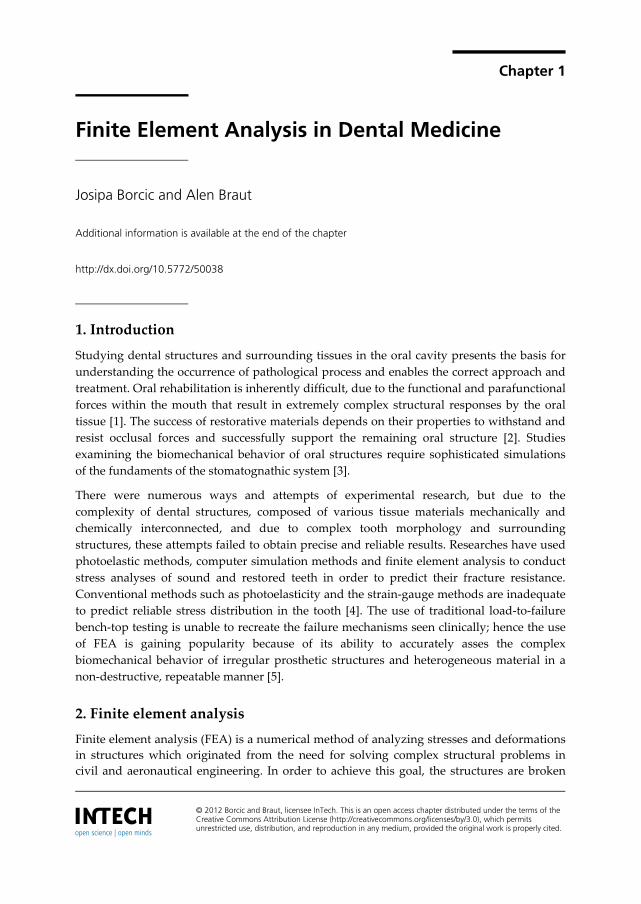
down into many small simple segments or elements, each with specific physical properties (figure 1). Than, an operator uses a computer program in order to obtain a model of stresses produced by various loads [6,7]. A major advantage of finite element analysis (FEA) is its ability to solve complex biomechanical problems for witch other study methods are inadequate. Stress, strain and some other qualities can be calculated in every point throughout the structure. FEA is also being used as part of the design process to simulate possible structure failure, as a mean to reduce the need for making prototypes, and reducing a need for performing actual experiments, that are usually expensive and time-consuming [8]. This method allows researches to overcome some ethical and methodological limitations and enables them to verify how the stresses are transferred throughout the materials [9].
In the area of dentistry, FEA has been used to simulate the bone remodeling process, to study internal stresses in teeth and different dental materials, and to optimize the shape of restorations. Because of the large inherent variations in biological material properties and anatomy, mechanical testing involving biomaterials usually require a large number of samples. With FEA the necessity of traditional specimens can be avoided, and by using a mathematical model it also eliminates the need for large number of experimental teeth. It has been used to represent simulated tooth mechanical behavior under occlusal loads in details [8].
Figure 1. Elements of an FEA model.
Finite Element Analysis in Dental Medicine 5
2.1. Finite element model
The decision to use 2D or 3D models to investigate biomechanical behavior of complex structures, by FEA, depends on many inter-related factors, such as the complexity of the geometry, material, properties, mode of analysis, etc. Although 2D models are simpler, easier to build and less time consuming, they do not represent the complexity of the real problem. 2D model might be considered when studying the qualitative biomechanical behavior, but for the quantitative stress analysis the 2D models overestimate stress magnitudes and do not represent the realistic model. 3D model may provide more reliable data that more accurately represent non-linear and anisotropic materials. 3D models should be carefully created with appropriate mesh density [3]. Khera et al. were the pioneers in the utilization of 3D models. The models were obtained from sectional images of human mandible, but this is no longer required due to the use of a computerized tomography (CT) [10].
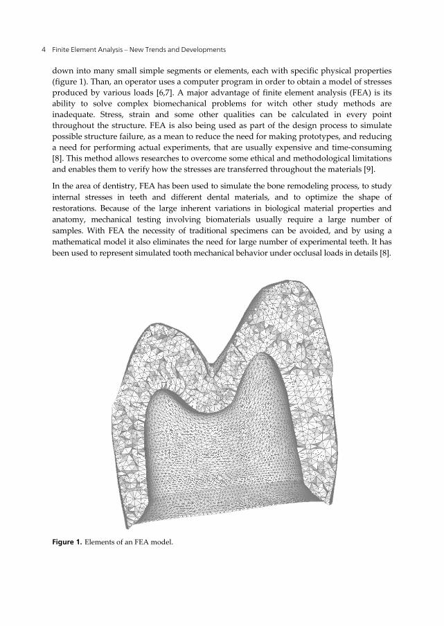
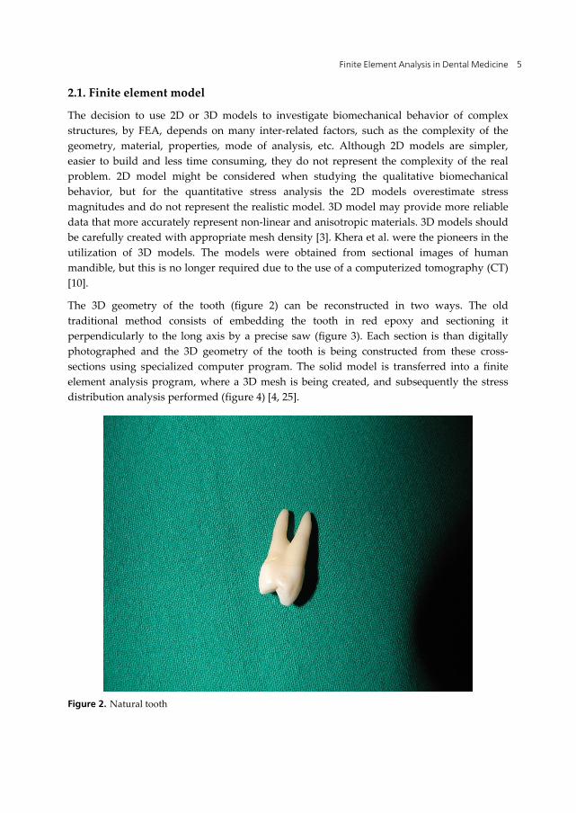
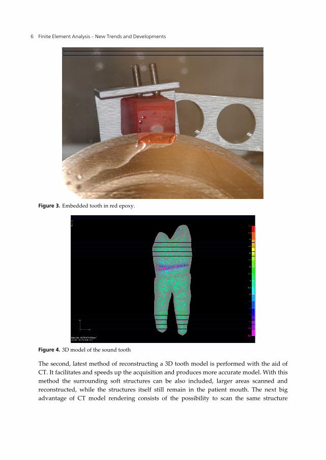
The 3D geometry of the tooth (figure 2) can be reconstructed in two ways. The old traditional method consists of embedding the tooth in red epoxy and sectioning it perpendicularly to the long axis by a precise saw (figure 3). Each section is than digitally photographed and the 3D geometry of the tooth is being constructed from these cross-sections using specialized computer program. The solid model is transferred into a finite element analysis program, where a 3D mesh is being created, and subsequently the stress distribution analysis performed (figure 4) [4, 25].
Figure 2. Natural tooth
Finite Element Analysis – New Trends and Developments 6
Figure 3. Embedded tooth in red epoxy.
Figure 4. 3D model of the sound tooth
The second, latest method of reconstructing a 3D tooth model is performed with the aid of CT. It facilitates and speeds up the acquisition and produces more accurate model. With this method the surrounding soft structures can be also included, larger areas scanned and reconstructed, while the structures itself still remain in the patient mouth. The next big advantage of CT model rendering consists of the possibility to scan the same structure
Finite Element Analysis in Dental Medicine 7
before and after the performed therapy procedures, and periodical follow-ups of the therapy success. Technologies such as micro-CT scanning open up the possibility for complex 3D modeling [11]. However, the process of going from image to mesh involves a number of processing steps, each with potential geometric errors [12].
2.2. Interpretation of the FEA results
The results obtained from a FEA on the restored system contain information about the stress distribution of each component of the restoration, instead of only a single value of failure load typical of in vitro results. A correct interpretation of FEA results should be based on the stresses and strength of each component of the system. To obtain accurate conclusions from these interpretations, three conditions must be fulfilled. First, FEA should adequately represent the real stress values; second, strength of the different materials must be known; third, an adequate failure criterion must be used [13].
It is not possible to implement the results from FEA directly into a clinical situation, but it has to design the model in such a way that is mimics the real situation as closely as possible. FEA analysis must be interpreted with a certain amount of caution. Most of the researches modeled dental structures as isotropic and not othotropic. The finite element model represents a static situation at the moment of load application and not an actual clinical situation. In reality, the loading of the structure is more dynamic and cyclic. The materials of the various tooth structures were assumed to be isotropic, homogenous and elastic, and that they remain such under applied loads. More precise measurements can be obtained if the material properties are set as anizotrophic and non-homogeneous, but such setup requires much more complex mathematical calculations. It is better to use a non-linear elastic-plastic material model than the linear models that are used in most FEA studies [14].
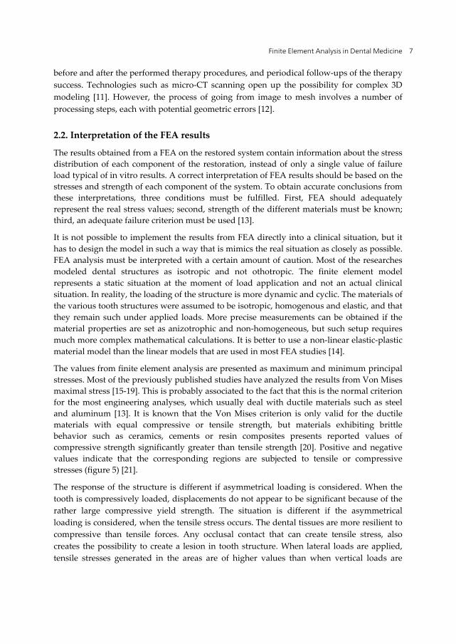
The values from finite element analysis are presented as maximum and minimum principal stresses. Most of the previously published studies have analyzed the results from Von Mises maximal stress [15-19]. This is probably associated to the fact that this is the normal criterion for the most engineering analyses, which usually deal with ductile materials such as steel and aluminum [13]. It is known that the Von Mises criterion is only valid for the ductile materials with equal compressive or tensile strength, but materials exhibiting brittle behavior such as ceramics, cements or resin composites presents reported values of compressive strength significantly greater than tensile strength [20]. Positive and negative values indicate that the corresponding regions are subjected to tensile or compressive stresses (figure 5) [21].
The response of the structure is different if asymmetrical loading is considered. When the tooth is compressively loaded, displacements do not appear to be significant because of the rather large compressive yield strength. The situation is different if the asymmetrical loading is considered, when the tensile stress occurs. The dental tissues are more resilient to compressive than tensile forces. Any occlusal contact that can create tensile stress, also creates the possibility to create a lesion in tooth structure. When lateral loads are applied, tensile stresses generated in the areas are of higher values than when vertical loads are
Finite Element Analysis – New Trends and Developments 8
applied onto the same areas. The increase in the load does not cause a change in the overall stress pattern, but increases the values. The loading, that the tooth is subjected to, may cause cracks in the tooth, but not necessarily its immediate failure. Most of the failures of dental materials used for tooth restorations are caused by tensile stress. Precise occlusal adjustments of teeth occlusal surfaces should be performed to prevent such events. The average chewing force varies between 11 and 150 N, whereas force peaks are 200N in the anterior, 350N in the posterior and 1000N with bruxism [22].
Figure 5. FEA model of a restored apicotomysed tooth
3. Materials and types of reconstructions in dental medicine
The use of different materials for restoration substantially modifies the stress distribution of an originally healthy tooth. The difference between the elastic modulus of tooth and restorative material may be a source of stress in the dental structures. If the stress exceeds the yield strength of the materials, fracture of the restorative materials or the tooth may occur. The occlusal force leaning against the tooth or dental implant axis causes the structure to bend, and the higher tensile stresses are produced. The oblique force loading on the dental structure is the major cause of dental damage and the further attention should be paid to the importance of the occlusal adjustment [4, 7, 25].
The way the chewing force application is much more important than the dentine and the enamel properties, or even the properties of the restorative materials. The consequences of the same chewing force for different teeth also need to be highlighted because structural
Finite Element Analysis in Dental Medicine 9
changes can occur depending upon the magnitude of the force, which can affect the tooth morphology in extreme (premature contacts) or repetitive cases (fatigue) [11].
3.1. Natural tooth
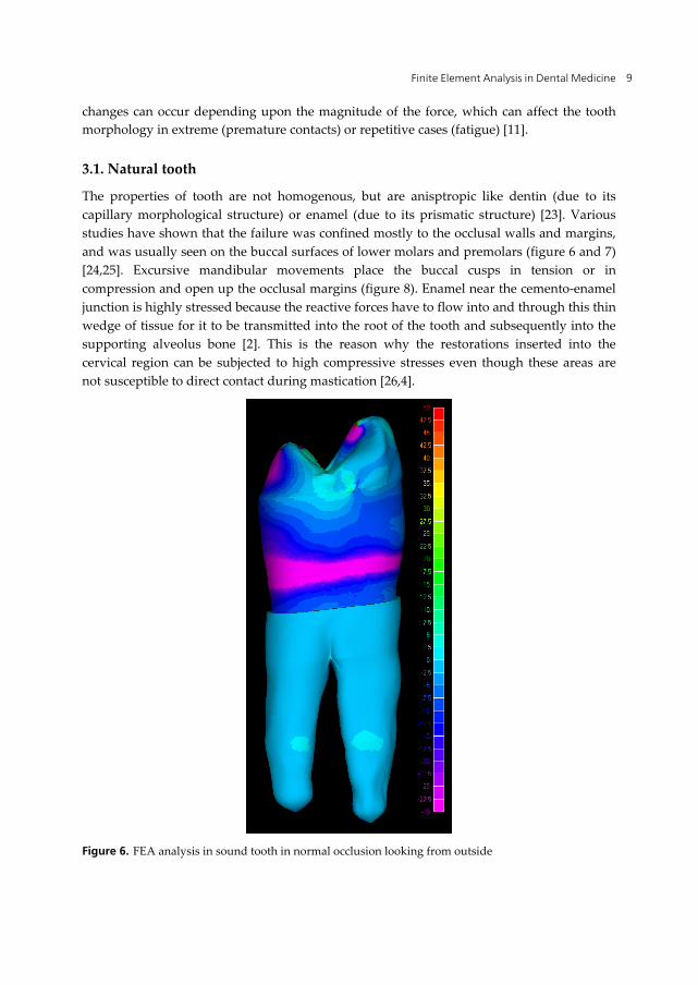
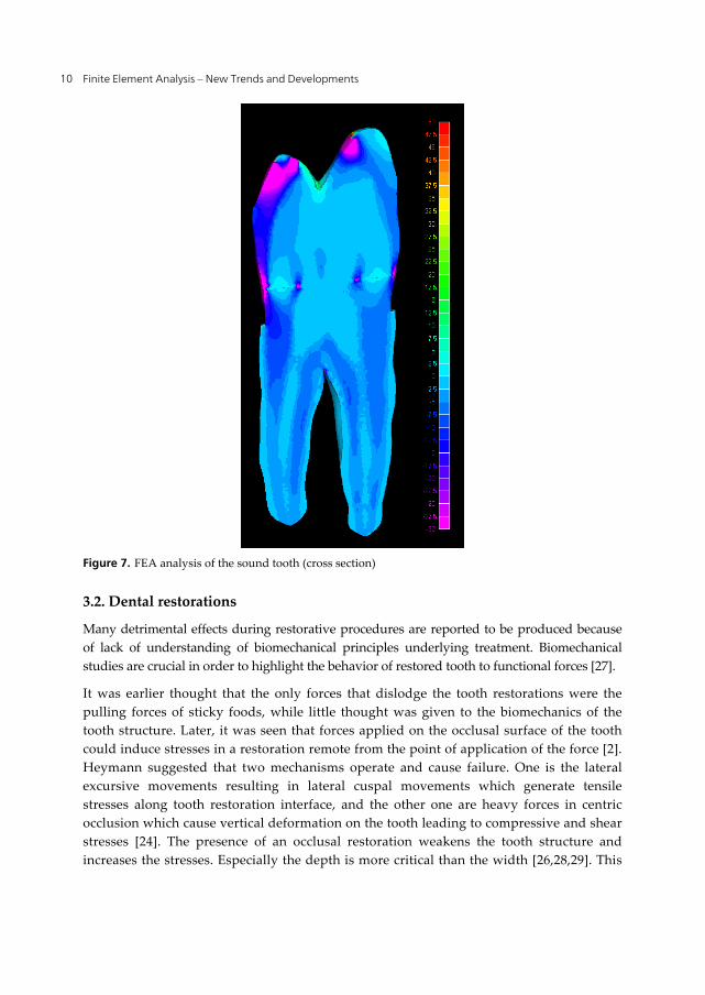
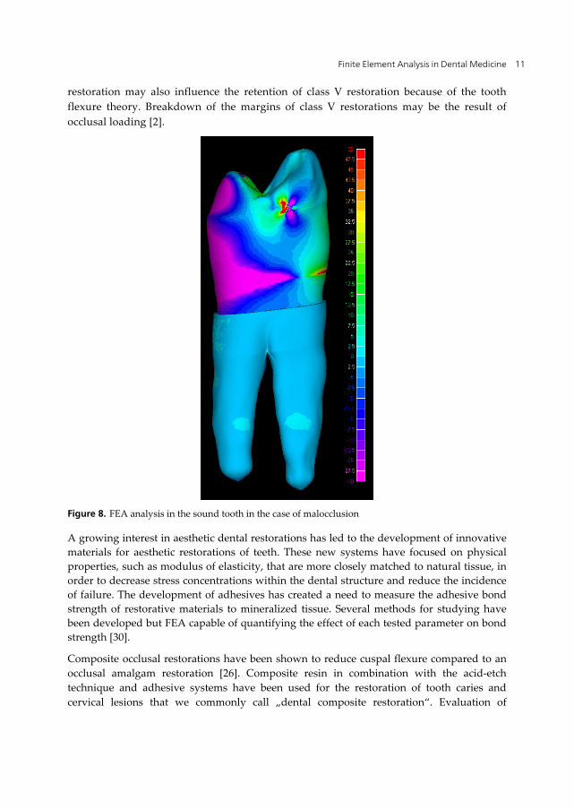
The properties of tooth are not homogenous, but are anisptropic like dentin (due to its capillary morphological structure) or enamel (due to its prismatic structure) [23]. Various studies have shown that the failure was confined mostly to the occlusal walls and margins, and was usually seen on the buccal surfaces of lower molars and premolars (figure 6 and 7) [24,25]. Excursive mandibular movements place the buccal cusps in tension or in compression and open up the occlusal margins (figure 8). Enamel near the cemento-enamel junction is highly stressed because the reactive forces have to flow into and through this thin wedge of tissue for it to be transmitted into the root of the tooth and subsequently into the supporting alveolus bone [2]. This is the reason why the restorations inserted into the cervical region can be subjected to high compressive stresses even though these areas are not susceptible to direct contact during mastication [26,4].
Figure 6. FEA analysis in sound tooth in normal occlusion looking from outside
Finite Element Analysis – New Trends and Developments 10
Figure 7. FEA analysis of the sound tooth (cross section)
3.2. Dental restorations
Many detrimental effects during restorative procedures are reported to be produced because of lack of understanding of biomechanical principles underlying treatment. Biomechanical studies are crucial in order to highlight the behavior of restored tooth to functional forces [27].
It was earlier thought that the only forces that dislodge the tooth restorations were the pulling forces of sticky foods, while little thought was given to the biomechanics of the tooth structure. Later, it was seen that forces applied on the occlusal surface of the tooth could induce stresses in a restoration remote from the point of application of the force [2]. Heymann suggested that two mechanisms operate and cause failure. One is the lateral excursive movements resulting in lateral cuspal movements which generate tensile stresses along tooth restoration interface, and the other one are heavy forces in centric occlusion which cause vertical deformation on the tooth leading to compressive and shear stresses [24]. The presence of an occlusal restoration weakens the tooth structure and increases the stresses. Especially the depth is more critical than the width [26,28,29]. This
Finite Element Analysis in Dental Medicine 11
restoration may also influence the retention of class V restoration because of the tooth flexure theory. Breakdown of the margins of class V restorations may be the result of occlusal loading [2].
Figure 8. FEA analysis in the sound tooth in the case of malocclusion
A growing interest in aesthetic dental restorations has led to the development of innovative materials for aesthetic restorations of teeth. These new systems have focused on physical properties, such as modulus of elasticity, that are more closely matched to natural tissue, in order to decrease stress concentrations within the dental structure and reduce the incidence of failure. The development of adhesives has created a need to measure the adhesive bond strength of restorative materials to mineralized tissue. Several methods for studying have been developed but FEA capable of quantifying the effect of each tested parameter on bond strength [30].
Composite occlusal restorations have been shown to reduce cuspal flexure compared to an occlusal amalgam restoration [26]. Composite resin in combination with the acid-etch technique and adhesive systems have been used for the restoration of tooth caries and cervical lesions that we commonly call „dental composite restoration“. Evaluation of
Finite Element Analysis – New Trends and Developments 12
marginal integrity at the composite resin-tooth interface is required for clinically successful restorations. Polymerization contraction occurs during light curing and may cause marginal disintegration [31]. The maximum stresses due to the shrinkage of the cement layer may cause debonding of the cement layer. This debonding on one side will cause relaxation of stresses at the other side of the restoration and will cause (micro) leakage with all its detrimental effects [14].
The fracture load of the final restoration is the result of the combined effects of bonding between the underlying tooth, the ceramic restoration, and the resin composite cement. Clinical stress distribution in ceramic dental restorations may be quite complex. Several factors are associated with crack initiation and propagation, including the shape, microstructural no homogeneities, the size and distribution of surface flows, residual stresses, ceramic-cement interfacial features, thickness of restorations, different elastic modulus and the magnitude and orientation of the applied load. On the structural factors, the connector areas are the most influential in failure [22]. Traditional load-to–failure testing has proved irrelevant in predicting the clinical performance of ceramics, largely because they cannot recreate the failure mechanisms seen in clinical specimens [5]. The FEA was used to determine the optimal stress distribution in the ceramics bridges that would reduce the risk of connector fracture. The points of greater stress were found within, or near the connector [22].
The FEA demonstrated that with the use of an idealized inlay preparation form and an optimized bridge design emphasizing a broadening of the gingival embrasure, the forces derived from mastication can be adequately distributed to levels which are within the fracture strength of current ceramics [5]. Tensile stresses tend to be more critical than compressive stresses for ceramic materials. The strength of ceramic restorations is significantly affected by the presence of flows or other microscopic defects [32]. Tensile stress concentration at cementation surface of the ceramic layer was suggested to be the predominant factor controlling ceramic failure [33]. Fea showed lower tensile stress levels at the cementation surface than in the area under and between the load points, which could explain the occlusal to cervical direction of fracture seen in the fractographic analysis. Although the polymer crown had a higher fracture resistance than ceramics, a larger amount of the occlusal load was transferred through the tooth, resulting in catastrophic fracture of the tooth. This fracture behavior can limit the use of polymer crowns when compared to ceramic systems [32]. Molar crowns made of stiffer materials are less prone to debonding and crowns made of higher elastic modulus material protect the tooth structures from damage [23].
Veneers used in restorative rehabilitations for anterior teeth are retained by the adhesive systems and resin cements. These restorations are mechanically not strong, because they are made up of a brittle material, but they have good retention due to the resin-dentine bonding. The remaining tooth tissue is the most important factor for the longevity of the veneers where the buccal, cervical region is the most critical region. Teeth totally recover their properties when veneers are placed as a partial enamel substitute. The use of ceramic
Finite Element Analysis in Dental Medicine 13
was a key element since the elasticity modulus of porcelains is matched well with enamel [11].
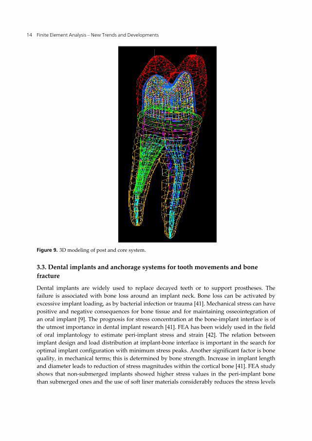
The widely used method for treatment of structurally weakened teeth is the post and core system. This system can be classified into two basic core system, metal posts and cores that are custom cast as a single piece, and two element designs composing a prefabricated post to which other materials core is subsequently adapted [34]. The difference between the elastic modulus of dentine and the post material may be a source of stress for root structures. Debonding of posts because of contraction stress of the cement was found as the most common mode of failure [27]. The effect of post design is also very important for dentinal stress distribution since the placement of a post can create stresses that lead to root fracture (figure 9) [1]. Increased intracanal stresses below the level of crestal bone would explain the higher incidence of deep root fractures in teeth restored with post-retained crowns Horizontal loads generate more dentinal stress than vertical loads. Shorter posts are associated with more dentinal stress concentration around the post apex. Consequently, extending the apical post beyond the level of alveolar bone is essential to avoid stress concentration in the region of the post apex. However, very long posts are associated with higher intracanal stress values. A higher amount of radicular dentin around the post is important in order to reduce dentinal stress concentration within the root [35]. The use of post materials conflicts with the mechanical resistance of teeth because of mismatch in the stiffness with the residual dental structure [36]. Many studies have shown that fiberglass posts give better biomechanical performance. Titanium posts concentrate stress close to the post-cement interface, promoting weakness of restored tooth. Akkayan [37] observed that the fractures occurring with the use of fiberglass and quartz posts systems could be repaired, whereas this was not the case with zirconium and titanium posts. Thus, fiberglass post can be considered a very good choice because they offer good biomechanical performance, provide excellent aesthetics, and exhibit good adhesion to cementing agents [38].
Clinicians generally agree that NiTi rotary files have good properties to produce desirable tapered root canal forms, but also have a risk of fracture during instrumentation. These instruments have been developed to overcome the rigidity of stainless steel instruments [39]. Design of an instrument is the main factor in their mechanical behavior. Cyclic fatigue, which is a failure process associated with repetitive stressing, and torsion have been reported as dominant factors in file fracture [40].
With the application of adhesive technology to endodontics, the term monoblock has become familiar. Monoblock units can be created in a root canal system either by adhesive root sealers in combination with a bondable root filling material or adhesive post systems. The concept of creating mechanically homogenous units within the root dentine is excellent in theory, but accomplishing these ideal monoblock in the canal space is challenging because bonding to dentine is compromised by volumetric changes in resin-based materials, high cavity configuration factors, debris on canal walls, and differences in regional bond strengths [27].
Finite Element Analysis – New Trends and Developments 14
Figure 9. 3D modeling of post and core system.
3.3. Dental implants and anchorage systems for tooth movements and bone fracture
Dental implants are widely used to replace decayed teeth or to support prostheses. The failure is associated with bone loss around an implant neck. Bone loss can be activated by excessive implant loading, as by bacterial infection or trauma [41]. Mechanical stress can have positive and negative consequences for bone tissue and for maintaining osseointegration of an oral implant [9]. The prognosis for stress concentration at the bone-implant interface is of the utmost importance in dental implant research [41]. FEA has been widely used in the field of oral implantology to estimate peri-implant stress and strain [42]. The relation between implant design and load distribution at implant-bone interface is important in the search for optimal implant configuration with minimum stress peaks. Another significant factor is bone quality, in mechanical terms; this is determined by bone strength. Increase in implant length and diameter leads to reduction of stress magnitudes within the cortical bone [41]. FEA study shows that non-submerged implants showed higher stress values in the peri-implant bone than submerged ones and the use of soft liner materials considerably reduces the stress levels
Finite Element Analysis in Dental Medicine 15
in the peri-impant bone interfaces. Different heights and the use of soft liners were relevant in the stress distribution to the bone adjacent to the implants. Better distribution of the stresses will provide a more predictable osseointegration [9].
Prosthesis retention remains a much debated topic in the implant literature. Clinical studies comparing cement- and screw- retained implant restorations reveal no differences in outcomes. There is evidence from laboratory and FEA studies that implants with an internal-type connection exhibit better stress distribution with off-axis loading [43]. The combined use of implants and teeth has been questioned because of the differences of mobility between the abutments. Several authors have concluded that the tooth-implant bond does not have a negative influence on the marginal bone and soft tissues, but special care must be taken in planning in order to compensate for the differences in biomechanical responses between the implant and the tooth [44].
The biomechanical background of orthodontic tooth movement has been explored by many authors, and orthodontic movement principally depends on stress and strain in periodontal ligament (PDL). PDL is a thin connective tissue between the root and bone and play a key role in tooth mobility [12]. Accurate FEA model creation of a tooth and PDL is possible due to the use of micro-CT. Anchorage control in orthodontic treatment is an important factor in treatments outcome. Miniscrews and miniplates are being widely used because of their small size and superiority over endosseous implants due to the fact that they can be immediately loaded. Miniplates have the same features with the plates used in maxillofacial surgery [45]. Good treatment results have been reported by using miniscrews for orthodontic anchorage in various malocclusions, but major problem is their high failure rate. Unlike dental implants, mechanical interdigitation at the cortical bone rather than osseointegration is required for the stability of miniscrews. The placement angle, the type of miniscrews, and the direction of forces significantly affect the distribution area and the amount of stress [46]. Inadequate design and non-homogenous force distribution can cause stress directly effecting on the screws and may impair screws stability. Mobile plates can irritate the surrounding tissue and may cause inflammation. The FEA study revealed that the new miniplates are highly efficient in reducing stress on the fixation screws [45].
Fractures of the mandibular angle are the most problematic in the facial region because of the high frequency of complications and difficult surgical access to the site [47]. Infection and nonunion are commonly reported after rigid internal fixation of these fractures [48]. The stress analyses obtained from FEA modeling can provide information regarding interactions between hardware and bone during normal patient functioning. A single tension band on the superior borders provided more angle fracture stability than a single bicortical plate placed inferiorly. This results support the use of the single tension band configuration as a less invasive fixation approach to fractures [47].
4. General guidelines
The results of the finite element analysis must be interpreted with a certain amount of caution. Most of the researches modeled dental structures as isotropic and not othotropic.
Finite Element Analysis – New Trends and Developments 16
The finite element model represented a static situation at the moment of load application and not an actual clinical situation. In reality, the loading of the structure is more dynamic and cyclic. More precise measurements could be obtained if the material properties are set as anisotropic and non-homogeneous, but such setup requires much more complex mathematical calculations.
To obtain better understanding of the tooth lesions, which is important for the clinical treatment and restoration of damage, analyses of stress distribution in the oral cavity under various loading condition are highly desirable. FEA is a valuable tool for investigation of stress distribution within various types of reconstructions and prosthodontic appliances in dental medicine.
The dental profession is influenced by various sources of information, which may be considered as “evidence-based” (controlled clinical studies) and “expert opinion”. A realistic approach is to identify the strengths and weaknesses of the available clinical data and combine it with clinical experience [43]. Most researchers in FEA assumed that all materials used were homogenous, isotropic and linearly elastic. However, this assumption does not reflect the exact situation. The periodontal ligament has nonlinear mechanical properties and the bone is inhomogeneous [9,35]. The 3D analysis permits high efficiency when the biomechanical behavior of the structure needs to be evaluated under different loading conditions. In the last four decades many studies have shown how the Finite Element Analysis applied to dental mechanics has become a popular numerical method to investigate the critical aspects related to stress distribution. The use of more detailed 3D models could be helpful in understanding critical problems related to the restorative material choice and optimal application procedures. Improved computer and modeling techniques render the FEA a very reliable and accurate approach in biomechanical applications [9].
The results from FEA confirm the concept that the interfaces of materials with different module of elasticity represent the weak point of restorative systems. Restorations with material having a similar elastic modulus to tooth can save and strengthen the remaining tooth structure [27]. Combining fatigue experiments with FEA may eliminate, or at least minimize, experimental limitations by correlating fatigue failure to stress instead of specific testing configuration.
5. Conclusions
There are numerous ways and attempts of experimental research, but due to complexity of dental structures, composed of various tissue materials mechanically and chemically interconnected, and due to complex tooth morphology and surrounding structures, most of these attempts fail to present precise and reliable results.
The 3D analysis permits high efficiency when the biomechanical behavior of the structure should be evaluated under different loading conditions. In the biomedical fields, the FEA is an important tool since it can avoid the necessity of traditional specimens, and by using a
Finite Element Analysis in Dental Medicine 17
mathematical model it eliminates the need of large number of teeth. The use of more detailed 3D models is helpful in understand critical problems related to the restorative material choice and optimal application procedures. Improved computer and modeling techniques render the FEA a very reliable and accurate approach in biomechanical applications.
When the tooth is compressively loaded, displacements do not appear to be significant because of its rather large compressive yield strength. The situation is different when asymmetrical loading is considered and tensile stress occurs. The dental tissues are more resilient to compressive than tensile forces. Any occlusal contact that can create tensile stress, also creates the possibility to create a lesion in tooth structure. Most of the failures of dental materials used for tooth restorations are caused by tensile stress. Precise occlusal adjustments of teeth occlusal surfaces should be performed to prevent such events. The difference between the elastic modulus of tooth and restorative material may be a source of stress in the dental structures. If the stress exceeds the yield strength of the materials, fracture of the restorative materials or the tooth may occur.
The FEA helps to improve preparation designs, indicates the right material or combination of materials to be used in various load and stress conditions in order to reduce material and/or tooth failure in clinical practice.
Author details
Josipa Borcic* Department of Oral and Maxillofacial Surgery, Medical Faculty, School of Dental Medicine, University of Rijeka, Rijeka, Croatia
Alen Braut Department of Restorative Dentistry and Endodontics, Medical Faculty, School of Dental Medicine, University of Rijeka, Rijeka, Croatia
6. References
[1] Silva NR, Castro CG, Santos-Filho PCF, Silva GR, Campos RE, Soares OV, Soares CJ. Influence of different post design and composition on stress distribution in maxillary central incisor: Finite element analysis. Indian J Dent Res 2009;20:153-158.
[2] Vasudeva G, Bogra P, Nikhil V, Singh V. Effect of occlusal restoration on stresses around class V restoration interface: A finite-element study. Indian J Dent Res 2011;22:295-302.
[3] Poiate IAVP, Vasconcellos AB, Mori M, Poiate E Jr. 2D and 3D finite element analysis of central incisor generated by computerized tomography. Computer method and programs in biomedicine 2011;104:292-299.
[4] Borcic J, Antonic R, Muhvic Urek M, Petricevic N, Nola-Fuchs P, Catic A , Smojver I. 3-D Stress Analysis in Premolar, Coll. Antropol. 31 (2007) 4: 315–319.
* Corresponding Author
Finite Element Analysis – New Trends and Developments 18
[5] Thompson MC, Field CJ, Swain MV. The all-ceramic, inlay supported fixed partial denture. Part 2. Fixed partial denture design: a finite element analysis. Australian Dental Journal 2011;56:301-311.
[6] Ding X, Zhu XH, Liao SH, Zhang XH, Chen H. Implant-bone interfaces stress distribution in immediately loaded implants of different diameters: a three-dimensional finite element analysis. J Prosthodont 2009;18:393-402.
[7] Zienkiewicz OC, Taylor RL. The Finite Element Method. 5th ed. Oxford, England: Butterworth-Heinemann 2000;1:1-20.
[8] Haiyan L, Jianying L, Zhenmin Z, Fok ASL. Fracture simulation of restored teeth using a continuum damage mechanics failure model. Dental Materials 2011;27:e125-e133.
[9] Santos MBF, Silva Neto JP, Consani RLX, Mesquita MF. Three-dimensional finite element analysis of stress distribution in peri-implant bone with relined dentures and different heights of healing caps. Journal of Oral Rehabilitation 2011;38:691-6.
[10] Khera SC, Goel VK, Chen RCS, Gurusami SA. A three-dimensional element model. Operative Dentistry 1988;13:128-137.
[11] Matson MR, Lewgoy HR, Barros Filho DA, Amore R, Anido-Anido A, Alonso RCB, Carrilho MRO, Ansuate-Netto C. Finite element analysis of stress distribution in intact and porcelain veneer restored teeth. Computer Methods in Biomechanics and Biomedical Engineering 2011;iFirst article:1-6.
[12] Hohmann A, Kober C, Young P, Dorow C, Geiger M, Boryor A, Sander FM, Sander C, Sander FG. Influence of different modeling strategies for the periodontal ligament on finite element simulation results. American Journal of Orthodontics and Dentofacial Orthopedics 2011;139:775-783.
[13] Perez-Gonzalez A, Iserte-Vilar JL, Gonzalez-Lluch C. Biomedical Engineering http://www.biomedical-engineering-online.com/content/10/1/44 (accessed 10 March 2012).
[14] Jongsma LA, Jager Ir. N, Kleverlaan CJ, Feilzer AJ. Reduced contraction stress formation obtained by a two-step cementation procedure for fiber posts. Dental Materials 2011;27:670-676.
[15] Pegoretti A, Fambri L, Zappini G, Biachetti M. Finite element analysis of glass fibre reinforced composite endodontic post. Biomaterials 2002;23:2667-2682.
[16] Asmussen E, Peutzfeldt A, Sahafi A. Finite element analysis of stresses in endodontically treated, dowel-restored teeth. Journal of Prosthetics Dentistry 2005;94:321-329.
[17] Genocese K, lamberti L, pappalettere C. Finite element analysis of a new customized composite post system for endodontically treated teeth. Journal of Biomechanics 2005;38:2375-2389.
[18] Sorrentino R, Aversa R, Ferro V, Auriemma T, Zarone F, Ferrari M et al. Three-dimensional finite element analysis of strain and stress distributions in endodontically treated maxillary central incisors restored with different post, core and crown materials. Dental Materials 2007;23:983-993.
[19] Gonzalez-Lluch C, Rodriguez-Cervantes PJ, Sancho-Bru JL, perez-Gonzalez A, barjau-Escribano A, Vergara-Monedero M et al. Influence of material and diameter of pre-
Finite Element Analysis in Dental Medicine 19
fabricated posts on maxillary central incisors restored with crown. Journal of Oral Rehabilitation 2009;36:737-747.
[20] Craig R, Powers JM. Restorative Dental Materials. St Louis Mosby 2002;11 [21] Lang L, Wang RF. Validation of finite element analysis in dental ceramics research.
Journal of Prosthetics Dentistry 2001;86:650-654. [22] Rezaei SMM, Heidarifar H, Arezodar FF, Azary A, Mokhtarykhoee S. Influence of
Connector Width on the Stress Distribution of Posterior Bridges under Loading. Journal of Dentistry 2011;8:67-74.
[23] (N-23) Dejak B, Mlotkowski A, Langot C. Three-dimensional finite element analysis of molars with thin-walled prosthetic crowns made of various materials. Dental Materials 2012;28:433-441.
[24] Heymann HO, Sturdevant JR, Bayne S. Examining tooth flexure effects on cervical restorations.: a two year clinical study. Journal of American Dental Assosiations 1991;122:41-47.
[25] Borcic J, Anic I, Smojver I, Catic A, Miletic I, Pezelj Ribaric S. 3D finite element model and cervical lesion formation in normal occlusion and in malocclusion. Journal of Oral Rehabilitation 2005;32:504-510.
[26] Lee MR, Cho BH, Son HH, Um CM, Lee IB. Influence of cavity dimension and restoration methods on the cusps deflection of premolars in composite restoration. Dental Materials 2007;23:288-295.
[27] Belli S, Eraslan O, Eskitascioglu G, Karbhari V. Monoblocks in root canals: a finite elemental stress analysis study. International Endodontic Journal 2011;44:817-826.
[28] Hood JA. Biomechanical of the intact, prepared and restored tooth. Some implication an adaptive finite-element approach fo the analysis of dental restorations. International Dental Journal 1991;41:25-32.
[29] Goel VK, Khera SC, Gurusami S, Chen RC. Effect of cavity depth on stresses in restored tooth. Journal of Prosthetic Dentistry 1992;67:174-183.
[30] Ferreira RC, Caldas J, Paula GA, Albuquerque RC, Almeida CM, Vasconcellos WA, Caldas RB. Influence of surface area and geometry of specimens on bond strength in a microtensile test: an analysis by the three-dimensional finite element method. Journal of Prosthodontics 2011;20:456-463.
[31] Choi NS, Gu JU, Arakawa K. Acoustic emission characterization of the marginal disintegration of dental composite restoration. Composites 2011;42:604-611.
[32] Campos RE, Soares CJ, Quagliatto PS, Soares PV, Batista de Oliviera O, Santos-Filho PCF, Salazar-Marocho SM. In vitro study of fracture load and fracture pattern of ceramic crowns: a finite element and fractography analysis. Journal of Prosthodontics 2011;20:447-455.
[33] Dong XD, Darvell B. Stress distribution and failure mode of dental ceramic structures under Hertzian indentation. Dental Materials 2003;19:542-51.
[34] Adanir N, Belli S. Stress analysis of a maxillary central incisor restored with different posts. European Journal of Dentistry 2007;2:67-71.
Finite Element Analysis – New Trends and Developments 20
[35] Al-Omiri MK, Rayyan MR, Abu-Hammad O. Stress analysis of endodontically treated teeth restored with post-retained crowns: A finite element analysis study. The Journal of the American Dental Association 2011;142:289-300.
[36] Ausiello P, Franciosa P, Martorelli M, Watts D. Mechanical behaviour of post-restored upper canine teeth: A 3D FE analysis. Dental Materials 2011;27:1285-1294.
[37] Akkayan B, Gulmez T. Resistance to fracture of endodontically treated teeth restored with different post systems. Journal of Prosthetic Dentistry 2002;23:2667-2682.
[38] Cooney JP, Caputo AA, Trabert KC. Retention and stress distribution of tapered-end endodontic posts. Journal of Prosthetics Dentistry 1986;55:540-546.
[39] LeeMH, Versluis A, Kim BM, Lee CJ, Hur B, Kom HC. Journal of Endodontics 2011;37:1152-1157.
[40] Sattapan B, Nervo GJ, palamara JE, Messer HH. Defects in rotary nickel-titanium files after clinical use. Jounal of Endodontics 2000;26:161-165.
[41] Demenko V, Linetskiy I, Nesvit K, Shevchenko A. Ulitmate masticatory force as a criterion in implant selection. Journal of Dental Research 2011;90:1211-1215.
[42] Geng JP, Tan KB, Liu GR. Application of finite element analysis in implant dentistry: a review of the literature. Journal of Prosthetic Dentistry 2001;85:585-598.
[43] Lewis MB, Klineberg I. Prosthodontic considerations designed to optimize outcomes for single-tooth implants. A review of the literature. Australian Dental Journal 2011;56:181-192.
[44] Lanza MDS, Seraidarian PI, Jansen WC, Lanza MD. Stress analysis of a fixed implant-supported denture by the finite element method (FEM) when varying the number of teeth used as abutments. J Appl Oral Sci 2011;19:655-661.
[45] Nalbantgil D, Tozlu M, Ozdemir F, Oztoprak MO, Arun T. FEM analysis of a new miniplate: stress distribution on the plate, screws and the bone. European Journal of Dentistry 2012;6:9-15.
[46] Suzuki A, Masuda T, Takahashi I, Deguchi T, Suzuki O, Takana-Yamamoto T. Changes in stress distribution of orthodontic miniscrews and surrounding bone evaluated by 3-dimensional finite element analysis. Americam Journal of Orthodontics and Dentofacial Orthopedics 2011;140:e273-e280.
[47] Kimsal J, Baack B, Candelaria L, Khraishi T, Lovald S. Biomechanical analysis of mandibular angle fractures. Journal of Oral and Maxillofacial Surgery 2011;69:3010-3014.
[48] Mathog RH, Toma V, Clayman L et al. Nonunion of the mandible: An analysis of contributing factors. Journal of Oral and Maxillofacial Surgery 2000;59:746.