Embed Size (px)
Citation preview
Fine Structure of Rat Pituitary Cilia' ESTHER CARPENTER Department of Biological Sciences,2 Clark Science Center, Smith College, Northampton, Massachusetts 01 060
ABSTRACT Pituitary glands of 30 0 rats from one to seven and one-half months old were fixed in glutaraldehyde followed by osmium tetroxide. One five-month gland was fixed in Karnovsky's paraformaldehyde mixture.
Cilia in the residual cleft at all ages and in the electron lucid vesicles found in the pars distalis during the first two months occur in great numbers per cell and show the 9 -k 2 pattern (9 peripheral doublets around 2 central tubules slightly separated from each other) characteristic of kinocilia. The spaces into which these cilia project may contain disintegrating cells as well as colloid-like material of variabsle density.
Cilia found in or between parenchymal cells of the pars distalis usually occur one per cell and lack central tubules. One of the doublets is often displaced to- ward the center. Cilia have been seen in three types of granular as well as in non-granular cells. A pair of cilia enclosed in the same membrane was found once within and twice between parenchymal cells.
Central tubules appear to develop only in locations (cleft and vesicles) where motility is possible. The single cilia found in parenchymal cells may be consid- ered immature arid are presumably non-functional.
Observations by Fawcett and Porter ( '52) and by Manton ('52) of the similar- ity in number and arrangement of fila- ments or cylinders in cilia of organisms widely separated phylogenetically have been amply confirmed in a great variety of organisms by these and subsequent investi- gators (see reviews by Rivera, '62; Sleigh, '62). In electron micrographs cilia have been found since then in many locations where the function served is not obvious including the endocrine glands of mam- mals (Coupland, '65; Kagayama, '65; Dahl, '67).
Cilia reported by Kurosumi et al. ('61) in d' rats on the pars intermedia side of the residual cleft of the pituitary gland have the characteristic structure associated with motile cilia : two central tubules sur- rounded by a ring of nine double tubules. The motility of these cilia in the rat has been verified with the phase microscope by the present author in both males and fe- males of a wide range of ages (Carpenter, '62). Motility was olclserved also in cilia found in bits of fresh tissue from the parenchyma of the anterior lobe.
A N ~ T . REC., 169: 637-650.
The present study is an effort to locate the cells of origin of cilia more precisely and to determine whether variations in fine structure of these organelles can be cor- related with their location in the gland or the type of cells in which they arise. A brief note appeared in 1968 (Carpenter and Mada).
MATERIALS AND METHODS
The pituitary glands used were taken from 0 Sprague-Dawley rats raised in our laboratory. Thirty were from virgin females one to six months old plus one additional gland from a lactating rat seven and one- half months old.
During removal from the sella turcica, each gland was flooded with cold, buffered 2 or 3% glutaraldehyde. After a median sagittal cut the gland was left not less than two hours in fresh glutaraldehyde solution, then washed and stored in cold phosphate buffer solution from one to several days. Additional cuts were made and the loca- tion of each piece noted before post-fixa-
Received June 11, '70. Accepted Sept. 28, '70. 1 Supported in part by USPHS grant AM-5261 2 Contribution 26.
637
638 ESTHER CARPENTER
tion in 1% buffered osmium tetroxide for one to two hours. One five-month gland was fixed by Karnovsky's ('65) paraformal- dehyde method. After dehydration and epon embedding of the bits of tissue, thin sections were cut on an MT-2 Porter-Blum ultratome and stained with uranyl acetate followed by lead citrate. Micrographs were taken with a Philips 200 electron micro- scope at 60 kv. The majority of the sections were from the median regions of the pars anterior and toward the ventral side.
Micrographs were examined for cilia ex- tending into the residual cleft from either the intermedia or pars distalis cells; for cilia arising from cells lining the small vesicles numerous during the first two months in the pars distalis; and for single or double cilia lying in or between paren- chymal cells of the pars distalis.
OBSERVATIONS
Residual cleft and vesicle cilia In the lining of the residual cleft, cilia-
bearing cells are more often found on the intermedia surface than on that of the pars distalis. When the cilia are cut longitudin- ally, as in figure 1, the tubules appear to be symmetrically arranged and frequently can be followed for nearly half their length. Living cilia measured under the phase microscope reach a length of 7 (Carpen- ter, '62). When striated lateral rootlets can be seen in adjacent cilia as in figure 1 they are oriented in the same direction. Cilia- bearing cells in this region are thickly studded near the surface with parts of marly cilia as in figure 2. No secretory granules are present. In transverse sections of cilia (figs. 3, 4) the typical arrangement associated with functional capacity is ap- parent : nine peripherally located doublets enclosing two central tubules. Central tubules are not only present but aligned in the same direction (fig. 3).
The small, electron-lucid vesicles usually present in the ventral half of the pars distalis during the first two months are lined with cells of variable height. Some are rounded distally and bear both cilia and microvilli (fig. 5). Microtubules, mito- chondria and lysosome-like bodies are fre- quent (fig. 6). Lateral extensions from the bases of the cilia are shown in two of those in figure 6 and one in figure 7. Those in
figure 6 are oriented in the same direction. The 9 + 2 pattern of tubules associated with functional capability is clearly shown in the transverse section of a cilium in figure 8.
Thus cilia present in either the residual cleft or in the lining of vesicles found in the pars distalis during the first two months have in common large numbers of cilia per cell and the 9 + 2 pattern of tubules which is characteristic of functional organelles found in other locations. When many transverse sections appear in the same field it is apparent also that the central pairs of tubules show a directional orientation characteristic of mature kinocilia.
Parenchymal cell cilia of the pars distalis
Single cilia have been found both in and between the granular cells of the pars distalis as well as arising from non-granu- lar cells of this region. Infrequently a pair of cilia has been found embedded within a cell or lying within the same membrane ex tr acellul arly .
The longitudinal sections of cilia shown in figures 9, 10 and 11 were found in cells characterized in each case by granules of a different maximum diameter: 150 mp; 250 mp; and 350 mp. In figures 9 and 10 the ciliary tubules appear to run a straight course, while in figure 11 there is some dis- placement not far from the base of the cilium. In figure 9 there is some indication of a partition between the cilium and the basal body. A deep invagination of the plasma membrane with a sac-like swelling around the base of the cilium (figs. 10, 11) is not uncommon. Striated lateral rootlets are apparent in figure 11. A centriole lying at an angle to the base of the cilium is shown in both figures 10 and 11.
The pattern of the ciliary tubules in par- enchymal cells is more readily studied in sections where these organelles have been cut transversely (fig. 12-15). Whether lo- cated intracellularly (figs. 12, 13) or be- tween cells (figs. 14, 15) one of the peri- pheral doublets is frequently displaced centrally. Occasionally, however, one does find a symmetrical ring of nine doublets with no central tubules as shown in figure 15. No typical central tubules such as those in figure 8 have been found in par-
F[NE STRUCTURE OF RAT PITUITARY CILIA 639
enchymal cell cilia fixed in glutaraldehyde followed by osmium tetroxide. In a five month gland fixed in Karnovsky's para- formaldehyde mixture, however, a cilium was found (fig. 16) with a less homo- genous central area which could be inter- preted as containing the beginnings of cen- tral tubules. In figure 117, showing a pair of cilia, the central area of the one with nine peripheral doublets is more uniform in texture.
In cilia arising from parenchymal cells of the pars distalis, the almost invariable lack of anything resembling typical central tubules is associated with conditions in which there is little if any room for move- ment of these organelles. In the cleft and in the vesicles where conditions for motil- ity are present, on the other hand, we find the full development of the two central tubules of the 9 + 2 pattern.
Irregularities in ciliary pattern Irregularities in number or arrange-
ment of tubules other than those already described are occasionally found when one examines large numbers of transverse sec- tions of cilia. In the upper half of figure 2 extra doublets can be seen lying near two of the cilia. In figure 4 the upper left cilium has apparently normal central tu- bules but one of the peripheral doublets is missing leaving a gap jn the circle formed by the other eight.
DISCU S8ION
Mature kinocilia such as are found in the trachea (Rhodin and Dalhamn, '56) and the oviduct (Yasuzumi and Wakisaka, '56) show in cross-section nine peripherally arranged double tubules around a central pair which do not share a common wall. When fully developed the central tubules of cilia in the same region tend to become oriented in the same direction. Frisch and Farbman ('68) studying the nasal epithe- lium in mouse embryos have shown that the arrangement of central tubules and of basal feet is still ranclom up to 15 to 18 days of gestation.
Cilia found in the residual cleft and in the vesicles of the pars distalis of the rat
cilia in these two locations have the char- acteristics of motile cilia and probably function in moving the material found in these spaces. There is no doubt about the motility of cilia found on intermedia cells since this region can be isolated fairly easily and examined with phase optics in the living state (Carpenter, '62). As in other regions where motile cilia are found there are a great many cilia arising from each cell.
Unlike the cilia-bearing cells of the cleft or of the vesicles, parenchymal cells of the pars distalis seldom produce more than one cilium. In material fixed in glutarlde- hyde followed by osmium tetroxide no trace of central tubules has been found, although one of the doublets was frequently displaced centrally. Wheatley ('67) ob- served the 9 + 0 pattern in a 50-day old 0 rat of the Sprague-Dawley strain and rear- rangement of doublets as the cilia tapered. As seen in figure 11 of the present study, displacement of a peripheral doublet can begin quite close to the base of the cilium. Dahl ('67) noticed a similar displacement in some of the cilia found in the mouse adenohypophysis. He suggests also that lack of central filaments represents a de- velopmental stage rather than a mecia1 adaptation as intimated by Barnes ('61).
Wheatley ('67) in the 50-day 0 rat ob- served a pair of cilia lying in the same in- dentation of the plasma membrane. We have seen pairs of cilia in our material three times: once in a five-month gland and twice in the gland of a seven and one- half month lactating rat.
In 1954 Fawcett and Porter reported that separating cross-walls are absent be- tween the cilium and basal body in the cilia of mammals (mouse and man). Some of our figures (plate 3) suggest that this re- gion should be reexamined using aldehyde fixation.
Heavily ciliated mucous cysts such as have been seen with the light microscope in pituitary glands of 8 rats of different ages (Carpenter et al., '64) have not been encountered in the thin sections of female glands.
ACKNOWLEDGMENTS
have a pair of central tubules and in adja- The author is greatly indebted to Mrs. cent cilia these are oriented in the same Eleanor Mada for her constant interest and direction. Morphologically, therefore, the for her expert technical assistance.
640 ESTHER CARPENTER
Thanks are also due Mr. Ed Russell for his careful breeding records and his han- dling of the animals.
LITERATURE CITED Barnes, B. G. 1961 Ciliated secretory cells in
the pars distalis of the mouse hypophysis. J. ‘L‘ltrastruc. Res., 5: 453-467.
Carpenter, E. 1962 Motile cilia in the rat pi- tuitary gland. Am. Zool., 2: 397.
Carpenter, E., D. Pitochelli and G . Barton 1964 “Colloid” vesicles, mucous cysts and cleft col- loid in the male rat pituitary at different ages from 25 days to 20 months. Am. Zool., 4: 400- 4131.
Carpenter, E., and E. Mada 1968 Fine structure of pituitary cilia in the female albino rat. Anat. Rec., 160: 462.
Coupland, R. E. 1965 Electron microscopic ob- servations on the structure of the rat adrenal medulla. J. Anat., 99: 231-254.
Dahl, H. A. 1967 On the cilium cell relation- ship in the adenohypophysis of the mouse. Zeitschr. f. Zellforsch., 83: 169-177.
Fawcett, D. W., and K. R. Porter 1952 A study of the fine structure of ciliated epithelial cells with the electron microscope. Anat. Rec., 113: 539.
__- 1954 A study of the fine structure of ciliated epithelia. J. Morph., 94: 221-282.
Kagayama, M. 1965 The follicular cells i n the pars distalis of the dog pituitary gland: an electron microscope study. Endocrinology, 77:
Karnovsky, M. J. 1965 A formaldehyde-glutar- aldehyde fixative of high osmolality for use in electron microscopy. J. Cell Biol., 27: 137A- 138A.
Kurosumi, K., T. Matsuzawa and S. Shibasaki 1961 Electron microscope studies on the fine structures of the pars nervosa and pars inter- media and their morphological interrelation in the normal rat hypophysis. Gen. and Comp. Endocrin., 1: 433-452.
Manton, I. 1952 The fine structure of plant cilia. Symp. SOC. Exp. Biol., 6: 306-319.
Rhodin, J., and T. Dalhamn 1956 Electron microscopy of the tracheal ciliated mucosa in the rat. Zeitscher. f. Zellforsch. mikros. Anat., 44: 345-412.
Rivera, J. A. 1962 Cilia, ciliated epithelium and ciliary activity. Pergamon Press.
Sleigh, M. A. 1962 The biology of cilia and flagella. Pergamon Press, The Macmillan Co., New York.
Wheatley, D. N. 1967 Cells with two cilia in the rat adenohypophysis. J. Anat., 101: 479-485.
Yasuzumi, G., and I. Wakisaka 1956 A com- parative study of kinocilia and stereocilia as revealed by electron microscopy. Cytologia, 21 : 157-164.
1053-1060.
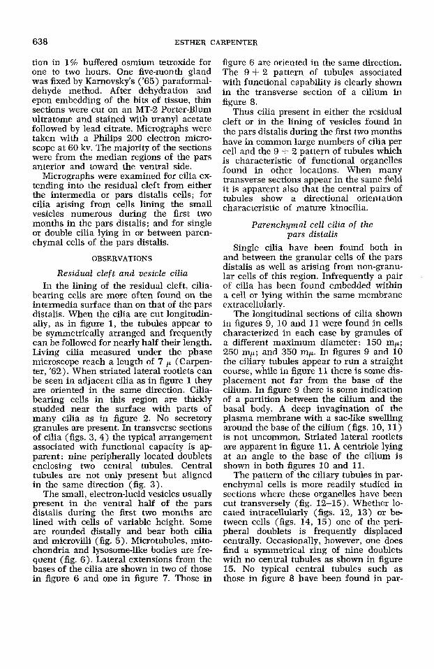
PLATE 1
EXPLANATION OF FIGURES
A longitudinal section through residual cleft cilia of a two-month 0 rat pituitary. Central tubules are evident. Two of the cilia show striated lateral extensions from the basal region. x 27,600.
Figs. 2-4
Parts of many cilia appear in the same cell. Extra doublets are present near two of the cilia in the upper half of the figure. No secretory granules are found. x 27,300.
Cross-sections of cleft cilia show the characteristic 9 + 2 pattern of tubules with the central pair slanted in the same direction. x 46,800.
The difference between the central pair of tubules and the doublets is seen clearly here. Note that the cilium at the upper left lacks one of the peripheral doublets but the central pair appears to be normal. x 80,700.
Cilia from the cleft region of a three-month old 0 rat.
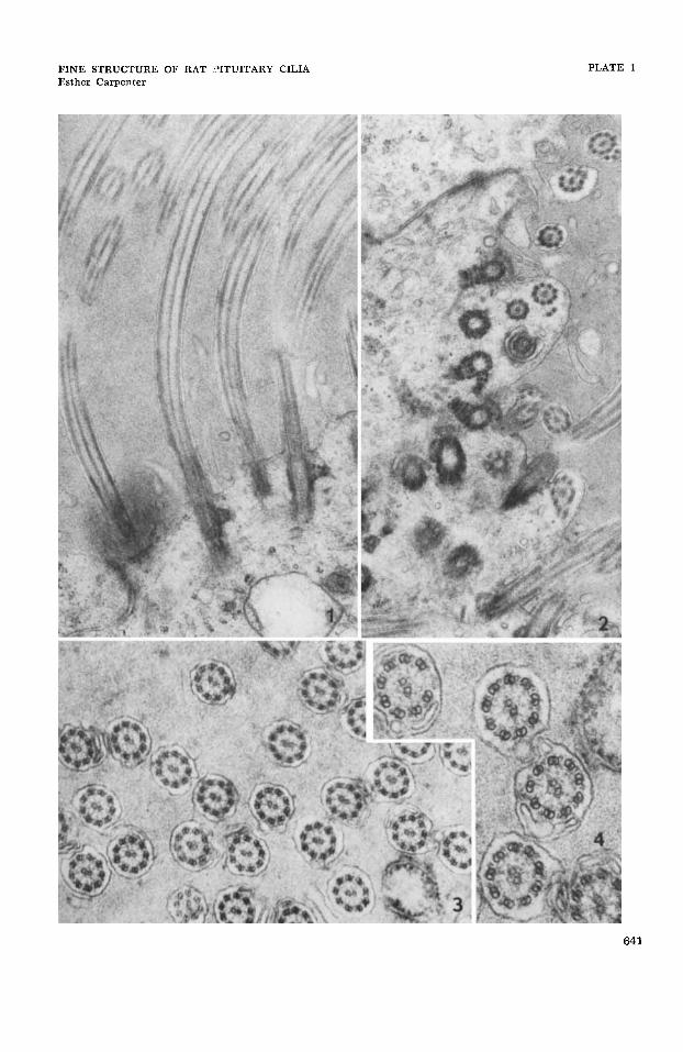
FINE STRUCTURE OF RAT I?ITUITARY CILIA Esther Carpenter
PLATE 1
641
PLATE 2
EXPLANATION OF FIGURES
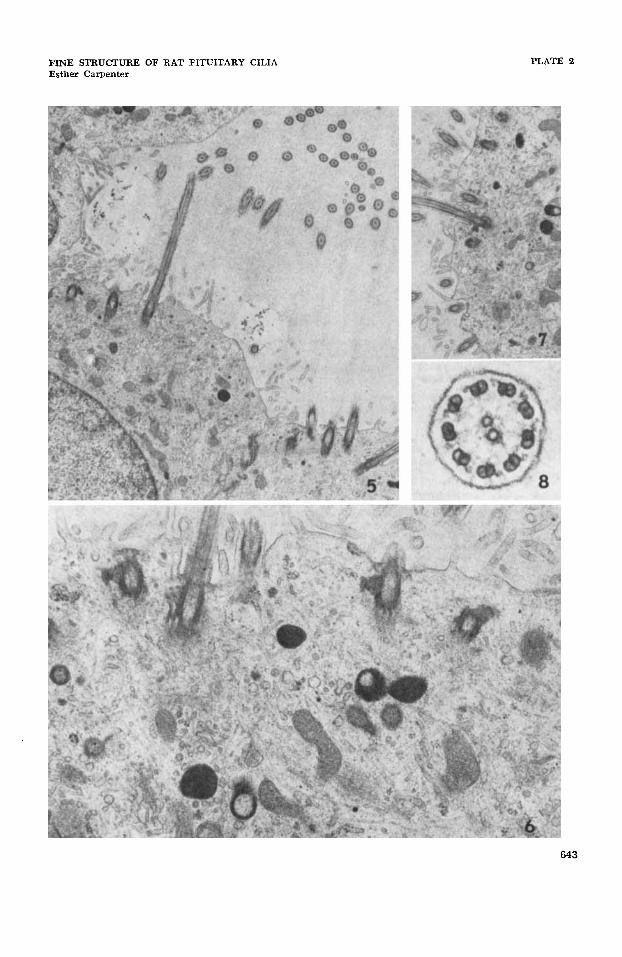
Figs. 5-8 Cilia found in vesicles of the pars distalis of a two-month p rat pituitary gland.
Note the rounded surface of the cells, the microvilli and the numerous mitochondria. In the upper part of the figure central tubules in ad- jacent cilia are oriented in the same direction. x 10,080.
Lateral rootlets project from the base of some of the cilia. Note also the frequency of microtubules, minute vesicles of various kinds and the presence of lysosome-like bodies. x 27,300.
This shows another cilium with a lateral projection from the base and the complexity of the surrounding cytoplasm. As in figures 5 and 6 cilia and microvilli arise from the same cell. x 10,080.
The 9 + 2 pattern of kinocilia is particularly well revealed in this cross-section of a cilium from one of the vesicles. x 134,500.
642
FINE STRUCTURE OF RAT PITUITARY CILIA Esther Carpenter
PLATE 2
643
PLATE 3
EXPLANATION O F FIGURES
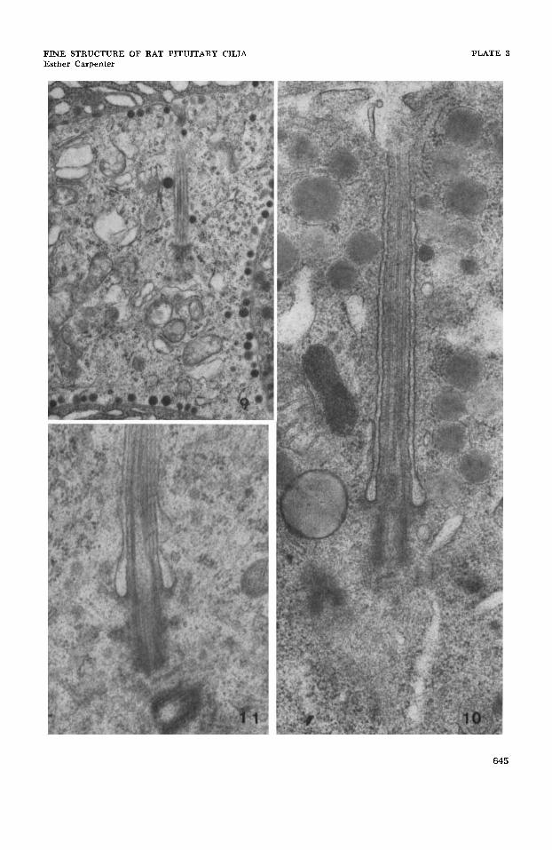
Figures 9-11 are from 5-month 0 ra t pituitaries. Figures 10 and 11 are from the same rat.
9
10
11
A longitudinal section through the lateral region of a cilium showing the boundary between the cilium and the basal body. The granules in this cell do not exceed 150 m p i n diameter. x 16,560.
A median longitudinal section of a cilium in which the tubules appear to run a straight course. Note the deeply invaginated plasma mem- brane surrounding the cilium and the sac-like swelling in the region where the cilium joins the basal body. The adjacent centriole lies on the left a t a slight angle to the basal body. Granules in this cell reach a diameter of 250 mF. x 45,500.
A median longitudinal section of a cilium in which one of the pe- ripheral doublets has been displaced centrally. The basal body shows a striated lateral extension on the left. The adjacent centriole lies be- low the basal body. Granules in this cell (not shown) reach a diam- eter of 350 mp. x 46,800.
644
FINE STRUCTURE OF RAT PITUITARY CILIA Esther Carpenter
PLATE 3
645
PLATE 4
EXPLANATION O F FIGURES
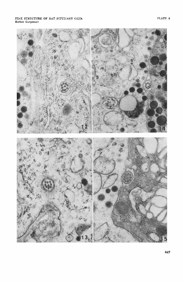
12 A typical transverse section of a cilium embedded i n a parenchymal cell of the pars distalis of a five-month old 0 rat. The centrally placed doublet is similar to those at the periphery and unlike the central tubules shown in figure 4 and figure 8. x 45,500.
A similar cross-section of a cilium in a parenchymal cell of a six- month ? rat. x 45,500.
A transverse section of a cilium lying between granular cells of the anterior lobe of the pituitary of another six-month 0 rat. As in the previous figures one of the doublets is displaced toward the center. x 27,600.
A cilium lying between granular cells in the same six-month rat as figure 14. 'The nine doublets are arranged peripherally around a homogeneous central region lacking tubules. x 45,500.
13
14
15
646
FINE STRUCTURE OF RAT PfTUITARY CILIA Esther Carpenter
PLATE 4
647
PLATE 5
EXPLANATION OF FIGURES
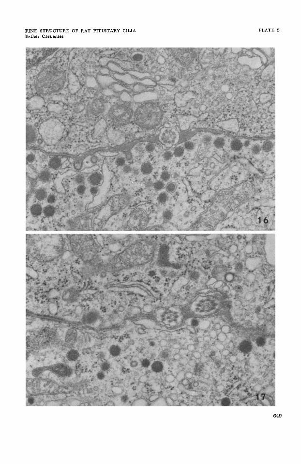
Both figures are taken from the pars distalis of a five-month old 0 rat. The gland was fixed by Karnovsky’s (’65) paraformaldehyde method recommended for microtubules. Both are x 45,500.
16 In the cilium lying between the two cells all doublets are peripherally located. The central area is not as homogeneous as that in figure 15 from a gland fixed in glutaraldehyde followed by osmium tetroxide and could be interpreted as showing some indication of central tubules.
Two cilia are shown lying in the same intercellular space. In one a doublet is displaced centrally; in the other all doublets are peripherally located and there is no indication of central tubules.
17
648
FINE !STRUCTURE OF RAT PITUITARY CILIA Esther Carpenter
PLATE 5
649