Embed Size (px)
Citation preview

HBA 531 - THE BODYFinal Written Examination - December 22, 2010
Name: _______________________________________
1. As a result of an automobile accident, a woman suffers a fracture of the right superior pubic ramus of her pelvis. What nerve passing superiorly to the ramus is in jeopardy from such an injury? (1.25)
List the muscles that are innervated by this nerve:
For each of these muscles, list its primary action at the hip and/or knee:
2. What muscles comprise the hamstrings? (1.5)
What is their innervation?
What is(are) their principle action(s)?
3. The human lower limb is characterized by physiologic genu valgus. What does this mean in laymen’s terms?(2)
Describe how this condition is expressed quantitatively:
What useful purpose does it serve?
Explain how it can also have negative consequences:
4. Describe in anatomical terms what the femoral canal and femoral ring are, and explain their clinical significance.(1)
5. What anatomical structures are most important in helping to prevent the following undesirable motions: (2.5)
a) Lateral patellar displacement
b) Rotation of the tibia on the femur
c) Collapse of the pedal longitudinal arch
6. For the following symptoms or signs, name the nerve and muscle(s) that are most likely to be associated with thefollowing deficits: (1.5)a) Severe weakness in eversion of the foot
b) Severe weakness in thigh abduction
1
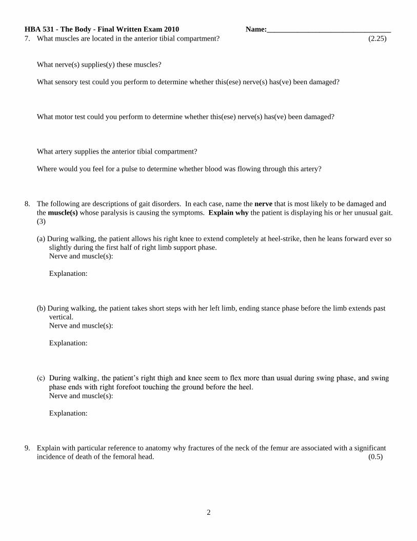
HBA 531 - The Body - Final Written Exam 2010 Name:_________________________________7. What muscles are located in the anterior tibial compartment? (2.25)
What nerve(s) supplies(y) these muscles?
What sensory test could you perform to determine whether this(ese) nerve(s) has(ve) been damaged?
What motor test could you perform to determine whether this(ese) nerve(s) has(ve) been damaged?
What artery supplies the anterior tibial compartment?
Where would you feel for a pulse to determine whether blood was flowing through this artery?
8. The following are descriptions of gait disorders. In each case, name the nerve that is most likely to be damaged andthe muscle(s) whose paralysis is causing the symptoms. Explain why the patient is displaying his or her unusual gait.(3)
(a) During walking, the patient allows his right knee to extend completely at heel-strike, then he leans forward ever soslightly during the first half of right limb support phase. Nerve and muscle(s):
Explanation:
(b) During walking, the patient takes short steps with her left limb, ending stance phase before the limb extends pastvertical.Nerve and muscle(s):
Explanation:
(c) During walking, the patient’s right thigh and knee seem to flex more than usual during swing phase, and swingphase ends with right forefoot touching the ground before the heel.Nerve and muscle(s):
Explanation:
9. Explain with particular reference to anatomy why fractures of the neck of the femur are associated with a significantincidence of death of the femoral head. (0.5)
2

HBA 531 - The Body - Final Written Exam 2010 Name:_________________________________
10. Name the muscle(s) that is(are) attached to the following sites: (1)(a) anterior superior iliac spine
(b) adductor tubercle
(c) tibial tuberosity
(d) lesser trochanter
11. Label this oblique axial t1-weighted MRI section near the ankle. The plane of the section is shownin the small image. (2.5)
12. A woman suffered a pelvic fracture in an automobile accident. Ina follow-up exam she displayed a positive Trendelenburg sign when standing on her right limb. (1)
(a) Describe the appearance of this sign
(b) What has been damaged?
13. What anatomical structures are most important in helping to prevent the following undesirable motions: (0.5)
(a) Distal displacement of the radius relative to the ulna
(b) Shoulder separation (dislocation of acromioclavicular joint)
14. An angiogram of the vasculature of a patient’s upper limb reveals plaque build up that completely obstructs the firstpart of the axillary artery, yet he suffers no symptoms associated with ischemia of the limb. Trace one alternativeroute of blood flow by-passing the obstruction. (1)
3

HBA 531 - The Body - Final Written Exam 2010 Name:_________________________________15. Due to a fall from her bicycle, a young woman suffers a fracture of the surgical neck of her humerus. What nerve is
in jeopardy from the fracture? (1)
If this nerve was injured, what muscles would be affected?
What motor test could you perform to test for damage to this nerve?
16. If the musculocutaneous nerve was damaged at the point were it comes off of the brachial plexus, what muscles wouldbe affected? (1.25)
Describe the motor deficits you would expect to see.
Where would you test for loss of sensation due to damage of the musculocutaneous nerve?
17. A man stumbles and falls breaking the medial epicondyle of his humerus. What nerve is in jeopardy from thisfracture? (1)
What impact would this injury have on the man’s ability to flex his elbow?
What impact would this injury have on the man’s ability to flex his wrist?
What impact would this injury have on the man’s ability to abduct/adduct his fingers
18. For the following symptoms or signs, name the nerve and muscle(s) that are most likely to be associated with thefollowing deficits: (1.25)
(a) Marked weakness of lateral rotation of the upper arm
(b) Inability to extend the elbow against resistance
19. The man in the photograph below has suffered a nerve injury. He was asked to make an “O” with his thumb andindex finger and then press them together. (1)
Is the injury on the right or left side?
What nerve has been damaged?
Explain your diagnosis:
4
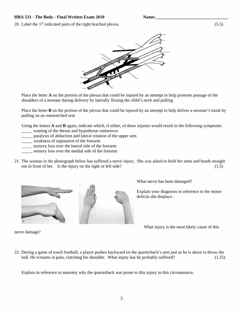
HBA 531 - The Body - Final Written Exam 2010 Name:_________________________________20. Label the 17 indicated parts of the right brachial plexus. (5.5)
Place the letter A on the portion of the plexus that could be injured by an attempt to help promote passage of the shoulders of a neonate during delivery by laterally flexing the child’s neck and pulling
Place the letter B on the portion of the plexus that could be injured by an attempt to help deliver a neonate’s trunk bypulling on an outstretched arm
Using the letters A and B again, indicate which, if either, of these injuries would result in the following symptoms:_____ wasting of the thenar and hypothenar eminences_____ paralysis of abduction and lateral rotation of the upper arm_____ weakness of supination of the forearm_____ sensory loss over the lateral side of the forearm_____ sensory loss over the medial side of the forearm
21. The woman in the photograph below has suffered a nerve injury. She was asked to hold her arms and hands straightout in front of her. Is the injury on the right or left side? (1.5)
What nerve has been damaged?
Explain your diagnosis in reference to the motordeficits she displays :
What injury is the most likely cause of thisnerve damage?
22. During a game of touch football, a player pushes backward on the quarterback’s arm just as he is about to throw theball. He screams in pain, clutching his shoulder. What injury has he probably suffered? (1.25)
Explain in reference to anatomy why the quarterback was prone to this injury in this circumstance.
5

HBA 531 - The Body - Final Written Exam 2010 Name:_________________________________23. The photograph on the right illustrates a nerve injury. (0.75)
What nerve has been damaged?
List two observations from the photograph that support your diagnosis:
24. Below is a series of t1-weighted MRIs of the shoulder. The sections are inthe plane of the scapular blade. The small image in the upper left corner of each section indicates the plane of thatslice. First identify each labeled structure, then enter the letters that satisfy the last two statements. (4)
A ______________________________ B ______________________________ C ______________________________
D ______________________________ E ______________________________ F ______________________________
G ______________________________ H ______________________________ I _______________________________
J _______________________________ Letters representing medial rotators of the upper arm __________________
Letters representing muscles used in typical elevation of the arm above the head ____________________
6

HBA 531 - The Body - Final Written Exam 2010 Name:_________________________________25. To the right is a 3D rendering of a patient’s thoracic aorta (white
indicates wall pathology) and an axial CT through the lower half ofthe manubrium. The patient presented with hoarseness. What would you see if you looked at her larynx with a laryngoscpeor mirror? (2.5)
Identify three other laryngeal symptoms and explain why theyarise:
(a)
(b)
(c)
If you asked her to stick out her tongue, what would notice? Give a reason for your answer.
26. Below is a representation of your face. Draw it as it will appear to a patient facing you at a distance of 4 feet who hasdamage to the optic chiasm. (1.5)
Name one struc- ture that when enlarged can damage the optic chiasm:____________________________
Draw your face as it will appear to a patient facing you at a distance of 4 feet who has complete interruption of herright optic tract.
27. What aspects of skull development mitigate against placing forceps posterior the ear during delivery of a baby? Why?(1)
28. To the right are a superior and lateral view of the head of a young child. What is the name ofthis condition and what has gone wrong? (1)
7
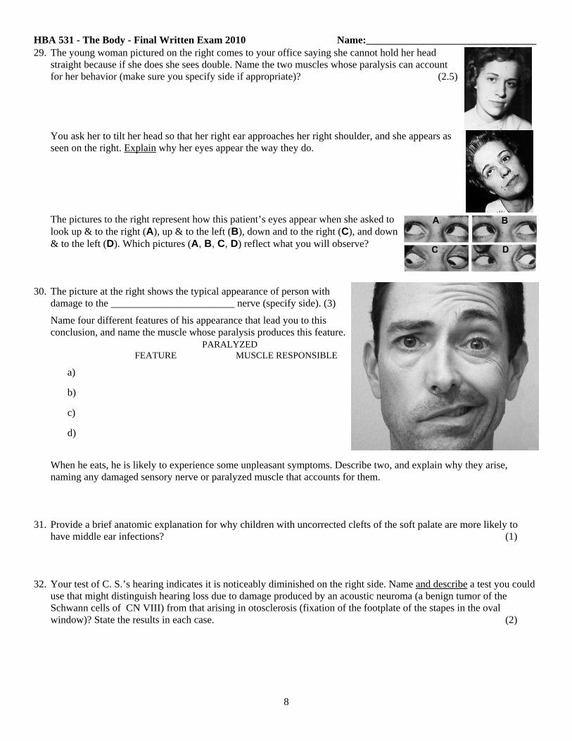
HBA 531 - The Body - Final Written Exam 2010 Name:_________________________________29. The young woman pictured on the right comes to your office saying she cannot hold her head
straight because if she does she sees double. Name the two muscles whose paralysis can accountfor her behavior (make sure you specify side if appropriate)? (2.5)
You ask her to tilt her head so that her right ear approaches her right shoulder, and she appears asseen on the right. Explain why her eyes appear the way they do.
The pictures to the right represent how this patient’s eyes appear when she asked tolook up & to the right (A), up & to the left (B), down and to the right (C), and down& to the left (D). Which pictures (A, B, C, D) reflect what you will observe?
30. The picture at the right shows the typical appearance of person withdamage to the ________________________ nerve (specify side). (3)
Name four different features of his appearance that lead you to thisconclusion, and name the muscle whose paralysis produces this feature. PARALYZED
FEATURE MUSCLE RESPONSIBLE
a)
b)
c)
d)
When he eats, he is likely to experience some unpleasant symptoms. Describe two, and explain why they arise,naming any damaged sensory nerve or paralyzed muscle that accounts for them.
31. Provide a brief anatomic explanation for why children with uncorrected clefts of the soft palate are more likely tohave middle ear infections? (1)
32. Your test of C. S.’s hearing indicates it is noticeably diminished on the right side. Name and describe a test you coulduse that might distinguish hearing loss due to damage produced by an acoustic neuroma (a benign tumor of theSchwann cells of CN VIII) from that arising in otosclerosis (fixation of the footplate of the stapes in the ovalwindow)? State the results in each case. (2)
8

HBA 531 - The Body - Final Written Exam 2010 Name:_________________________________33. This patient has nervous system pathology. What has been injured and
what is the evidence for your conclusion? (2)
What test could you perform to give you a better idea of the location of theproblem. Explain how the results of the test provide useful information.
34. D. W. comes into your office complaining that her teeth don’t seem to fit together well. You notice that her righttemple looks sunken, and the right side of her face looks less full than the left side. What do you expect to observewhen she opens her mouth. Explain why this occurs. (2)
From these observations alone, what nerve are you certain is not doing its job? _____________________________
Describe a test you could perform to determine if the damage was proximal to the beginning of this nerve?
35. The patient shown in the image at the top on the right was asked to look at you.Paralysis of what muscle is obvious? _______________________________ (3)
In the bottom picture the patient was asked to look to his right. Draw the appear-ance of the left iris and pupil.
In addition to the muscle identified above, name at least 6 muscles(smooth or striated) that would also be paralyzed:
Name and give the locations of glands that are likely to have lost their secretoryinnervation:
36. What nerves mediate the pupillary light reflex? (1)
You shine a penlight in a patient’s right eye and both pupils constrict. You then quickly move the penlight so that itshines in the patient’s left eye. You observe that both pupils dilate. What do you conclude?
9

HBA 531 - The Body - Final Written Exam 2010 Name:_________________________________37. The pictures to the right depict the open mouth of a woman asked
to say “Aaah”, and then show her head when asked to turn it to theright and to the left.. (1.5)
Which of the pictures shown above (A, B, C) is likely to depict her appearance when asked to shrug her shoulders?Give a reason for your answer.
Name the muscles whose actions can be visualized with the naked eye that are innervated by nerve(s) that has(ve)been damaged?
38. Name the structures that satisfy each of the following statements (there may be more than one answer per blank). (5)
(a) lies in the labyrinthine wall of middle ear immediately superior to the stapes footplate _________________________
(b) opens into the nasopharynx posterior to the inferior nasal meatus __________________________________________
(c) crosses the lateral surface of the internal carotid artery, and bathed on all sides by venous blood, in the posterior part of the cavernous sinus ____________________________________
(d) up against the lateral wall of the cavernous sinus _______________________________________________________
(e) the spinal nerve that is tested when you assess sensation from the earlobe ___________________________________
(f) when torn, leads to an epidural hematoma _____________________________________________________________
(g) passes transversely 1 finger’s breadth below the zygomatic arch ___________________________________________
(h) two muscles innervated by the marginal mandibular n. __________________________________________________
(i) the laryngeal structure that needs to be penetratedin emergency access to the subglottic airway ___________________________________________________
(j) is (are) the kind of teeth not preceded by deciduous precursors ____________________________________________
(k) is (are) the kind of teeth for which there are two in each jaw on each side ___________________________________
(l) lies immediately anterior to the 2nd- 4th tracheal rings (be specific) _______________________________________
(m) the dural sinuses through which venous blood passes on its way from the pituitary gland to the IJV ___________________________________________________
(n) is a structure that can be palpated to tell you about intracranial pressure in an infant ___________________________
39. Your patient, an inveterate cigar smoker, develops cancer of the lower lip near the corner of the mouth. Trace thelymphatic drainage from the site of cancer into the venous system. (1)
10

HBA 531 - The Body - Final Written Exam 2010 Name:_________________________________40. P. W. is a 60 year old former smoker who experiences severe diffuse abdominal pain 15 - 30 minutes after eating a
full meal. You suspect intestinal angina (pain arising from ischemia) and order an angiogram to look for blockage inthe major artery that feeds most the small intestine. What is this artery? (2.5)
You find that it is almost completely blocked for a length of 3 mm at a site about one centimeter from its origin, yetthe small intestine is obviously still alive. Trace two substantially different (but not incredibly circuitous) routes bywhich arterial blood can reach most of the small intestine.
(a)
(b)
41. A 45-year old premenopausal woman is scheduled to undergo uterine artery embolization to treat uterine fibroids.You are obligated to inform her that the procedure may induce menopause. What is the anatomic reason for this? (1)
42. Below are listed various coronary arteries, each associated with a letter of the alphabet. Following this list arestatements asking you to identify arteries that supply certain regions of the heart. For each statement, enter into theblank the letters corresponding to all arteries that satisfy the statement. Make sure you enter the letters for all correctarteries, even if some are only the parent vessels of others that you list. (4)
A = left main coronary B = right coronary C = LAD D = circumflex coronaryE = acute marginal F = diagonal G = obtuse marginal H = PDA
(a) supplies left ventricle in a left coronary dominant heart _______________________
(b) supplies interventricular septum in a right coronary dominant heart _______________________
(c) supplies right atrium in a right coronary dominant heart ______________________
(d) supplies right ventricle in a right coronary dominant heart ______________________
43. Name the first group(s) of lymph nodes to which each of the following structures drain (3.5)
(a) mammary gland ___________________________,_________________________________
(b) testis _________________________________
(c) ovary __________________________________,_______________________________
(d) uterus __________________________________
(e) transverse colon __________________________________
(f) vulva ____________________________________
(g) skin of hallux ____________________________________
(h) skin of pollex ____________________________________
(i) skin of glans penis ____________________________________
Circle the names of nodes that do not drain into the part of the thoracic duct that lies in the abdomen and thorax.
11

HBA 531 - The Body - Final Written Exam 2010 Name:_________________________________44. What effect does the parasympathetic nervous system have on (3.5)
(a) heart rate - (b) the pyloric sphincter - (c) the urinary bladder -(d) the vasculature of the corpus cavernosum -(e) the sphincter urethrovaginalis -(f) tear production -(g) saliva production -
Next to the effect you listed, state the name of the nerve(s) that carries(y) preganglionic parasympathetic axons out ofthe CNS for production of that effect.
45. On the pictures shown below, draw the following and label with the letter following the description : (4.5)
a circle where you ought to place a stethoscope to hear the aortic valve - Aa circle where you ought to place a stethoscope to hear the middle lobe of the lung - Ba line indicating the path of the inferior epigastric artery - Ca + where you ought to insert a needle to withdraw CSF - Da circle where you ought to place a stethoscope to hear the superior segment of the lower lobe - Ea circle where you ought to place a stethoscope to hear the apical segment of the left lung - Fa circle where you ought to place a stehoscope to hear the mitral valve - Ga + where you should palpate for an abdiminal aortic aneurysm just above the aortic bifurcation - Ha circle where you should palpate to feel the emergence of an indirect inguinal hernia - I
46. What is the relationship between the ureter and the (1.25)
(a) psoas major -(b) iliac arteries -(c) infundibulopelvic ligament - (d) lateral fornix of the vagina (or lateral wall of cervix) - (e) uterine artery -
12

HBA 531 - The Body - Final Written Exam 2010 Name:_________________________________47. A patient presents with a vague epigastric pain and jaundice. At first you expect gall bladder disease. Where should
you attempt to palpate the gall bladder to see if it is painful? (1.5)
When you do feel here, you discover a greatly dilated gall bladder but it is NOT painful. You now fear pancreaticcancer. How can this disease account for the patient’s jaundice?
Abdominal CT confirms your diagnosis. The tumor is small and appears resectable, but you worry about blood-borne metastases. Where on the CT should you look for them?
48. Provide a brief anatomical explanation for the following: (2.5)(a) a patient that underwent resection of the rectum for treatment of cancer is impotent following the surgery.
(b) an anesthesiologist specially trained to monitor spinal cord function is invaluable during an operation to repair ananeurysm of the aorta just distal to the left subclavian artery
(c) cirrhosis of the liver may cause death by hemorrhage
(d) it is acceptable to treat internal hemorrhoids, but not external hemorrhoids, by rubber band ligation
(e) gonorrhea has a much greater likelihood of spreading to the peritoneal cavity of women than men
49. Identify the structures (A - I) on the three sequential (superior to inferior, 5mm apart) CT images shown below.(2.25)
A _______________________________ B __________________________ C ___________________________
D ____________________________ E __________________________ F____________________________
G ____________________________ H __________________________ I____________________________
13

HBA 531 - The Body - Final Written Exam 2010 Name:_________________________________50. Presented below is a series of eight coronal MR angiograms of the chest. The most anterior of the sections is at the
upper left. The sections that follow (indicated by higher numbers in the upper left corner) are progressively moreposterior, but not by equal increments (a unit change in image number corresponds to 8 mm). Identify the structures (A- N) to which lines are drawn. (3.5)
A: ________________________________
B: ________________________________
C: ________________________________
D: ________________________________
E: ________________________________
F: ________________________________
G: ________________________________
H: ________________________________
I: ________________________________ J: _______________________________ K:_________________________________
L: _______________________________ M: _______________________________ N: ________________________________
14