Embed Size (px)
Citation preview

Final Draft of the original manuscript: Mathew, S.; Lendlein, A.; Wischke, C.: Characterization of protein-adjuvant coencapsulation in microparticles for vaccine delivery In: European Journal of Pharmaceutics and Biopharmaceutics (2014) Elsevier DOI: 10.1016/j.ejpb.2014.04.003

Article type: Notes Characterization of protein-adjuvant coencapsulation in microparticles for vaccine delivery Simi Mathew, Andreas Lendlein and Christian Wischke * Simi Mathew, Prof. Dr. Andreas Lendlein, and Dr. Christian Wischke
Institute of Biomaterial Science and Berlin-Brandenburg Center for Regenerative Therapies,
Helmholtz-Zentrum Geesthacht, 14153 Teltow, Germany
*Corresponding author: Tel.: + 49 3328 352 450; Fax: + 49 3328 352 452
E- mail address: [email protected]
Abstract
Protein antigens encapsulated as vaccines in poly[(rac-lactide)-co-gylcolide] (PLGA)
microparticle carriers can induce immune responses. The intensity and directions of this response
can be controlled by coloading the microparticles with immunomodulatory adjuvants, e.g.,
muramyl dipeptide (MDP) as adjuvant combined with ovalbumin (Ova) as protein antigen. In
this study, methodologies for an individual quantification of both encapsulated substances should
be reported, which comprise i) a separation process to isolate and determine MDP as intact
molecule and ii) a simultaneous degradation of both analytes with subsequent specific
quantification of Ova fragments. It was shown that coloading of both substances resulted in a
substantially reduced encapsulation efficiency of MDP. This illustrates that correct conclusions
on dose-response relationships in future vaccination studies can only be drawn, if a selective
method for adjuvant and protein quantification will be applied.
1

Keywords: amino acid analysis, co-encapsulation, microparticles, ovalbumin, poly[(rac-lactide)-co-gylcolide]
Introduction
Degradable polymeric microparticles of phagocytizable size (<10 µm) have been explored as
carrier systems for antigenic vaccine components. In most cases, increased immune responses
compared to soluble antigen were observed, which may be partly attributed to the enhanced
engulfment of theses carriers by immune cells due to their resemblance to microorganisms in
terms of size [1]. Furthermore, it was indicated that microparticles can modulate antigen
presentation pathways of exogenous antigens towards a cross presentation [2]. For further
enhancing the immunogenic potency of such particles, immunomodulatory substances
(adjuvants) can be coencapsulated with the antigenic proteins to elicit superior immune
responses [2-4].
The specific combination of adjuvant/antigen mixtures finally present in the microparticles may
crucially affect the cell activation status due to mutual interference and enhancement of
responses. Therefore, it will be essential to quantify the final microparticle payload properly.
Generally, the coencapsulation of the different types of molecules could occur at different
efficiencies in case of different solubilities, hydrophilicities, and hydrodynamic radii. The
capability to quantify the protein and the coloaded adjuvant independently strongly depends on
the nature of the coencapsulated compounds. In the past, most of the coloaded vaccine
microparticles contained nucleotide-derived adjuvants [2, 3], which are chemically different
from proteins and may be detected without interference. Recently, polymer microparticles
encapsulating peptidoglycan-based agonists of receptors sensing nucleotide and oligomerization
domains (NOD) were reported, which illustrated their capacity to induce immunoactivation and
2
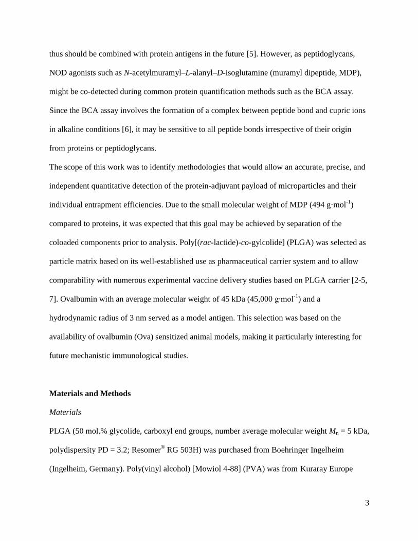
thus should be combined with protein antigens in the future [5]. However, as peptidoglycans,
NOD agonists such as N-acetylmuramyl–L-alanyl–D-isoglutamine (muramyl dipeptide, MDP),
might be co-detected during common protein quantification methods such as the BCA assay.
Since the BCA assay involves the formation of a complex between peptide bond and cupric ions
in alkaline conditions [6], it may be sensitive to all peptide bonds irrespective of their origin
from proteins or peptidoglycans.
The scope of this work was to identify methodologies that would allow an accurate, precise, and
independent quantitative detection of the protein-adjuvant payload of microparticles and their
individual entrapment efficiencies. Due to the small molecular weight of MDP (494 g·mol-1)
compared to proteins, it was expected that this goal may be achieved by separation of the
coloaded components prior to analysis. Poly[(rac-lactide)-co-gylcolide] (PLGA) was selected as
particle matrix based on its well-established use as pharmaceutical carrier system and to allow
comparability with numerous experimental vaccine delivery studies based on PLGA carrier [2-5,
7]. Ovalbumin with an average molecular weight of 45 kDa (45,000 g∙mol-1) and a
hydrodynamic radius of 3 nm served as a model antigen. This selection was based on the
availability of ovalbumin (Ova) sensitized animal models, making it particularly interesting for
future mechanistic immunological studies.
Materials and Methods
Materials
PLGA (50 mol.% glycolide, carboxyl end groups, number average molecular weight Mn = 5 kDa,
polydispersity PD = 3.2; Resomer® RG 503H) was purchased from Boehringer Ingelheim
(Ingelheim, Germany). Poly(vinyl alcohol) [Mowiol 4-88] (PVA) was from Kuraray Europe
3

GmbH (Frankfurt, Germany), MDP was procured from Invivogen (San Diego, CA, USA), and
Endograde Ovalbumin was from Hyglos GmbH (Bernried, Germany). The bicinchoninic acid
(BCA) assay kit was purchased from Sigma-Aldrich (Taufkirchen, Germany). All other
chemicals including HPLC solvent were of analytical grade.
Preparation and characterization of microparticle size and surface morphology
Microparticles (MP) were prepared in a biological safety cabinet under laminar air flow by the
water-in-oil-in-water (w/o/w) double emulsion/solvent evaporation method as reported before
[5]. Briefly, 75 µL 0.1% (w/v) MDP and/or 1.3% (w/v) Ova in water containing 1% (w/v)
sodium bicarbonate and 5% (w/v) sucrose were added to a 19 wt.% PLGA solution in
dichloromethane and emulsified by sonication. This emulsion was then added to 2% (w/v) PVA
solution, homogenized by rotor-stator homogenization, and further treated as reported [5].
Particle size analysis was performed by laser diffraction using a Mastersizer 2000 (Hydro 2000S
dispersion unit, Malvern Instruments, Herrenberg, Germany). The morphology of lyophilized
microparticles was studied with a Gemini SupraTM 40 VP SEM (Carl Zeiss NTS GmbH,
Oberkochen, Germany) without sputtering to avoid artefacts.
Determination of encapsulation efficiency
The MDP content of microparticles was determined after polymer extraction (3x) from 10 mg
samples with 1 mL acetonitrile, dissolution of the pellet after centrifugation in 10 mg·mL-1
aqueous SDS solution, and separation by Amicon Ultra-0.5 mL centrifugal filters (Millipore
GmbH, Schwalbach am Taunus, Germany) at 14,000 g (Heraeus Biofuge Primo R, Hanau,
Germany). MDP was quantified by HPLC using a Lichrosphere 100 RP 18 5 µm column
4

(250 x 4 mm) with UV detection at 240 nm and 0.01 M phosphate buffer pH 3/methanol (98/2
v/v) as eluents (25 °C, 1.5 mL·min-1) [5].
The encapsulated protein was preferentially quantified by amino acid analysis. Briefly, 10 mg of
microparticles and encapsulated protein were treated with 1 mL of 7.5 N NaOH at 106 °C for
12 h (PMC block heater, Germany) for basic hydrolysis. Subsequently, samples were neutralized
and subjected to HPLC analysis on a C18 column (EC 250/4 Nucleosil 100-5 C18 HD) with pre-
column derivatization using 50 µL sample and 50 µL o-phthaldialdehyde (10 mg·mL-1 in
methanol/ borate buffer (90/10 v/v)). Acetonitrile/water (80/20 v/v) were used as mobile phase
(35 °C, 1 mL·min-1) with fluorescence detection (ex. 335 nm, em. 450 nm). Alternatively, the
Ova content of only Ova loaded microparticles could be determined after polymer extraction by
the BCA assay performed on microtiter plates (Carl Roth GmbH, Karlsruhe, Germany).
Determination of osmolality
The osmolality of the different w1 phases were determined by freezing point depression using
Knauer Semi-Micro Osmometer A-0300 (Knauer GmbH, Berlin, Germany).
Results and discussion
Microparticles were prepared using the water-in-oil-in-water (w1/o/w2) emulsion/solvent
evaporation technique. Ova and MDP were coencapsulated by their dissolution in the w1 phase,
which also contained additives such as sodium bicarbonate to neutralize acidic polymer
degradation products and sucrose as lyoprotectant [8]. The obtained microparticles, as
exemplarily shown for coloaded microparticles (Ova−MDP MP) (Fig. 1A), showed a narrow
particle size distribution in all cases with d(0.5) particle sizes below 10 µm (Table 1).
5

As illustrated in Figure 1B, the microparticles were of spherical shape and exhibited small open
pores. The pore formation can be assigned to the hydrophilic substances in the w1 phase, which
build up osmotic pressure in the nascent particles as indicated by the determined osmolality of
the w1 solutions (Table 1). For Ova, surface active properties in emulsions are known [9]. It may
be speculated on whether its leaching to the w2-phase, particularly in combination with MDP,
supported the stabilization of the o/w2 interphases in addition to PVA employed as steric
stabilizer.
Figure 1: (A) Size distribution and (B) morphology of particles coencapsulating MDP and
Ova.
During the particle preparation process, not only the o-phase solvent but also the desired payload
from the w1 phase may partially diffuse to the external aqueous phase. This is supported by an
influx of water into the polymer matrix due to the osmolality differences of the w1-phase and w2-
phase.
The encapsulation efficiency of microparticles loaded with MDP only (MDP MP) was
determined by i) removing PLGA by repeatedly extracting the samples with acetonitrile, ii)
dissolving MDP into an aqueous medium, and iii) quantifying MDP by reverse phase HPLC.
6

Although being a small hydrophilic molecule, MDP was successfully loaded in the
microparticles with 70 wt.% efficiency. Similarly, microparticles loaded only with Ova (Ova
MP) could be characterized after similar sample pretreatment (polymer extraction, protein
dissolution in aqueous medium) followed by protein quantification with the BCA assay method.
Ova loaded microparticles showed high encapsulation efficiencies of 94 wt.%.
Table 1: Characteristics of different microparticle (MP) compositions.
Sample ID Content Particle sizeb)
w1-phase osmolality
Theoretical payload
Encapsulation efficiency
d(0.5) Span
- [µm] [mOsm∙Kg-1] [µg·mg-1] [wt.%]
MDP MPa) Muramyl dipeptide 5.5 1.6 578 3 70 ± 1
Ova MP Ova 6.7 1.1 614 40 94 ± 3
Ova–MDP MP Ova MDP 4.2 1.6 720 40
3 95 ± 3 48 ± 4
a) Data reproduced from [5], Copyright 2012, with permission from Elsevier. b) Data derived from an average of three measurements In principle, the individual encapsulation efficiencies may be altered for coloaded systems due to
the increased osmotic pressure and different hydrodynamic radii of the two encapsulated
substances. For coloaded particles, the previously applied standard strategy with PLGA
extraction and direct analysis of Ova and MDP could not be followed, because: i) Ova as a larger
molecule would block HPLC columns suitable for MDP quantification, thus prohibiting the
analysis of MDP + OVA mixtures, and ii) MDP with its dipeptide segment interfered with the
Ova quantification by the BCA assay as identified by a standard addition experiment (Figure
2A). Therefore, a separation of the two hydrophilic molecules should be applied before
individual analysis.
7

Figure 2: Evaluation of experimental methods and their shortcomings in the quantification of
MDP and Ova. (A) Interference of MDP (20 µg∙mL-1) in the Ova determination by
the BCA assay. (B) Effect of absorption on filter membranes on the individual
recovery of Ova (250 µg∙mL-1; BCA assay) and MDP (25 µg∙mL-1; HPLC)
standards subjected to size exclusion centrifugation for their separation.
Based on the substantial differences in the number of amino acids in the protein and adjuvant
(Ova: 385; MDP: 2), a selective precipitation behavior using trichloroacetic acid was assumed.
However, despite successful removal of the protein and subsequent qualitative detection of MDP
in the supernatant, the peak of the α-anomer of MDP (see HPLC chromatogram, inset of
Figure 3) could surprisingly not be detected by HPLC. This suggests that MDP may not have
quantitatively remained in solution.
8

Figure 3: Scheme presenting the different analytical strategies followed for the quantification of
the single and coencapsulated components.
As an alternative approach, the substantial differences of hydrodynamic radii of the analytes
should be used to separate MDP from Ova by ultra-filtration. For this purpose, centrifugal filters
with different types of membrane materials were explored. The centrifugal filter equipped with
polyethersulfone membranes showed considerable adsorption of MDP, which was avoided with
a subsequently employed low-binding regenerated cellulose membrane having a molecular
weight cut-off of 3 kDa. Importantly, adsorption was reduced by first washing and hydrating the
filters several times with water (Figure 2B). This methodology was found useful in case of
MDP, which could be quantitatively recovered in control experiments with standard solutions.
9

By applying this method, the MDP encapsulation efficiency in Ova−MDP loaded particles was
identified to be 48 wt.%, which is low compared to the MDP loaded particles (Table 1).
In contrast to MDP, Ova could not be quantitatively recovered in all cases and apparently
remained partially adsorbed inside the centrifugal filter tubes, which may possibly assigned to its
relatively higher hydrophobicity. Accordingly, the low encapsulation efficiency of Ova
determined by this procedure (26 ± 5 wt.%) is erroneous and an artifact resulting from the
unsuitable separation process.
Therefore, a method for Ova detection was required which would not be disturbed by the
presence of MDP. In principle, antibody-based assays would be suitable, but always depend on
the availability of the respective protein-specific antibody. Therefore, amino acid analysis was
selected for Ova quantification since it may be widely applicable to different proteins. This
method should be applied after hydrolysis of the entire microparticles rather than the multi-step
extraction/separation of the protein. The resulting mixture of PLGA degradation products and
amino acids should be quantified by HPLC analysis, where each amino acid in the mixture can
be separated and quantified individually. In this way, alanine and isoglutamine partially
originating from MDP can be separated from other amino acids and excluded in data evaluation
for Ova quantification.
Acidic hydrolysis, especially using aqueous hydrochloric acid, is known as preferable method for
peptide bond cleavage since it ensures complete hydrolysis of proteins with less destruction of
amino acids [10]. However, when particles were subjected to acidic hydrolysis using 6 N HCl at
115 ºC for 24 hours, a brown discoloration of the hydrolysate was observed. In contrast, this was
not observed for standards of the pure protein subjected to the same procedure. Apparently,
Maillard products were formed by reaction of amino acids with reducing sugars. The same
10

products were observed when spiking blank microparticles with soluble ovalbumin, but not for
pure PLGA with Ova. This observation suggested that sucrose used as lyoprotectant was
converted under strong acidic conditions to glucose and fructose, which then can react with
amino acids. The contribution of MDP with its N-acetylmuramyl moiety in this reaction was
considered to be negligible.
Since glycosidic bonds should be more stable in basic conditions, the alkaline hydrolysis of the
loaded PLGA particles in 7.5 N aqueous NaOH solution at 106 ºC for 12 h was evaluated. Under
these conditions, the microparticles and Ova was completely hydrolyzed without interference by
the Maillard reaction, thus allowing subsequent amino acid analysis for Ova quantification. The
encapsulation efficiency of Ova in coloaded microparticles very well agreed with that of the
particles encapsulating only Ova and showed high values of > 90 wt.% (Table 1).
Overall, it can be concluded that the presence of MDP as small molecule did not affect the
encapsulation process of Ova as a large protein in the investigated particles. In contrast, the MDP
encapsulation efficiency was diminished in coloaded microparticles. This may probably be due
to its much smaller hydrodynamic radius, which makes it more prone to osmotic pressure
mediated leakage during preparation such as through water-rich domains acting as diffusion
channels. Ova and MDP did not appear to exhibit strong physical interactions, which could
promote MDP entrapment in the polymer matrix. Importantly, the substantial differences of
theoretical and final payload observed particularly for the immunostimulating adjuvant
illustrated that correct conclusions on dose-response relationships in future vaccination studies
demand the application of selective methods for adjuvant and protein quantification. In case of
MDP and Ova studied in here, an accurate, precise and independent quantitative detection was
possible by combining i) a separation process to isolate and determine MDP as intact molecule
11

and ii) a procedure of simultaneous fragmentation of both analytes with subsequent specific
quantification of Ova.
Acknowledgements
Technical support from Andrea Pfeiffer is gratefully acknowledged. S.M. is grateful to the
Berlin-Brandenburg School for Regenerative Therapies (DFG-GSC 203) for a fellowship.
References
[1] J.F. Correia-Pinto, N. Csaba, M.J. Alonso, Vaccine delivery carriers: Insights and future perspectives, Int J Pharm, 440 (2013) 27-38. [2] A. Heit, F. Schmitz, T. Haas, D.H. Busch, H. Wagner, Antigen co-encapsulated with adjuvants efficiently drive protective T cell immunity, Eur. J. Immunol., 37 (2007) 2063-2074. [3] S. Fischer, E. Schlosser, M. Mueller, N. Csaba, H.P. Merkle, M. Groettrup, B. Gander, Concomitant delivery of a CTL-restricted peptide antigen and CpG ODN by PLGA microparticles induces cellular immune response, J. Drug Target., 17 (2009) 652-661. [4] E. Schlosser, M. Mueller, S. Fischer, S. Basta, D.H. Busch, B. Gander, M. Groettrup, TLR ligands and antigen need to be coencapsulated into the same biodegradable microsphere for the generation of potent cytotoxic T lymphocyte responses, Vaccine, 26 (2008) 1626-1637. [5] C. Wischke, S. Mathew, T. Roch, M. Frentsch, A. Lendlein, Potential of NOD receptor ligands as immunomodulators in particulate vaccine carriers, J. Control. Release, 164 (2012) 299-306. [6] P.K. Smith, R.I. Krohn, G.T. Hermanson, A.K. Mallia, F.H. Gartner, M.D. Provenzano, E.K. Fujimoto, N.M. Goeke, B.J. Olson, D.C. Klenk, Measurement of protein using bicinchoninic acid, Anal. Biochem., 150 (1985) 76-85. [7] C. Wischke, J. Zimmermann, B. Wessinger, A. Schendler, H.-H. Borchert, J.H. Peters, T. Nesselhut, D.R. Lorenzen, Poly(I:C) coated PLGA microparticles induce dendritic cell maturation, International journal of pharmaceutics, 365 (2009) 61-68. [8] Y.K. Katare, T. Muthukumaran, A.K. Panda, Influence of particle size, antigen load, dose and additional adjuvant on the immune response from antigen loaded PLA microparticles, Int. J. Pharm., 301 (2005) 149-160. [9] N.-P.K. Humblet-Hua, E. van der Linden, L.M.C. Sagis, Surface rheological properties of liquid-liquid interfaces stabilized by protein fibrillar aggregates and protein-polysaccharide complexes, Soft Matter, 9 (2013) 2154-2165. [10] M.G. Davies, A.J. Thomas, Investigation of Hydrolytic Techniques for Amino-Acid Analysis of Foodstuffs, Journal of the Science of Food and Agriculture, 24 (1973) 1525-1540.
12


![pET Express & Purify Kits User Manual - Takara Bio Manual/PT5018-1.pdf15 µl pET6xHN-C Vector (In-Fusion Ready) [100 ng/µl] 10 µl pET6xHN-GFPuv Vector [500 ng/µl] 15 µl 1.1 kb](https://img.pdfslide.us/doc/110x75/5e7b57982623d66a901d15a7/pet-express-purify-kits-user-manual-takara-bio-manualpt5018-1pdf-15-l.jpg)