Embed Size (px)
Citation preview
Film Formation of Poly (methyl methacrylate) Latex WithPyrene Functional Poly (divinylbenzene) MicrospheresPrepared by Click Chemistry
S�aziye Ugur,1 Onder Yargı,1 Yasemin Yuksel Durmaz,2 Bunyamin Karagoz,2 Niyazi Bıcak,2
Yusuf Yagcı,2 Onder Pekcan3
1Department of Physics, Istanbul Technical University, Maslak, 34469, Istanbul, Turkey
2Department of Chemistry, Istanbul Technical University, Maslak, 34469, Istanbul, Turkey
3Faculty of Arts and Science, Kadir Has University, Cibali, 34320, Istanbul, Turkey
This work reports on the application of steady statefluorescence (SSF) technique for studying film forma-tion from poly(methyl methacrylate) (PMMA) latex andpoly(divinylbenzene) (PDVB) microsphere composites.Pyrene (P) functionalized PDVB cross-linked sphericalmicrospheres with diameters of 2.5 lm were synthe-sized by using precipitation polymerization techniquefollowed by click coupling reaction. The diameter ofthe PMMA particles prepared by emulsion polymeriza-tion were in the range of 0.5–0.7 lm. PMMA/PDVBcomposite films were then prepared by physicallyblending of PMMA latex with PDVB microspheres atvarious composition (0, 1, 3, 5, 10, 20, 40, and 60 wt%).After drying, films were annealed at elevated tempera-tures above Tg of PMMA ranging from 100 to 2708C for10 min time intervals. Evolution of transparency of thecomposite films was monitored by using photon trans-mission intensity, Itr. Monomer (IP) and excimer (IE) flu-orescence intensities from P were measured after eachannealing step. The possibility of using the excimer-to-monomer intensity ratio (IE/IP) from PDVB micropar-ticles as a measure of PMMA latex coalescence wasdemonstrated. Diffusion of the PMMA chains acrossthe particle–particle interfaces dilutes the dyes,increasing their separation. The film formation stagesof PMMA latexes were modeled by monitoring the IE/IPratios and related activation energies were determined.There was no observable change in activation energiesconfirming that film formation behavior is not affectedby varying the PDVB composition in the studied range.SEM images of PMMA/PDVB composites confirmedthat the PMMA particles undergo complete coales-cence forming a continuous phase in where PDVBmicrospheres are dispersed. POLYM. COMPOS., 32:869–881, 2011. ª 2011 Society of Plastics Engineers
INTRODUCTION
A number of methods including water-based emulsion,
seeded suspension, nonaqueous dispersion polymerization,
and precipitation polymerization were successfully
employed for the preparation of monodisperse micro-
spheres [1–4]. Among them, precipitation polymerization,
which can be performed in the absence of any added sur-
factant or stabilizer [5–12] appeared to be an attractive
route to obtain microspheres with uniform size and shape.
Typically, monodisperse and highly crosslinked poly(divi-
nylbenzene) (PDVB) surfactant-free microspheres (with
diameters between 2 and 5 lm) were prepared by using
only monomer (commercial divinylbenzene, DVB55), rad-
ical initiator (2, 20-azobisisobutronitrile, AIBN), and sol-
vent (acetonitrile) [5]. Interestingly, PDVB microspheres
formed in this method contained significant residual dou-
ble bonds in the particle and on the surface of the particle
[13]. The residual double bonds located at the surface per-
mitted further growth and modification of particles.
The ‘‘click reactions’’ [14, 15], in particular Cu(I)-cata-
lyzed 1,3-dipolar Huisgen cycloaddition reactions between
an azide and an alkyne, have gained a great deal of atten-
tion due to their high specificity and nearly quantitative
yields in the presence of many functional groups.
Recently, PDVB microspheres were functionalized [16]
by polymeric chains using two click reactions, namely
thiol-ene chemistry and azide-alkyne cycloaddition reac-
tion. We also reported functionalization of PDVB micro-
spheres by the copper-catalyzed Huisgen 1-3 dipolar
cycloaddition click reaction with a small fluorescent mol-
ecule-alkyne modified pyrene [17].
Polymer composites are often prepared by mixtures of
two or more different kinds of particles in the dispersed
state. Upon drying of the dispersion, both types of particles
Correspondence to: Saziye UGUR; e-mail: [email protected]
Contract grant sponsor: Turkish Academy of Sciences (TUBA).
DOI 10.1002/pc.21094
Published online in Wiley Online Library (wileyonlinelibrary.com).
VVC 2011 Society of Plastics Engineers
POLYMER COMPOSITES—-2011
contribute to the properties of the film that is formed. Com-
posite materials form a class of materials in which immisci-
ble compounds are combined in order to obtain new proper-
ties [18]. They are heterogeneous mixtures of their compo-
nents which are generally of quite different properties.
Their properties often depart very much from those of the
pure components, so that mixing two homogeneous materi-
als into a composite really results in the formation of a new
material. Properties depend much more on the morphology
and adhesion properties at the interfaces than on the volume
fraction of each phase as would happen in an ideal mixture.
Several authors have examined the properties of hard/soft
latex blends. Hard refers to particles consisting of a polymer
with a Tg above room temperature, and soft refers to par-
ticles of a low-Tg polymer. As a result of worldwide theo-
retical and experimental efforts, a very good understanding
of the mechanisms of latex film formation has been
achieved [19–23]. Traditionally, the film formation process
of pure polymer latex is considered in terms of three se-
quential steps: (i) water evaporation and subsequent pack-
ing of polymer particles (ii) deformation of the particles
and close contact between the particles if their Tg is less
than or close to the drying temperature (soft or low Tg la-
tex). Latex with a Tg above the drying temperature (hard or
high Tg latex) stays undeformed at this stage. In the anneal-
ing of hard latex system, deformation of particles first leads
to void closure [24, 25] and then after the voids disappear,
diffusion across particle-particle boundaries starts, i.e. the
mechanical properties of hard latex films evolve during
annealing, after all solvent has evaporated and all voids
have disappeared. (iii) Coalescence of the deformed par-
ticles to form a homogeneous film [25] where macromole-
cules belonging to different particles mix by interdiffusion
[26, 27]. However, the film formation process of composite
latex is more complicated than homogeneous one due to the
interactions between the different phases. In addition, sev-
eral factors such as molecular weight and its distribution,
synthetic methodology, morphologies of latex particles, sta-
bilizers, surfactants, annealing, film formation conditions,
etc. were experimentally shown to influence composite la-
tex film formation [28–32].
Commercial plastics and rubbers are often filled with
solid particles, either to enhance their mechanical proper-
ties or to reduce cost [33]. The properties of these materi-
als depend primarily on the interactions between the ma-
trix and the filler particles, although interparticle interac-
tions are also important [34]. Consequently, the influence
of physical interactions on the rheology of filled polymers
can be very complex [35, 36]. Strong interactions between
the matrix polymer and the filler particles can increase
the viscosity and the dynamic moduli, for example
through adsorption of the polymer on the filler surface
restricting chain mobility within the matrix. The nature
and surface composition of the particles, as well as matrix
properties such as the polarity and the molecular weight
influence the rheology of the mixtures. While much effort
has been devoted to investigating the influence of filler
surface treatment on the rheological behavior of filled
polymers, most studies have used commercial fillers such
as carbon black, calcium carbonate, mica, and talc [37–
39]. These fillers often have a complex structure and gen-
erally form aggregated suspensions with poorly character-
ized particle-matrix interactions impeding the interpreta-
tion of the rheological results. The influence on melt rhe-
ology of model cross-linked fillers has been investigated
by simple dispersion of the particles in different matrices
[40–43], by incorporation of the particles into the matrix
network through covalent bonding [44], and by adding a
shell to enhance filler compatibility with the matrix [45–
48]. The importance of gelled rubber, as a component in
rubber formulations for improved processing has
increased in the last decades. Improvements in processing,
however, are usually obtained at the expense of certain
physical properties of the finished product. This drawback
has been overcome by latex blending, where crosslinked
particles of colloidal size were reported to successfully
reinforce various rubbers and plastics [49]. Such a mixing
route results in a good degree of dispersion of the gel. It
has been pointed out that reinforcement practically occurs
only with latex blending.
Fluorescence technique provides useful information
about the environment of fluorophores on much smaller
length scale (a few nanometers) and, therefore, can be
used to establish whether two polymers are mixed on a
molecular level. In some applications of fluorescence, it is
often advantageous to design the experiment so that one
can monitor two separate emissions at different wave-
lengths. This approach provides increased accuracy
because one signal can act as a reference, or the two sig-
nals may be coupled in a well-understood way so that in-
formation can be obtained from the ratio of intensities. In
a series of articles from Winter’s group [50, 51] the
results of studies of blends containing polystyrene plus
poly(vinyl methyl ether) in a shear flow with a modified
rheometer with fluorescence measurement capabilities are
described. Morawetz et al. [52, 53] pioneered the applica-
tion of nonradiative energy transfer (ET) method to the
study of miscibility in polymer blends. In this technique,
one of the polymers is labeled with a fluorescence donor,
and the other is labeled with a fluorescence acceptor.
When the two polymers are miscible, the donors and
acceptors are spatially close, and ET from donor to
acceptor can take place. This process results in a decrease
of donor fluorescence intensity, accompanied by an
increase in the fluorescence intensity of the acceptor.
The fluorescence spectrum of pyrene consist of two
components; there is a structured emission band between
370 and 450 nm which is characteristic of excited mono-
mer molecules and a structureless red shifted, broad band.
This blue emission band originates from excited dimers
called excimer, formed by the association of excited and
unexcited monomer molecules [54]. As the concentration
of pyrene molecules is increased, the monomer intensity,
IP, of pyrene monomer decreases and the excimer inten-
870 POLYMER COMPOSITES—-2011 DOI 10.1002/pc
sity, IE, increases. The IE/IP is proportional to the pyrene
concentration [55]. The absorption spectrum is independ-
ent of pyrene concentration and is characteristic of the
monomer, showing that the dimers are not present in the
ground state. Pyrene excimer formation was used to probe
the end-to-end cyclization dynamics in polymers [56, 57].
The morphology of non-aqueous particles was studied by
using pyrene excimer formation method by labeling par-
ticles with pyrene molecules [58]. In our previous studies,
we have studied film formation behaviors of latex blends
with different Tg values which undergo mixing as a con-
sequence of polymer diffusion across the boundary
between neighboring cells in a blend film [59–62]. The
rate and extent of polymer diffusion was monitored
through fluorescence measurements, where the one of
latexes are labeled with pyrene.
The objective of this work was to study the film form-
ing ability of PMMA latex and PDVB microsphere com-
posite depending on their weight fraction by means of flu-
orescence and UVV techniques. Films were prepared by
physically blending of PMMA and PDVB particles, the
latter being crosslinked and labeled with pyrene (P). Dif-
ferent compositions of composites were prepared and
annealed above the glass transition temperature of PMMA
ranging from 100 to 2708C for 10 min. The evolution of
film formation from PMMA/PDVB composite was studied
by monitoring monomer (IP) and excimer (IE) emission
intensities from PDVB. Transmitted photon intensity, Itrwas also monitored to study the evolution of transparency
in composite films. The surface morphologies are exam-
ined with scanning electron microscopy (SEM). The
changes in Itr and IE/IP ratio by increasing the annealing
temperatures were attributed to the void-closure and inter-
diffusion processes. Film formation stages were modeled
and related activation energies were obtained.
EXPERIMENT
Particle Preparation
Synthesis of PMMA Latex. Unlabeled PMMA latex
particles were prepared in a two-step process [63–65].
First MMA was polymerized to low conversion in cyclo-
hexane in the presence of polyisobutylene (PIB) contain-
ing 2% isoprene units to promote grafting. The graft co-
polymer so produced served as a dispersant in the second
stage of polymerization, in which MMA was polymerized
in a cyclohexane solution of the polymer. This material,
the dispersant, was collected and purified by precipitation
with methanol. The dispersant was then added to a second
reaction vessel containing cyclohexane, AIBN, and
MMA. These solutions were refluxed overnight. The reac-
tion became increasingly turbid as it progressed. The latex
particles were separated from the solvent and unreacted
monomers by repeated cycles of centrifugation, the super-
natant liquid was decanted, and the latex particles were
redispersed in fresh solvent. These latex dispersions in
cyclohexane could be separately freeze-dried and stored
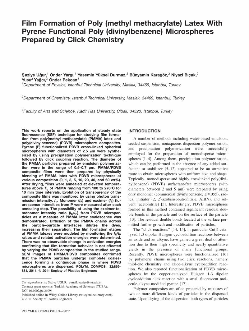
as a powder. The produced stable dispersions of spherical
polymer latexes range in radius from 0.5 to 0.7 lm (see
Fig. 1a). A combination of 1H NMR and UV analysis
indicates that these particles contain 4 mol% PIB (These
particles were prepared in Professor M. A.Winnik’s Labo-
ratory in Toronto). The molecular weight of graft PMMA
was measured as Mw ¼ 1.10 3 105 and the polydispersity
of the PMMA was 2.3.
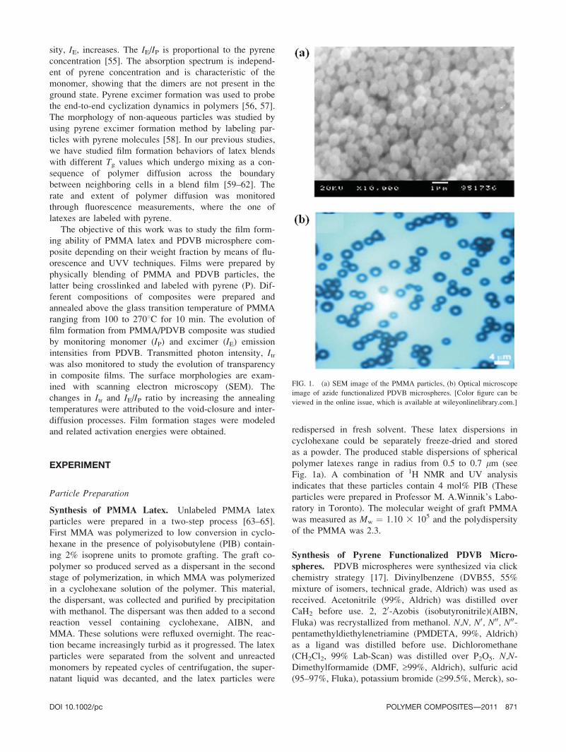
Synthesis of Pyrene Functionalized PDVB Micro-spheres. PDVB microspheres were synthesized via click
chemistry strategy [17]. Divinylbenzene (DVB55, 55%
mixture of isomers, technical grade, Aldrich) was used as
received. Acetonitrile (99%, Aldrich) was distilled over
CaH2 before use. 2, 20-Azobis (isobutyronitrile)(AIBN,
Fluka) was recrystallized from methanol. N,N, N0, N00, N00-pentamethyldiethylenetriamine (PMDETA, 99%, Aldrich)
as a ligand was distilled before use. Dichloromethane
(CH2Cl2, 99% Lab-Scan) was distilled over P2O5. N,N-Dimethylformamide (DMF, ‡99%, Aldrich), sulfuric acid
(95–97%, Fluka), potassium bromide (‡99.5%, Merck), so-
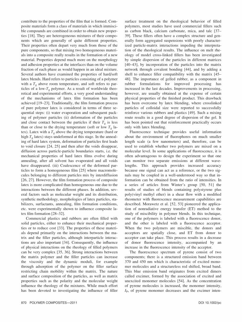
FIG. 1. (a) SEM image of the PMMA particles, (b) Optical microscope
image of azide functionalized PDVB microspheres. [Color figure can be
viewed in the online issue, which is available at wileyonlinelibrary.com.]
DOI 10.1002/pc POLYMER COMPOSITES—-2011 871
dium azide (‡99%, Merck), sodium chromate (Merck), sil-
ver nitrate (Merck), CuBr (98%, Acros), sodium hydride
(98%, Fluka), propargyl bromide (80 vol% in toluene,
Fluka), 1-pyrene methanol (98%, Aldrich), methanol (99%,
Reiden-de Haon), diethylether (98%, Carlo-Erba) and tetra-
hydrofuran (THF, 99% Lab-Scan) were used as received.
PDVB microspheres were prepared by precipitation po-
lymerization technique as described in the literature [5].
For this purpose, AIBN (0.2 g, 1.22 mmol, 4 wt% relative
to DVB55) was added to the solution of DVB55 (11 mL,
76.8 mmol, 4 vol% relative to total volume) and 274 mL
acetonitrile in dry 500 mL volume of three-necked flask
equipped with a mechanical stirrer and a nitrogen inlet.
The flask was placed in thermostated oil bath and the
temperature was adjusted to 708C. The nitrogen flow was
stopped and the reaction was conducted for 48 h at this
temperature under continuous stirring (32 rpm). The reac-
tion content was cooled to the room temperature and
polymer precipitated was filtered and successively washed
with tetrahydrofuran (20 mL), acetone (20 mL), and
methanol (20 mL). The product was dried at 458C under
vacuum for overnight. The yield was 3 g (30%).
The residual double bonds on the surface of nearly
monodisperse (�2.5 lm) (see Fig. 1b), PDVB micro-
spheres were activated to primary bromine [66, 67] and
then converted into azide functions by condensation with
NaN3. Finally, fluorescence labeled PDVB microspheres
were obtained by the click reaction [68] with propargyl
pyrene [69] through azide functions (Scheme 1).
Film Preparation
Composites were prepared by mixing of PMMA latex
and pyrene labeled crosslinked PDVB microsphere. For this
purpose, PMMA and PDVB particles were dispersed sepa-
rately in heptane solutions. The dispersion of PDVB in hep-
tane was mixed with the appropriate solutions of PMMA to
yield eventually composites with 0, 1, 3, 5, 10, 20, 40, and
60 wt% PDVB content. Each mixture was stirred for 30min
followed by sonication for 15 min at room temperature. The
mixed dispersion was then coated on a glass plates with
similar surface areas (0.8 3 2.5 cm2) and allowed to dry
under the ambient conditions of the laboratory. After dry-
ing, samples were separately annealed above Tg of PMMA
for 10 min at temperatures ranging from 100 to 2708C. Thetemperature was maintained within 628C during annealing.
After each annealing step, films were removed from the
oven and cooled down to room temperature.
Measurements
After annealing, each sample was placed in the solid
surface accessory of a Perkin-Elmer Model LS-50 fluores-
cence spectrometer. Pyrene (P) was excited at 345 nm
and monomer and excimer fluorescence emission spectra
were detected between 350 and 600 nm. All measure-
ments were carried out in the front-face position at room
temperature. Slit widths were kept at 8 nm during all SSF
measurements. The sample position, incident light, I0, IP,and IE emission intensities are shown in Fig. 2a.
Photon transmission experiments were carried out
using Variant Carry-100 UV-Visible (UVV) spectrometer.
The transmittances of the films were detected at 500 nm.
A glass plate was used as a standard for all UVV experi-
ments and measurements were carried out at room tem-
perature after each annealing process. The sample
position and the transmitted light intensity, Itr are pre-
sented in Fig. 2b.
Scanning electron microscope (SEM) images were
taken by using LEO Supra VP35 FESEM.
Scheme 1. Synthesis of pyrene functionalized PDVB microspheres via click chemistry.
872 POLYMER COMPOSITES—-2011 DOI 10.1002/pc
RESULTS AND DISCUSSIONS
Preformed Excimers in Composite Film
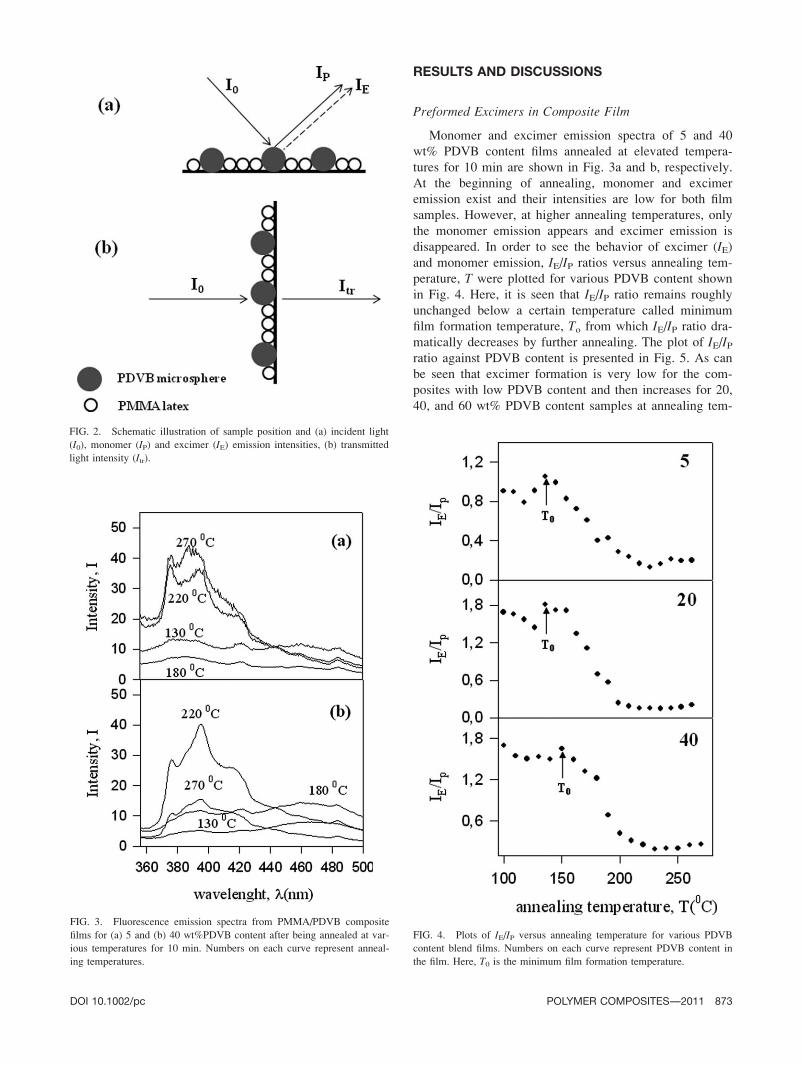
Monomer and excimer emission spectra of 5 and 40
wt% PDVB content films annealed at elevated tempera-
tures for 10 min are shown in Fig. 3a and b, respectively.
At the beginning of annealing, monomer and excimer
emission exist and their intensities are low for both film
samples. However, at higher annealing temperatures, only
the monomer emission appears and excimer emission is
disappeared. In order to see the behavior of excimer (IE)and monomer emission, IE/IP ratios versus annealing tem-
perature, T were plotted for various PDVB content shown
in Fig. 4. Here, it is seen that IE/IP ratio remains roughly
unchanged below a certain temperature called minimum
film formation temperature, To from which IE/IP ratio dra-
matically decreases by further annealing. The plot of IE/IPratio against PDVB content is presented in Fig. 5. As can
be seen that excimer formation is very low for the com-
posites with low PDVB content and then increases for 20,
40, and 60 wt% PDVB content samples at annealing tem-
FIG. 3. Fluorescence emission spectra from PMMA/PDVB composite
films for (a) 5 and (b) 40 wt%PDVB content after being annealed at var-
ious temperatures for 10 min. Numbers on each curve represent anneal-
ing temperatures.
FIG. 2. Schematic illustration of sample position and (a) incident light
(I0), monomer (IP) and excimer (IE) emission intensities, (b) transmitted
light intensity (Itr).
FIG. 4. Plots of IE/IP versus annealing temperature for various PDVB
content blend films. Numbers on each curve represent PDVB content in
the film. Here, T0 is the minimum film formation temperature.
DOI 10.1002/pc POLYMER COMPOSITES—-2011 873
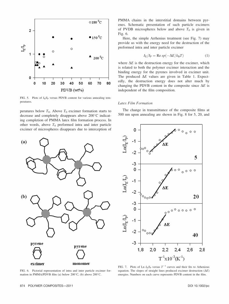
peratures below T0. Above T0 excimer formation starts to
decrease and completely disappears above 2008C indicat-
ing completion of PMMA latex film formation process. In
other words, above T0 preformed intra and inter particle
excimer of microspheres disappears due to interception of
PMMA chains in the interstitial domains between pyr-
enes. Schematic presentation of such particle excimers
of PVDB microspheres below and above T0 is given in
Fig. 6.
Here, the simple Arrhenius treatment (see Fig. 7) may
provide us with the energy need for the destruction of the
preformed intra and inter particle excimer
IE=IP ¼ Re xpð�DE=kBTÞ (1)
where DE is the destruction energy for the excimer, which
is related to both the polymer excimer interaction and the
binding energy for the pyrenes involved in excimer unit.
The produced DE values are given in Table 1. Expect-
edly, the destruction energy does not alter much by
changing the PDVB content in the composite since DE is
independent of the film composition.
Latex Film Formation
The change in transmittance of the composite films at
500 nm upon annealing are shown in Fig. 8 for 5, 20, and
FIG. 5. Plots of IE/IP versus PDVB content for various annealing tem-
peratures.
FIG. 6. Pictorial representation of intra and inter particle excimer for-
mation in PMMA/PDVB film (a) below 2008C, (b) above 2008C.
FIG. 7. Plots of Ln IE/IP versus T21 curves and their fits to Arhenious
equation. The slopes of straight lines produced excimer destruction (DE)energies. Numbers on each curve represents PDVB content in the film.
874 POLYMER COMPOSITES—-2011 DOI 10.1002/pc

40 wt% PDVB content. All the curves in Fig. 8 have the
same characteristic behavior. Upon annealing, the trans-
mitted light intensity Itr increases above the minimum
film formation temperature, T0 and then reaches a maxi-
mum. If the refractive indices of the two polymers are
close, little scattering is expected. In general, light scatter-
ing is a function not only of the refractive index differ-
ence of the components but also of the size and shape of
the dispersed-phase domains. Therefore, optical transmis-
sion measurements are a measure of the number and size
of air voids in a polymer film [70]. Regardless of the
void fraction, transmission decreases with increasing void
radius. On the basis of the change of transmittance of the
films, the structure evolution of the film could be
described as follow: at low annealing temperatures, de-
spite the refractive indices of two polymers are somewhat
different [71], the transparency of the film is related to
the size of the particles and voids inside the film which
leads to light scattering. As the annealing temperature is
increased, PMMA particles deform and flow into the
voids resulting in disappearance of interparticle voids and
flattening of the film. Therefore, transmittance of the film
increases upon annealing due to homogenization during
film formation [59–62]. Flow can also change the mor-
phology of the polymer composite, causing separation of
PDVB micro particles. It is shown that annealing the film
at temperature over T0 leads to deformation and coales-
cence of adjacent PMMA particles forming a continuous
matrix. Moreover, it was found that annealing seemed to
prevent coalescence at all of the crosslinked PDVB par-
ticles. After void closure is completed, further annealing
at higher temperatures causes healing and interdiffusion
processes between PMMA particles. In this case, PMMA
particles undergo coalescence and take part in film form-
ing process, and clearly acted as the continuous phase. On
the other hand, no coalescence of the PDVB particles is
expected as they are highly crosslinked. Apparently, they
do not contribute to latex film formation but disperse in
the PMMA continuous phase.
It has to be noted that Itr decreases dramatically with
increasing PDVB contents in the film. The low PDVB
content blend films are optically more transparent than
those with high PDVB content at all annealing tempera-
tures. The transparency of the latex film is mainly deter-
mined by the scattering of the light that is dependent on
the size of different polymer phases and the voids in the
film [70]. When the size of polymer phases and the voids
are bigger than the wavelength of irradiation light, the
light scatters, and as a result, the transparency decreases.
After the void closure process completed, scattering takes
place predominantly from the submicron crosslinked
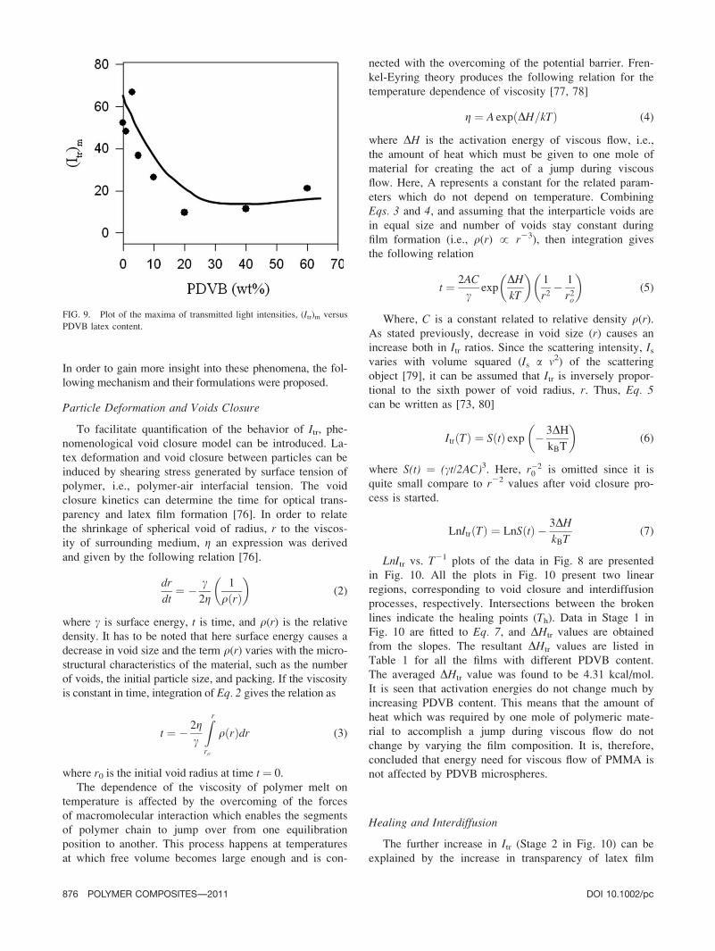
PDVB particles in PMMA matrix. The plots of the maxi-
mum values of Itr, (Itr)m versus PDVB content in Fig. 9
also confirms this hypothesis. As the PDVB content
increased, (Itr)m decreased, indicating that low transpar-
ency occurs at higher PDVB content. Since the size of
PDVB particles is large with respect to the wavelength of
the visible light, the light is scattered resulting in low
transparency in the film [72]. It should also be pointed
out that the curve in Fig. 9 is just the reverse mode of the
curve in Fig. 5.
The increase in Itr may be interpreted by the mechanisms
of void closure, healing and interdiffusion processes [73–75].
TABLE 1. Void closure (DHtr), excimer destruction(DE) and
interdiffusion (DEtr) energies.
PDVB (wt%) DE (kcal/mol) DHtr (kcal/mol) DEtr (kcal/mol)
0 – 1.58 11.04
1 14.02 8.34 10.57
3 7.68 9.33 11.78
5 9.66 2.66 8.30
10 11.12 4.4 7.21
20 16.29 2.89 6.06
40 16.27 4.22 7.48
60 8.69 1.04 17.55
Average 11.96 4.31 10.00
Standard deviations on; DE range from 60.61 to 61.33; DHtr range
from 60.16 to 60.35; DEtr range from 60.56 to 62.2.
FIG. 8. Plots of transmitted photon intensities, Itr vs. annealing temper-
atures, T, for the composite films contain 5, 20, and 40 wt%PDVB.
DOI 10.1002/pc POLYMER COMPOSITES—-2011 875
In order to gain more insight into these phenomena, the fol-
lowing mechanism and their formulations were proposed.
Particle Deformation and Voids Closure
To facilitate quantification of the behavior of Itr, phe-nomenological void closure model can be introduced. La-
tex deformation and void closure between particles can be
induced by shearing stress generated by surface tension of
polymer, i.e., polymer-air interfacial tension. The void
closure kinetics can determine the time for optical trans-
parency and latex film formation [76]. In order to relate
the shrinkage of spherical void of radius, r to the viscos-
ity of surrounding medium, g an expression was derived
and given by the following relation [76].
dr
dt¼ � c
2g1
qðrÞ� �
(2)
where c is surface energy, t is time, and q(r) is the relative
density. It has to be noted that here surface energy causes a
decrease in void size and the term q(r) varies with the micro-
structural characteristics of the material, such as the number
of voids, the initial particle size, and packing. If the viscosity
is constant in time, integration of Eq. 2 gives the relation as
t ¼ � 2gc
Zr
ro
qðrÞdr (3)
where r0 is the initial void radius at time t ¼ 0.
The dependence of the viscosity of polymer melt on
temperature is affected by the overcoming of the forces
of macromolecular interaction which enables the segments
of polymer chain to jump over from one equilibration
position to another. This process happens at temperatures
at which free volume becomes large enough and is con-
nected with the overcoming of the potential barrier. Fren-
kel-Eyring theory produces the following relation for the
temperature dependence of viscosity [77, 78]
g ¼ A expðDH=kTÞ (4)
where DH is the activation energy of viscous flow, i.e.,
the amount of heat which must be given to one mole of
material for creating the act of a jump during viscous
flow. Here, A represents a constant for the related param-
eters which do not depend on temperature. Combining
Eqs. 3 and 4, and assuming that the interparticle voids are
in equal size and number of voids stay constant during
film formation (i.e., q(r) ! r23), then integration gives
the following relation
t ¼ 2AC
cexp
DHkT
� �1
r2� 1
r2o
� �(5)
Where, C is a constant related to relative density q(r).As stated previously, decrease in void size (r) causes an
increase both in Itr ratios. Since the scattering intensity, Isvaries with volume squared (Is a v2) of the scattering
object [79], it can be assumed that Itr is inversely propor-
tional to the sixth power of void radius, r. Thus, Eq. 5can be written as [73, 80]
ItrðTÞ ¼ SðtÞ exp � 3DHkBT
� �(6)
where S(t) ¼ (ct/2AC)3. Here, r�20 is omitted since it is
quite small compare to r22 values after void closure pro-
cess is started.
LnItrðTÞ ¼ LnSðtÞ � 3DHkBT
(7)
LnItr vs. T21 plots of the data in Fig. 8 are presented
in Fig. 10. All the plots in Fig. 10 present two linear
regions, corresponding to void closure and interdiffusion
processes, respectively. Intersections between the broken
lines indicate the healing points (Th). Data in Stage 1 in
Fig. 10 are fitted to Eq. 7, and DHtr values are obtained
from the slopes. The resultant DHtr values are listed in
Table 1 for all the films with different PDVB content.
The averaged DHtr value was found to be 4.31 kcal/mol.
It is seen that activation energies do not change much by
increasing PDVB content. This means that the amount of
heat which was required by one mole of polymeric mate-
rial to accomplish a jump during viscous flow do not
change by varying the film composition. It is, therefore,
concluded that energy need for viscous flow of PMMA is
not affected by PDVB microspheres.
Healing and Interdiffusion
The further increase in Itr (Stage 2 in Fig. 10) can be
explained by the increase in transparency of latex film
FIG. 9. Plot of the maxima of transmitted light intensities, (Itr)m versus
PDVB latex content.
876 POLYMER COMPOSITES—-2011 DOI 10.1002/pc

due to the disappearance of particle–particle boundaries
known as interdiffusion. As the annealing temperature is
increased above Th, some part of the polymer chains may
cross the junction surface and particle boundaries disap-
pear, consequently, the transmitted photon intensity Itrincreases, whereas IE decreases due to the disappearing of
pyrene excimer. In order to quantify these results, the
Prager-Tirrell (PT) model [81, 82] for the chain crossing
density can be employed. These authors used de Gennes’s
‘‘reptation’’ model to explain configurational relaxation at
the polymer–polymer junction where each polymer chain
is considered to be confined to a tube in which executes a
random back and forth motion [82]. The total ‘‘crossing
density’’ r(t) (chains per unit area) at junction surface
was calculated by PT by taking into account of the contri-
butions r1(t) since chains still retain some portion of their
initial tubes, and also a reminder the r2(t) which comes
from chains which have relaxed at least once. In terms of
reduced time s ¼ 2mt/N2, the total crossing density can be
given as [81, 82]
rðsÞ=rð1Þ ¼ 2p�1=2s1=2 (8)
Here, N is the number of freely jointed segments and mis the linear diffusion coefficient given by the following
relation
m ¼ mo expð�DE=kTÞ (9)
where DE is defined as the activation energy for backbone
motion depending on the temperature interval. Combining
Eq. 8 and Eq. 9 a useful relation is obtained as
rðsÞ=rð1Þ ¼ Ro expð�DE=2kTÞ (10)
where Ro ¼ (8mot/pN2)1/2 is a temperature independent
coefficient. The increase in Itr at Stage 2 in Fig. 8 is al-
ready related to the disappearance of particle–particle
interface, i.e., as annealing temperature increased, more
chains relaxed across the junction surface and as a result
the crossing density increases. To explain the behavior of
Itr above Th, it can be assumed that Itr is directly propor-
tional to the crossing density r(T). Then, Eq. 10 can be
solved for Itr to interpret the results in Fig. 8 as follows:
ItrðTÞ=Itrð1Þ ¼ Re xpð�DEtr=2kTÞ (11)
The activation energy of backbone motion, DEtr is pro-
duced by least squares fitting the data in Fig. 10 (Stage 2)
to Eq. 11. DEtr values are listed in Table 1. The average
DEtr value is found to be as 10.0 kcal/mol for transmis-
sion which are much larger than the void closure activa-
tion energies. This result is understandable because a sin-
gle chain needs more energy to execute diffusion across
the polymer–polymer interface than to be accomplished
by the viscous flow process. The calculated activation
energies were found roughly independent of PDVB con-
tent in the film.
Morphological Studies
To support these findings, SEM was used to examine
the morphologies of PMMA/PDVB composite films. Fig-
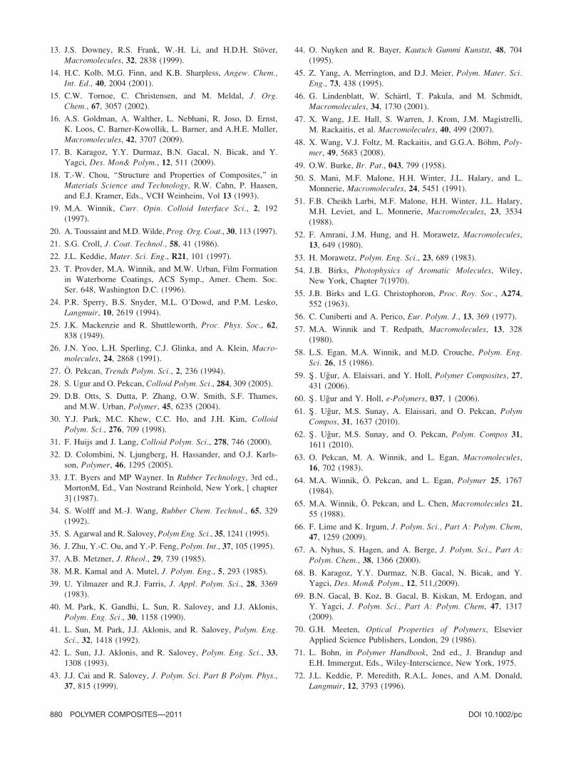
ures 11–13 present SEM photographs of the films which
have 0, 5, and 20 wt% PDVB content annealed at 100,
180, and 2708C, respectively. The SEM images of the
films annealed at 1008C (Figs. 11a, 12a, and 13a) show
that the surface of the composite films is very rough.
These results reveal that 1008C, close to the Tg of
PMMA, is too low to make the particles deform and
retain their spherical shapes. Most of the light scatters
due to the rough surface of the films which results low
transmitted light intensity. Here, the small particles are
PMMA latexes, while the large ones are crosslinked
PDVB microspheres.
FIG. 10. Plots of Ln Itr versus T21 curves and their fits to Eqs. 7 and
11. The slopes of straight lines produced void closure (DHtr) and inter-
diffusion (DEtr) energies. Stages 1 and 2 present the void closure and
interdiffusion processes. Numbers on each curve represents PDVB con-
tent in the film.
DOI 10.1002/pc POLYMER COMPOSITES—-2011 877
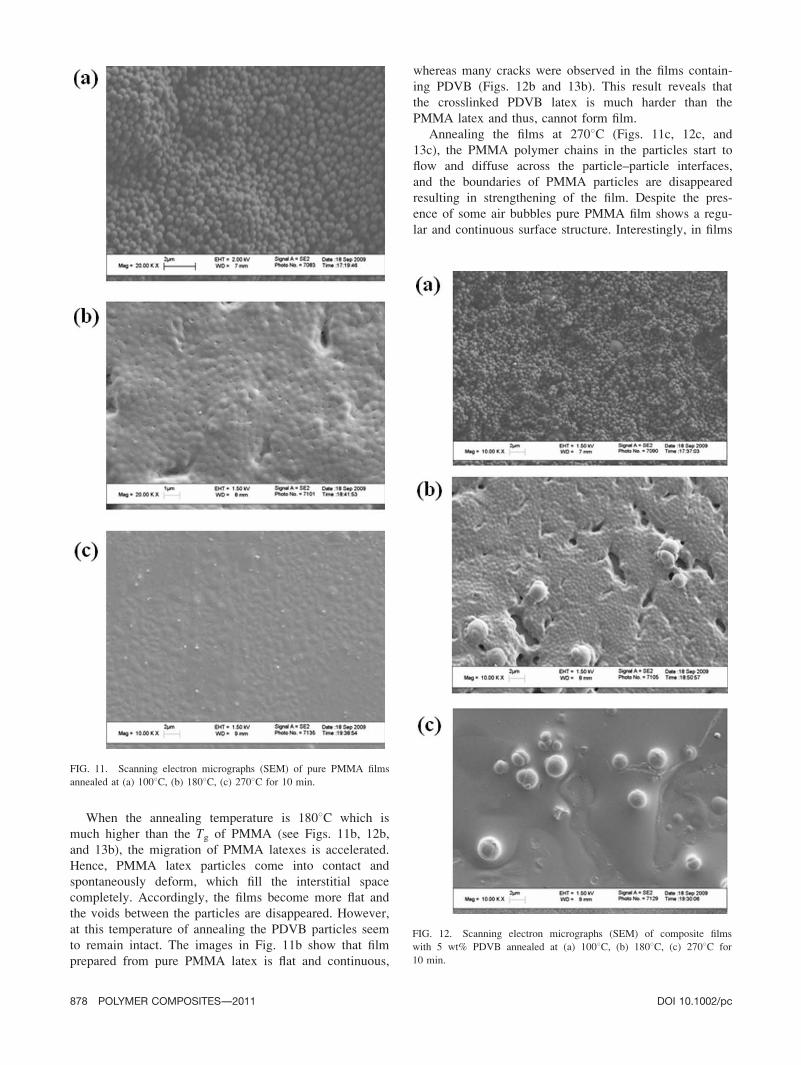
When the annealing temperature is 1808C which is
much higher than the Tg of PMMA (see Figs. 11b, 12b,
and 13b), the migration of PMMA latexes is accelerated.
Hence, PMMA latex particles come into contact and
spontaneously deform, which fill the interstitial space
completely. Accordingly, the films become more flat and
the voids between the particles are disappeared. However,
at this temperature of annealing the PDVB particles seem
to remain intact. The images in Fig. 11b show that film
prepared from pure PMMA latex is flat and continuous,
whereas many cracks were observed in the films contain-
ing PDVB (Figs. 12b and 13b). This result reveals that
the crosslinked PDVB latex is much harder than the
PMMA latex and thus, cannot form film.
Annealing the films at 2708C (Figs. 11c, 12c, and
13c), the PMMA polymer chains in the particles start to
flow and diffuse across the particle–particle interfaces,
and the boundaries of PMMA particles are disappeared
resulting in strengthening of the film. Despite the pres-
ence of some air bubbles pure PMMA film shows a regu-
lar and continuous surface structure. Interestingly, in films
FIG. 12. Scanning electron micrographs (SEM) of composite films
with 5 wt% PDVB annealed at (a) 1008C, (b) 1808C, (c) 2708C for
10 min.
FIG. 11. Scanning electron micrographs (SEM) of pure PMMA films
annealed at (a) 1008C, (b) 1808C, (c) 2708C for 10 min.
878 POLYMER COMPOSITES—-2011 DOI 10.1002/pc
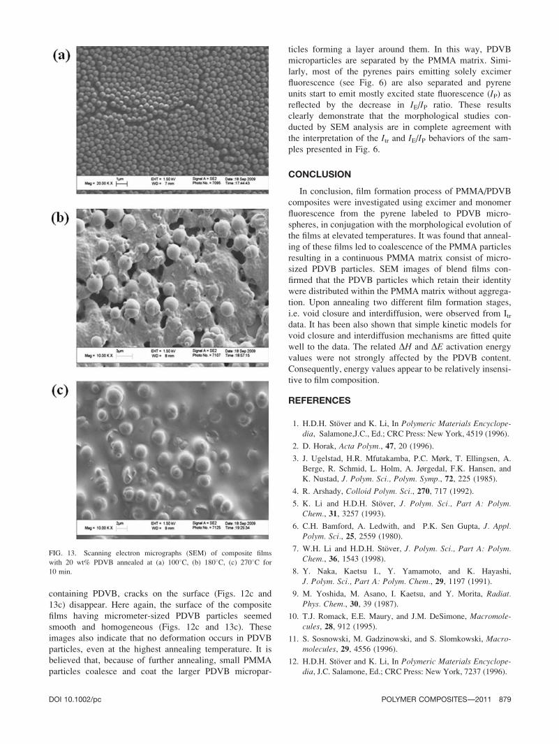
containing PDVB, cracks on the surface (Figs. 12c and
13c) disappear. Here again, the surface of the composite
films having micrometer-sized PDVB particles seemed
smooth and homogeneous (Figs. 12c and 13c). These
images also indicate that no deformation occurs in PDVB
particles, even at the highest annealing temperature. It is
believed that, because of further annealing, small PMMA
particles coalesce and coat the larger PDVB micropar-
ticles forming a layer around them. In this way, PDVB
microparticles are separated by the PMMA matrix. Simi-
larly, most of the pyrenes pairs emitting solely excimer
fluorescence (see Fig. 6) are also separated and pyrene
units start to emit mostly excited state fluorescence (IP) asreflected by the decrease in IE/IP ratio. These results
clearly demonstrate that the morphological studies con-
ducted by SEM analysis are in complete agreement with
the interpretation of the Itr and IE/IP behaviors of the sam-
ples presented in Fig. 6.
CONCLUSION
In conclusion, film formation process of PMMA/PDVB
composites were investigated using excimer and monomer
fluorescence from the pyrene labeled to PDVB micro-
spheres, in conjugation with the morphological evolution of
the films at elevated temperatures. It was found that anneal-
ing of these films led to coalescence of the PMMA particles
resulting in a continuous PMMA matrix consist of micro-
sized PDVB particles. SEM images of blend films con-
firmed that the PDVB particles which retain their identity
were distributed within the PMMA matrix without aggrega-
tion. Upon annealing two different film formation stages,
i.e. void closure and interdiffusion, were observed from Itrdata. It has been also shown that simple kinetic models for
void closure and interdiffusion mechanisms are fitted quite
well to the data. The related DH and DE activation energy
values were not strongly affected by the PDVB content.
Consequently, energy values appear to be relatively insensi-
tive to film composition.
REFERENCES
1. H.D.H. Stover and K. Li, In Polymeric Materials Encyclope-dia, Salamone,J.C., Ed.; CRC Press: New York, 4519 (1996).
2. D. Horak, Acta Polym., 47, 20 (1996).
3. J. Ugelstad, H.R. Mfutakamba, P.C. Mørk, T. Ellingsen, A.
Berge, R. Schmid, L. Holm, A. Jørgedal, F.K. Hansen, and
K. Nustad, J. Polym. Sci., Polym. Symp., 72, 225 (1985).
4. R. Arshady, Colloid Polym. Sci., 270, 717 (1992).
5. K. Li and H.D.H. Stover, J. Polym. Sci., Part A: Polym.Chem., 31, 3257 (1993).
6. C.H. Bamford, A. Ledwith, and P.K. Sen Gupta, J. Appl.Polym. Sci., 25, 2559 (1980).
7. W.H. Li and H.D.H. Stover, J. Polym. Sci., Part A: Polym.Chem., 36, 1543 (1998).
8. Y. Naka, Kaetsu I., Y. Yamamoto, and K. Hayashi,
J. Polym. Sci., Part A: Polym. Chem., 29, 1197 (1991).
9. M. Yoshida, M. Asano, I. Kaetsu, and Y. Morita, Radiat.Phys. Chem., 30, 39 (1987).
10. T.J. Romack, E.E. Maury, and J.M. DeSimone, Macromole-cules, 28, 912 (1995).
11. S. Sosnowski, M. Gadzinowski, and S. Slomkowski, Macro-molecules, 29, 4556 (1996).
12. H.D.H. Stover and K. Li, In Polymeric Materials Encyclope-dia, J.C. Salamone, Ed.; CRC Press: New York, 7237 (1996).
FIG. 13. Scanning electron micrographs (SEM) of composite films
with 20 wt% PDVB annealed at (a) 1008C, (b) 1808C, (c) 2708C for
10 min.
DOI 10.1002/pc POLYMER COMPOSITES—-2011 879
13. J.S. Downey, R.S. Frank, W.-H. Li, and H.D.H. Stover,
Macromolecules, 32, 2838 (1999).
14. H.C. Kolb, M.G. Finn, and K.B. Sharpless, Angew. Chem.,Int. Ed., 40, 2004 (2001).
15. C.W. Tornoe, C. Christensen, and M. Meldal, J. Org.Chem., 67, 3057 (2002).
16. A.S. Goldman, A. Walther, L. Nebhani, R. Joso, D. Ernst,
K. Loos, C. Barner-Kowollik, L. Barner, and A.H.E. Muller,
Macromolecules, 42, 3707 (2009).
17. B. Karagoz, Y.Y. Durmaz, B.N. Gacal, N. Bicak, and Y.
Yagci, Des. Mon& Polym., 12, 511 (2009).
18. T.-W. Chou, ‘‘Structure and Properties of Composites,’’ in
Materials Science and Technology, R.W. Cahn, P. Haasen,
and E.J. Kramer, Eds., VCH Weinheim, Vol 13 (1993).
19. M.A. Winnik, Curr. Opin. Colloid Interface Sci., 2, 192
(1997).
20. A. Toussaint and M.D. Wilde, Prog. Org. Coat., 30, 113 (1997).
21. S.G. Croll, J. Coat. Technol., 58, 41 (1986).
22. J.L. Keddie, Mater. Sci. Eng., R21, 101 (1997).
23. T. Provder, M.A. Winnik, and M.W. Urban, Film Formation
in Waterborne Coatings, ACS Symp., Amer. Chem. Soc.
Ser. 648, Washington D.C. (1996).
24. P.R. Sperry, B.S. Snyder, M.L. O’Dowd, and P.M. Lesko,
Langmuir, 10, 2619 (1994).
25. J.K. Mackenzie and R. Shuttleworth, Proc. Phys. Soc., 62,838 (1949).
26. J.N. Yoo, L.H. Sperling, C.J. Glinka, and A. Klein, Macro-molecules, 24, 2868 (1991).
27. O. Pekcan, Trends Polym. Sci., 2, 236 (1994).
28. S. Ugur and O. Pekcan, Colloid Polym. Sci., 284, 309 (2005).
29. D.B. Otts, S. Dutta, P. Zhang, O.W. Smith, S.F. Thames,
and M.W. Urban, Polymer, 45, 6235 (2004).
30. Y.J. Park, M.C. Khew, C.C. Ho, and J.H. Kim, ColloidPolym. Sci., 276, 709 (1998).
31. F. Huijs and J. Lang, Colloid Polym. Sci., 278, 746 (2000).
32. D. Colombini, N. Ljungberg, H. Hassander, and O.J. Karls-
son, Polymer, 46, 1295 (2005).
33. J.T. Byers and MP Wayner. In Rubber Technology, 3rd ed.,
MortonM, Ed., Van Nostrand Reinhold, New York, [ chapter
3] (1987).
34. S. Wolff and M.-J. Wang, Rubber Chem. Technol., 65, 329(1992).
35. S. Agarwal and R. Salovey, Polym Eng. Sci., 35, 1241 (1995).
36. J. Zhu, Y.-C. Ou, and Y.-P. Feng, Polym. Int., 37, 105 (1995).
37. A.B. Metzner, J. Rheol., 29, 739 (1985).
38. M.R. Kamal and A. Mutel, J. Polym. Eng., 5, 293 (1985).
39. U. Yilmazer and R.J. Farris, J. Appl. Polym. Sci., 28, 3369(1983).
40. M. Park, K. Gandhi, L. Sun, R. Salovey, and J.J. Aklonis,
Polym. Eng. Sci., 30, 1158 (1990).
41. L. Sun, M. Park, J.J. Aklonis, and R. Salovey, Polym. Eng.Sci., 32, 1418 (1992).
42. L. Sun, J.J. Aklonis, and R. Salovey, Polym. Eng. Sci., 33,1308 (1993).
43. J.J. Cai and R. Salovey, J. Polym. Sci. Part B Polym. Phys.,37, 815 (1999).
44. O. Nuyken and R. Bayer, Kautsch Gummi Kunstst, 48, 704(1995).
45. Z. Yang, A. Merrington, and D.J. Meier, Polym. Mater. Sci.Eng., 73, 438 (1995).
46. G. Lindenblatt, W. Schartl, T. Pakula, and M. Schmidt,
Macromolecules, 34, 1730 (2001).
47. X. Wang, J.E. Hall, S. Warren, J. Krom, J.M. Magistrelli,
M. Rackaitis, et al. Macromolecules, 40, 499 (2007).
48. X. Wang, V.J. Foltz, M. Rackaitis, and G.G.A. Bohm, Poly-mer, 49, 5683 (2008).
49. O.W. Burke, Br. Pat., 043, 799 (1958).
50. S. Mani, M.F. Malone, H.H. Winter, J.L. Halary, and L.
Monnerie, Macromolecules, 24, 5451 (1991).
51. F.B. Cheikh Larbi, M.F. Malone, H.H. Winter, J.L. Halary,
M.H. Leviet, and L. Monnerie, Macromolecules, 23, 3534
(1988).
52. F. Amrani, J.M. Hung, and H. Morawetz, Macromolecules,13, 649 (1980).
53. H. Morawetz, Polym. Eng. Sci., 23, 689 (1983).
54. J.B. Birks, Photophysics of Aromatic Molecules, Wiley,
New York, Chapter 7(1970).
55. J.B. Birks and L.G. Christophoron, Proc. Roy. Soc., A274,552 (1963).
56. C. Cuniberti and A. Perico, Eur. Polym. J., 13, 369 (1977).
57. M.A. Winnik and T. Redpath, Macromolecules, 13, 328
(1980).
58. L.S. Egan, M.A. Winnik, and M.D. Crouche, Polym. Eng.Sci. 26, 15 (1986).
59. S� . Ugur, A. Elaissari, and Y. Holl, Polymer Composites, 27,431 (2006).
60. S� . Ugur and Y. Holl, e-Polymers, 037, 1 (2006).
61. S� . Ugur, M.S. Sunay, A. Elaissari, and O. Pekcan, PolymCompos, 31, 1637 (2010).
62. S� . Ugur, M.S. Sunay, and O. Pekcan, Polym. Compos 31,1611 (2010).
63. O. Pekcan, M. A. Winnik, and L. Egan, Macromolecules,16, 702 (1983).
64. M.A. Winnik, O. Pekcan, and L. Egan, Polymer 25, 1767(1984).
65. M.A. Winnik, O. Pekcan, and L. Chen, Macromolecules 21,55 (1988).
66. F. Lime and K. Irgum, J. Polym. Sci., Part A: Polym. Chem,47, 1259 (2009).
67. A. Nyhus, S. Hagen, and A. Berge, J. Polym. Sci., Part A:Polym. Chem., 38, 1366 (2000).
68. B. Karagoz, Y.Y. Durmaz, N.B. Gacal, N. Bicak, and Y.
Yagci, Des. Mon& Polym., 12, 511,(2009).
69. B.N. Gacal, B. Koz, B. Gacal, B. Kiskan, M. Erdogan, and
Y. Yagci, J. Polym. Sci., Part A: Polym. Chem, 47, 1317(2009).
70. G.H. Meeten, Optical Properties of Polymers, Elsevier
Applied Science Publishers, London, 29 (1986).
71. L. Bohn, in Polymer Handbook, 2nd ed., J. Brandup and
E.H. Immergut, Eds., Wiley-Interscience, New York, 1975.
72. J.L. Keddie, P. Meredith, R.A.L. Jones, and A.M. Donald,
Langmuir, 12, 3793 (1996).
880 POLYMER COMPOSITES—-2011 DOI 10.1002/pc
73. S� . Ugur, A. Elaissari, and O. Pekcan, J. Coll. Int. Sci., 263,674 (2003).
74. S� . Ugur, A. Elaissari, and O. Pekcan, J. Coat. Technol.Res., 1(4), 305 (2004).
75. O. Pekcan and E. Arda, Colloids Suf. A, 153, 537 (1999).
76. J.L. Keddie, P. Meredith, R.A.L Jones, and A.M. Donald, in
Film Formation in Waterborne Coatings, T. Provder, M.A.
Winnik and M.W. Urban (Eds.), ACS Symp. Ser. 648,
American Chemical Society, 332 (1996).
77. G.S. Fulcher, J. Am. Ceram. Soc., 8, 339 (1925).
78. J. Frenkel, J. Phys. USSR, 9, 385 (1945).
79. S.S. Voyutskii, Collois Chemistry, MR Publisher, Mos-
cow(1978).
80. S� . Ugur, A. Elaissari, and O. Pekcan, Polym. Adv. Technol.,16, 405 (2005).
81. S. Prager and M. Tirrell, J. Chem. Phys., 75, 5194 (1981).
82. R.P. Wool, B.L. Yuan, and O.J. McGarel, J. Polym. Eng.Sci., 29, 1340 (1989).
DOI 10.1002/pc POLYMER COMPOSITES—-2011 881