Embed Size (px)
Citation preview
8/2/2019 Filling of Carbon Nanotubes With Silver, Gold, And
http://slidepdf.com/reader/full/filling-of-carbon-nanotubes-with-silver-gold-and 1/4
F i l li n g o f C a rb o n N a n o t u b e s w i t h S i lv e r , Go l d , a n d
Go ld C hlo ride
Andrew Chu,† J essica Cook,† Rufus J . R. Heesom,† J ohn L. Hutchison,‡
Malcolm L. H . Green ,*,† and J eremy Sloan †
In organ ic Ch em is tr y L ab ora tor y, S ou th Pa rk s R oad , Ox for d OX 1 3Q R , UK,and Department of Materials, Parks Road, Oxford OX1 3PH, UK
R eceiv ed Apr il 25 , 19 96 . R evi sed M an u scr ip t R eceiv ed J u ly 24 , 19 96 X
The synthesis of carbon nanotubes filled with silver, gold, and gold chloride is described.The resulting materials have been studied by high-resolution electron microscopy, X-raypowder diffraction, an d energy-dispersive spectroscopy (EDS). The EDS da ta wer e obta inedusing a high-resolution electron microscope equipped with a field emission gun which allowedfor X-ray analytical data to be obtained on individual particles located within the carbonnanotubes using a 0.7 nm probe.
I n t r o d u c t i o n
Theoretical studies on carbon nanotubes suggest thatthe introduction of foreign materials into hollow nano-tube cavities may have interesting effects on the physi-
cal and elect ronic pr opert ies of the encapsulatedmaterials.1-5 It is predicted that filled nan otubes mayfind pra ctical uses a s n an owires, composites an d n ovelcatalysts. Several different appr oaches to the problemof fi ll ing na notubes h ave been ta ken. Ear ly work onbismuth and lead relied upon capillary action to pullliquids into the hollow nanotube cavities.6,7 Certainlanthanide and transit ion-metal carbides have beentra pped in na notube cavities by the a rc-evapora tion of meta l-doped carbon r ods.8-13 Metal nitrates were en-capsulated by refluxing closed nanotubes in HNO 3 inthe pr esence of the soluble nitr ates.5 Herein we describethe filling of carbon nanotubes with Ag and Au crys-tal l i tes via a two-step method and the subsequent
characterization of the nanotube.
R e s u l t s a n d D i s c u s s i o n
A two-step method employing wet chemistry tech-niques was u sed to prepar e car bon nan otubes filled withAg and Au meta l. The nan otubes previously opened byoxidation with HNO3
5 were st irred overnight with aconcentra ted aqueous solution of either Ag(NO3) or
AuCl3. After concentr ation of the mixtur e and separ a-tion of the resulting tube samples from the excess of the solution, they were calcined in a furna ce at 250 °C(for Ag(NO3)) and at 160 °C (for the AuCl3 sam ple). At
these temperatures, Ag(NO3) (>160 °C) an d AuCl3 aredecomposed to give th e element s.14 AuCl3 decomposedat temperatures less than 150 °C to give AuCl whichdissociates into the element s at higher temper atu res.14
Alth ough it ha s been rep orted t ha t th e yields of filledtubes using a two-step preparation are lower than thoseobtained using a one-step method, in the cases of Agand Au, we observed a relatively h igh percentage of opened n an otubes being filled with the meta ls (∼70%).5,6
The overall yields of fi l led nanotubes were roughlyestimated by th e visual inspection of portions of th esample using TEM.
The an nealed Ag an d Au samples were examined byhigh-resolution transmission electron microscopy (HR-
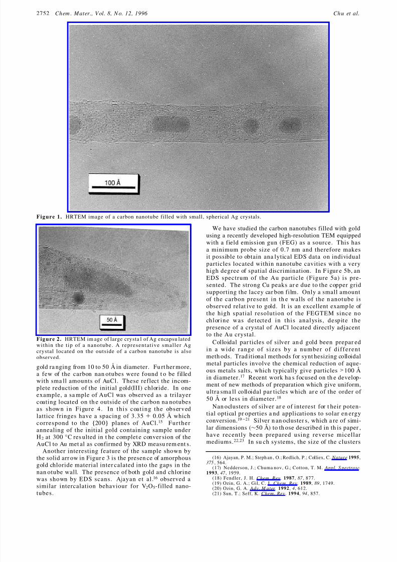
TEM), analytical energy-dispersive spectroscopy (EDS),an d X-ray powder diffraction (XRD). Pr eliminar y HR-TEM images of the Ag sample showed that smal lcrystallites of Ag met al were pres ent ins ide and outs ideof the carbon nan otubes. A typical carbon nanotubecontaining several small Ag crystals is shown in Figure1. The crystals observed in F igur e 1 ran ge in size from35 to 85 Å. An HRTEM image of the tip of a n anotubein which an unusually large single crystal of Ag (190Å) is encapsulated is shown in Figure 2. This Figure 2also shows a smaller crystal of Ag located on the outsidewall . The d spacing of the lat tice for the encapsulatedlarge crystal and the small spherical crystal was mea-sured to be 2.361 ( 0.05 and 2.369 ( 0.05 Å, respec-
tively, which both correspond to the reported value forthe {111} plan e of 2.359 Å.15
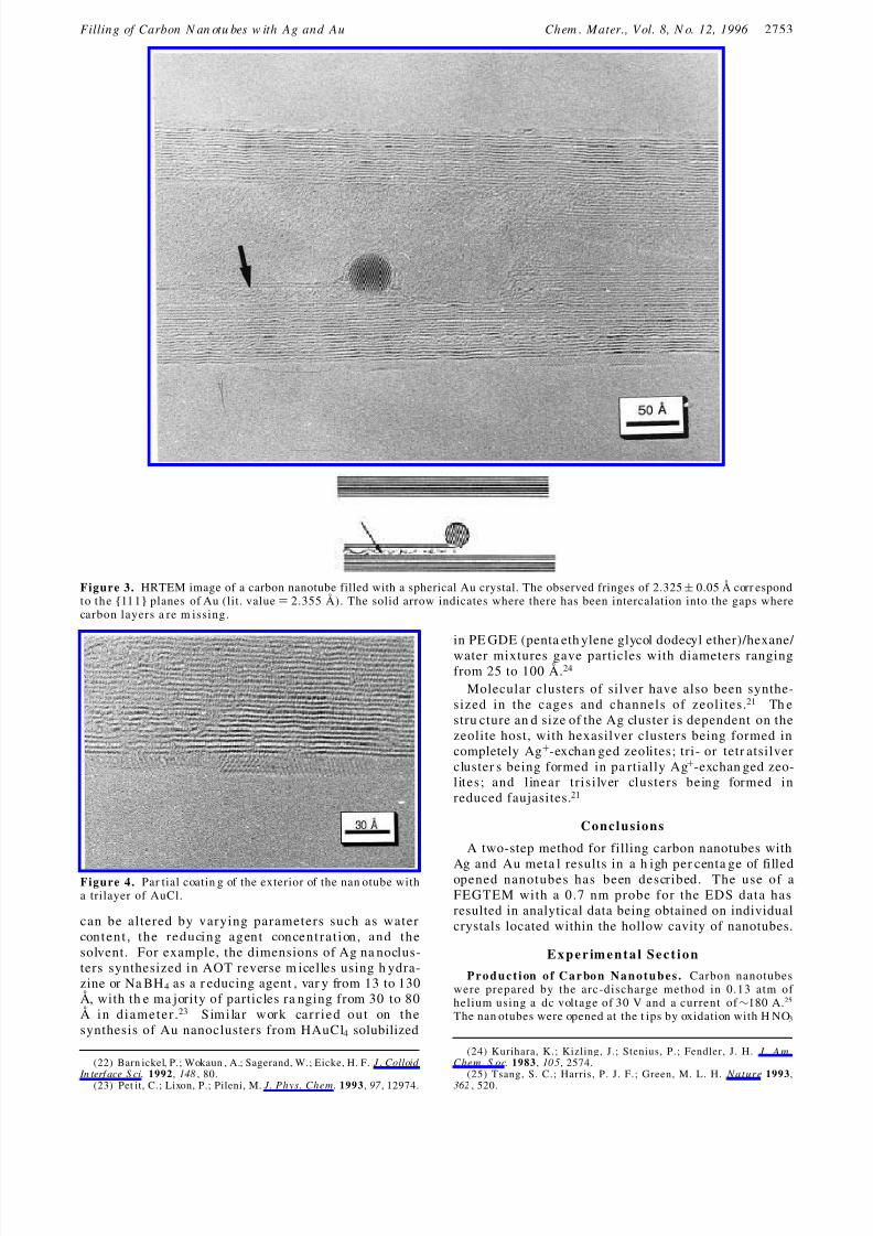
Figure 3 shows the HRTEM image of a sma ll goldcrystal located within a nanotube which, a ssuming aperfect sph ere, is estimat ed to conta in 3000 ( 200 atomsof gold. The measu red regular d spa cing of the observedplan es of th e latt ice is 2.325 ( 0.05 Å, which corr espondsto the {111} plan es of Au (lit. value ) 2.355 Å).15 Mostof the gold encapsulated within the carbon nanotubeswas found to consist of these spherical crystallites of
† Inorganic Chemistry Laboratory.‡ Department of Materials.X Abstract published in Advance ACS Abstracts, Septem ber 1, 1996.(1) La asonen, K.; Andreoni, W.; Par rinello, M. S cience 1992, 58 ,
1916.(2) Mintmir e, J. W.; Dunlap, B. I.; White, C. T. Phys. Rev. Lett. 1992,
68 , 631.(3) Sait o, R.; Fijita , M.; Dresselhau s, G.; Dresselh aus , M. S. Mater.Sci. Eng. 1993, B 19 , 185.
(4) Hamada, N.; Sawada, S.; Oshitama, A. Phys. Rev. L ett. 1992,68 , 1579.
(5) Tsang, S. C.; Chen, Y. K.; Harris, P. J . F.; Green, M. L. H. Nature1994, 372 , 159.
(6) Ajayan, P. M.; Iijima, S. Na t u r e 1993, 36 1, 333.(7) Seshadri, R.; Govindaraj, A.; Aiyer, H. N.; Sen, R.; Subbann a,
G. N.; Raju, A. R.; Rao, C. N. R. Curr. S ci. 1994, 66 , 839.(8) Subra money, S.; Ruoff, R. S.; Lorent s, D. C.; Chan, B.; Malhotra,
R.; Dyer, M. J .; Parvin, K. Carbon 1994, 32 , 507.(9) Yosida, Y. Appl. Phys. L ett. 1994, 22 , 64.(10) Sait o, Y.; Yoshikawa , T.; Okuda , M.; Fujim oto, N.; Sumiyam a,
K.; Suzuki, K.; Kasuya, A.; Nishina, Y. J. Phys. Chem. Solids 1994,54 , 1849.
(11) Liu, M.; Cowley, J. M. Carbon 1995, 33 , 225.(12) Liu, M.; Cowley, J. M. Carbon 1993, 33 , 749.(13) Cowley, J. M.; Liu, M. Micron 1994, 25 , 53.
(14) Greenwood, N. N.; Ear nshaw, A. Chemistry of the E lements;Pergamon Pr ess: Oxford, 1989; pp 1368-1374.
(15) Powder Dffraction Data File 38-1364, Inorganic Phases, J CPDSIntern ational Centr e for Diffraction Dat a, Swath more, PA, 1990.
2751Chem. Mater. 1996, 8, 2751-2754
S0897-4756(96)00246-3 CCC: $12.00 © 1996 American Chem ical Society
8/2/2019 Filling of Carbon Nanotubes With Silver, Gold, And
http://slidepdf.com/reader/full/filling-of-carbon-nanotubes-with-silver-gold-and 2/4
gold ra nging from 10 to 50 Å in diameter. Fu rt her more,a few of the carbon nan otubes were found t o be filledwith sma ll amounts of AuCl. These reflect the incom-plete reduction of the initial gold(III) chloride. In one
example, a sa mple of AuCl was observed as a trilayercoating located on th e outside of the carbon na notubesas shown in Figure 4. In this coat ing the observedlattice fringes have a spacing of 3.35 ( 0.05 Å whichcorrespond to the {200} planes of AuCl.15 Furtherannealing of the initial gold containing sample underH 2 at 300 °C res ulted in t he complet e conver sion of theAuCl to Au met al as confirmed by XRD measu rem ent s.
Another interesting feature of the sample shown bythe solid arr ow in Figur e 3 is the presen ce of amorphousgold chloride material inter calated into the gaps in thenan otube wall. The presence of both gold and chlorinewas shown by EDS scans. Ajayan et al .16 observed asimilar intercalation behaviour for V2O5-filled nano-
tubes.
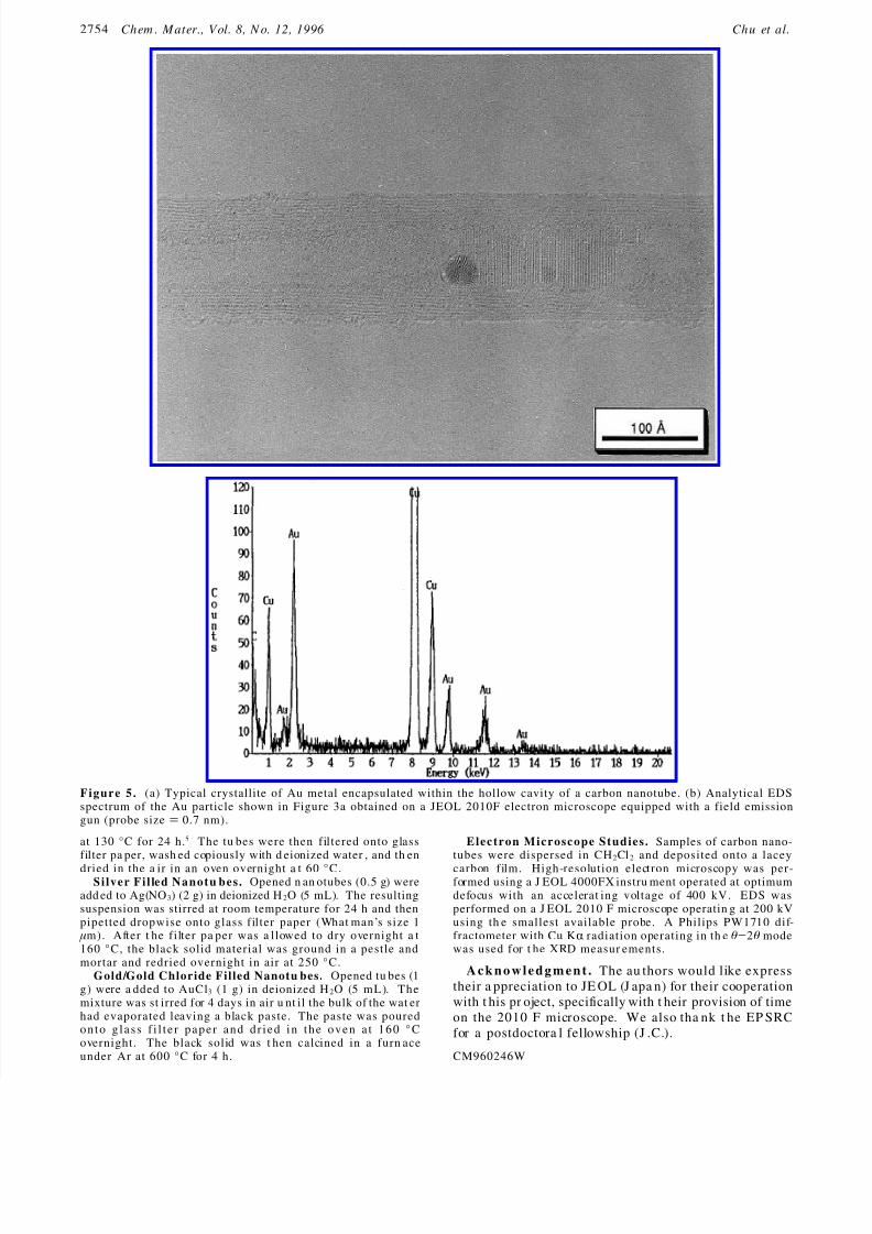
We have studied the carbon nanotubes filled with goldusing a recently developed high-resolution TEM equippedwith a field emission gun (FEG) as a source. This hasa minimum probe size of 0.7 nm and therefore makesit possible to obtain ana lytical EDS data on individualparticles located within nanotube cavities with a veryhigh degree of spatial discrimination. In F igur e 5b, anEDS spectrum of the Au particle (Figure 5a) is pre-sented. The strong Cu peaks ar e due to the copper gridsupporting the lacey car bon film. Only a small amountof the carbon present in th e walls of the n anotube isobserved relat ive to gold. It is an excellent examp le of the high spatial resolution of the FEGTEM since nochlorine was detected in this analysis , despite thepresence of a crystal of AuCl located directly adjacentto the Au crystal.
Colloidal par ticles of silver an d gold been prepar edin a wide range of sizes by a number of di fferentmeth ods. Trad itiona l methods for synt hesizing colloidalmetal particles involve the chemical reduction of aque-ous metals salts, which typically give particles >100 Åin diameter.17 Recent work ha s focused on th e develop-ment of new methods of preparation which give uniform,
ultra sma ll colloidal par ticles which ar e of the order of 50 Å or less in diameter. 18
Nan oclusters of silver ar e of interest for t heir poten-tial optical pr operties a nd applications to solar en ergyconversion.19-21 Silver n an ocluster s, which a re of simi-lar dimensions (∼50 Å) to th ose described in th is paper ,have recently been prepared using reverse micellarmediums.22,23 In su ch systems, the size of the clusters
(16) Ajayan, P. M.; Steph an , O.; Redlich, P.; Colliex, C. Nature 1995,375 , 564.
(17) Nedderson, J.; Chuma nov, G.; Cotton, T. M. Appl. S pectrosc.1993, 47 , 1959.
(18) Fendler, J. H. Chem. Rev. 1987, 87 , 877.(19) Ozin, G. A.; Gil, C. J. Chem. Rev. 1989, 89 , 1749.(20) Ozin, G. A. Adv. M ater. 1992, 4, 612.
(21) Sun, T.; Seff, K. Chem. Rev. 1994, 94 , 857.
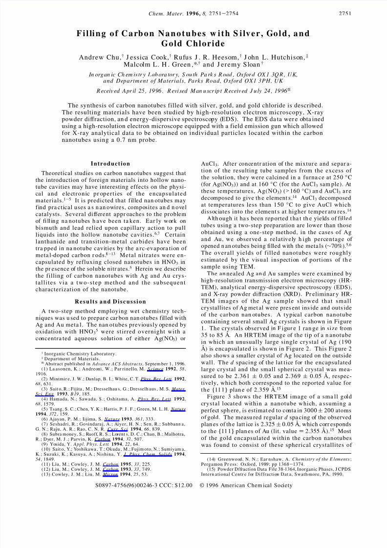
F i g u r e 1 . HRTEM image of a carbon nanotube filled with small, spherical Ag crystals.
F i g u r e 2 . HRTEM im age of large crysta l of Ag encapsu latedwithin the t ip of a nanotube. A representative smaller Agcrystal located on the outside of a carbon nanotube is alsoobserved.
2752 Ch em . M ater., Vol. 8, N o. 12, 1996 Ch u et al.
8/2/2019 Filling of Carbon Nanotubes With Silver, Gold, And
http://slidepdf.com/reader/full/filling-of-carbon-nanotubes-with-silver-gold-and 3/4
can be altered by varying parameters such as watercontent , the reducing agent concentrat ion, and thesolvent. For example, the dimensions of Ag na noclus-ters synthesized in AOT reverse m icelles using h ydra-zine or Na BH 4 as a r educing agent , var y from 13 to 130Å, with th e ma jority of particles ra nging from 30 to 80Å in diameter.23 Similar work carried out on thesynthesis of Au nanoclusters from HAuCl4 solubilized
in PE GDE (penta eth ylene glycol dodecyl ether)/hexane/ water mixtures gave particles with diameters rangingfrom 25 to 100 Å.24
Molecular clusters of silver have also been synthe-sized in the cages and channels of zeoli tes.21 Th estru cture an d size of the Ag cluster is dependent on thezeolite host, with hexasilver clusters being formed incompletely Ag+-exchan ged zeolites; tri- or tetr atsilvercluster s being formed in pa rtially Ag+-exchan ged zeo-lites; and l inear trisi lver clusters being formed inreduced faujasites.21
C o n c l u s i o n s
A two-step method for filling carbon nanotubes with
Ag and Au meta l results in a h igh per centa ge of filledopened nanotubes has been described. The use of aFEGTEM with a 0.7 nm probe for the EDS data hasresulted in analytical data being obtained on individualcrystals located within the hollow cavity of nanotubes.
E x p e r im e n t a l S e c t i o n
P r o d u c t i on o f C a r bo n N a n o t u b e s . Carbon nanotubeswere prepared by the arc-discharge method in 0.13 atm of helium using a dc voltage of 30 V and a current of ∼180 A.25
The nan otubes were opened at the t ips by oxidation with H NO3
(22) Barn ickel, P.; Wokaun , A.; Sagerand, W.; Eicke, H. F. J . Colloid In terf ace S ci. 1992, 148 , 80.
(23) Pet it, C.; Lixon, P.; Pileni, M. J. Phys. Chem. 1993, 97 , 12974.
(24) Kurihara, K.; Kizling, J.; Stenius, P.; Fendler, J. H. J . A m .Chem. S oc. 1983, 10 5, 2574.
(25) Tsang, S. C.; Harris, P. J. F.; Green, M. L. H. Na t u r e 1993,362 , 520.
Figure 3. HRTEM image of a carbon nanotube filled with a spherical Au crystal. The observed fringes of 2.325 ( 0.05 Å corr espondto the {111} planes of Au (lit. value ) 2.355 Å). The solid arrow indicates where there has been intercalation into the gaps wherecarbon layers a re m issing.
Figure 4. Par tial coatin g of the exterior of the nan otube witha trilayer of AuCl.
Fillin g of Carbon N an otu bes w ith Ag an d Au Ch em . M ater., Vol. 8, N o. 12, 1996 2753
8/2/2019 Filling of Carbon Nanotubes With Silver, Gold, And
http://slidepdf.com/reader/full/filling-of-carbon-nanotubes-with-silver-gold-and 4/4
at 130 °C for 24 h.5 The tu bes were then filtered onto glassfilter pa per, wash ed copiously with d eionized water , and th en
dried in the a ir in an oven overnight a t 60 °C.S i l v e r F i l le d N a n o t u b e s . Opened n an otubes (0.5 g) were
add ed to Ag(NO 3) (2 g) in deionized H 2O (5 mL). The resultingsuspension was stirred at room temperature for 24 h and thenpipetted dropwise onto glass filter paper (What man’s size 1 µm). After t he filter pa per was a llowed to dry overnight a t160 °C, the black solid material was ground in a pestle andmortar and redried overnight in air at 250 °C.
Gold/Gold Chloride Filled Nanotu bes. Opened tu bes (1g) were a dded to AuCl3 (1 g) in deionized H 2O (5 mL). Themixture was st irred for 4 days in air u nt il the bulk of the wat erhad evaporated leaving a black paste. The paste was pouredonto g la ss f i l te r pa pe r a nd dr ie d in the ove n a t 160 °Covernight. The black solid was t hen calcined in a furn aceunder Ar at 600 °C for 4 h.
E l e c t r o n M i c r o s c o p e S t u d i e s . Samples of carbon nano-tubes were dispersed in CH 2Cl2 and deposited onto a lacey
carbon film. High-resolution electron microscopy was per-formed using a J EOL 4000FX instru ment operated at optimumdefocus with an accelerat ing voltage of 400 kV. EDS wasperformed on a J EOL 2010 F microscope operatin g at 200 kVusing th e smallest available probe. A Philips PW1710 dif-fractometer with Cu KR radiation operating in th e θ-2θ modewas used for t he XRD measur ements.
A c k n o w l e d g m e n t . The au thors would like expresstheir a ppreciation to JE OL (J apa n) for their cooperationwith t his pr oject, specifically with t heir provision of timeon the 2010 F microscope. We also tha nk t he EP SRCfor a postdoctora l fellowship (J .C.).
CM960246W
F i g u r e 5 . (a) Typical crystallite of Au metal encapsulated within the hollow cavity of a carbon nanotube. (b) Analytical EDSspectrum of the Au particle shown in Figure 3a obtained on a JEOL 2010F electron microscope equipped with a field emissiongun (probe size ) 0.7 nm).
2754 Ch em . M ater., Vol. 8, N o. 12, 1996 Ch u et al.


![Synergic effect of silver nanoparticles and carbon ...cdmf.org.br/wp-content/uploads/2018/04/Synergic-effect-of-silver... · [1]. Multi-walled carbon nanotubes (MWCNTs) are widely](https://img.pdfslide.us/doc/110x75/605c8c7b3370d963e51a696b/synergic-effect-of-silver-nanoparticles-and-carbon-cdmforgbrwp-contentuploads201804synergic-effect-of-silver.jpg)