Embed Size (px)
Citation preview
Eastern Illinois UniversityThe Keep
Masters Theses Student Theses & Publications
1991
Filamentous Fungi Present in the External Mucusof Gizzard Shad (Dorosoma cepedianum)Kenneth C. StetinaThis research is a product of the graduate program in Botany at Eastern Illinois University. Find out moreabout the program.
This is brought to you for free and open access by the Student Theses & Publications at The Keep. It has been accepted for inclusion in Masters Thesesby an authorized administrator of The Keep. For more information, please contact [email protected].
Recommended CitationStetina, Kenneth C., "Filamentous Fungi Present in the External Mucus of Gizzard Shad (Dorosoma cepedianum)" (1991). MastersTheses. 2213.https://thekeep.eiu.edu/theses/2213
THESIS REPRODUCTION CERTIFICATE
TO: Graduate Degree Candidates who have written formal theses.
SUBJECT: Permission to reproduce theses.
The University Library is receiving a number of requests from other institutions asking permission to reproduce dissertations for inclusion in their library holdings. Although no copyright laws are involved, we feel that professional courtesy demands that permission be obtained from the author before we allow theses to be copied.
Please sign one of the following statements:
Booth Library of Eastern Illinois University has my permission to lend my thesis to a reputable college or university for the purpose of copying it for inclusion in that institution's library or research holdings.
I /1 /qt Date
I respectfully request Booth Library of Eastern Illinois University not allow my thesis be reproduced because
~~~~~~~~~~~~~
Date Author
m
Filamentous Fungi Present in the External Mucus
of Gizzard Shad (Dorosoma cepedianum) (TITLE)
BY
Kenneth C. Stetina
THESIS
SUBMITIED IN PARTIAL FULFILLMENT OF THE REQUIREMENTS FOR THE DEGREE OF
Master of Science
IN THE GRADUATE SCHOOL, EASTERN ILLINOIS UNIVERSITY CHARLESTON, ILLINOIS
1991 YEAR
I HEREBY RECOMMEND THIS THESIS BE ACCEPTED AS FULFILLING
THIS PART OF THE GRADUATE DEGREE CITED ABOVE
DATE
ABSTRACT
The mucus layers of 30 gizzard shad (~orosoma
~_e_Uianum) from the Lake Charleston side channel reservoir
were sampled to identify what fungi or fungal spores were
present. The fish sampled had a mean length of 171 mm, a
mean weight of 41 g, and a mean age of 2.1 years. Hemp
seed, fish scale, and polycell-gei substrates were examined,
and four genera of fungi were identified. Fusarium was most
frequently encountered, followed by PYthium. Allomyce~. and
Saprolegnia, respectively. Older fish (2.5 and 3.5 yrs) had
a greater diversity of associated fungi than did younger
fish (1.5 yrs), probably due to their benthic feeding habits
and greater reproductive stress.
ACKNOWLEOQEMENTS
I w6uld like to extend special gratitude to Dr. Andrew
Methven, my advisor, for his guidance and assistance during
this study.
Thanks also go to Dr. William Scott, who sparked my
interest in aquatic fungi, helped in species identification,
and served as a member of my committee.
Thanks are also extended to Dr. John Speer, who
provided constructive criticism throughout this study and
served as a member of my committee.
Several others also deserve a special note of thanks:
To Mark Christ, for helping out with the extraction of
the otoliths and aging techniques.
To Dr. Clay Pierce, for the use of the fiber optic
light sourde needed for aging the fish.
To Salliana Erwin, for her help with the collection of
the gizzard shad. Also, a special thanks for the help that
she gave with the writing and typing of this manuscript, and
the helpful ideas that tie this whole study together.
TABLE or CONTENTS
INTRODUCTION . . . . . . . . . . . . . . . . . . . . . . . . . . . . . . . . . . . . . . . . . . . 1
LITERATURE REVIEW . . . . . . . . . . . . . . . . . . . . . . . . . . . . . . . . . . . . . . 3 Gizzard shad natural history . . . . . . . . . . . . . . . . . 3 Mucus layer . . . . . . . . . . . . . . . . . . . . . . . . . . . . . . . . . . 5 Fungi associated with fish . . . . . . . . . . . . . . . . . . . 5 Fusariym natural history ..................... 6 PYthium natural history . . . . . . . . . . . . . . . . . . . . . . 7 Saprolegnia ferax natural history ............ 8 Allomyces arbuscula natural history . . . . . . . . . . 9
MATERIALS AND METHODS .................................. 10
RESULTS . . . . . . . . . . . . . . . . . . . . . . . . . . . . . . . . . . . . . . . . . . . . . . . . 11
DISCUSSION . . . . . . . . . . . . . . . . . . . . . . . . . . . . . . . . . . . . . . . . . . . . . 12 ·
TABLES . . . . . . . . . . . . . . . . . . . . . . . . . . . . . . . . . . . . . . . . . . . . . . . . . 15 Table I .............................. ·. . . . . . . . 15 Table II ..................................... 16 Table III .................................... 17 Table IV ..................................... 18
FIGURES . . . . . . . . . . . . . . . . . . . . . . . . . . . . . . . . . . . . . . . . . . . . . . . . 19 Fi.gure 1 . . . . . . . . . . . . . . . . . . . . . . . . . . . . . . . . . . . . . 19
APPENDIX I. Photographs of fungi identified ............ 20
REFERENCES ............................................. 25
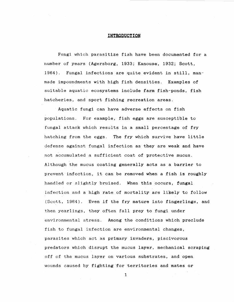
INTRODUCTION
Fungi which parasitize fish have been documented for a
number of years (Agersborg, 1933; Kanouse, 1932; Scott,
1964). Fungal infections are quite evident in still, man
made impoundments with high fish densities. Examples of
suitable aquatic ecosystems include farm fish-ponds, fish
hatcheries, and sport fishing recreation areas.
· Aquatic fungi can have adverse effects on fish
populations. For example, fish eggs are susceptible to
fungal attack which results in a small percentage of fry
hatching from the eggs. The fry which survive have little
def ense against fungal infection as they are weak and have
not accumulated a sufficient coat of protective mucus.
Although th.e mucus coating generally acts as a barrier to
prevent infection, it can be removed when a fish is roughly
handled or slightly bruised. When this occurs, fungal
infection and a high rate of mortality. are li:\tely to follow
(Scott, 1964). Even if the fry mature int6 fingerlings, and
then yearlings, they often fall prey to fungi under
environmental stress. Among the conditions which preclude
fish to fungal infection are environmental changes,
parasites which act as primary invaders, piscivorous
predators which disrupt the mucus layer, mechanical scraping
off of the mucus layer on various substrates, and open
wounds caused by fighting.for territories and mates or
1
fishermen whci extensively handle the fish and then throw
them ba-ck.
This study was de~igned to sample the mucus layer of 30
gizzard shad (Dorosoma cepedianum Lesneur) from the side
channel reservoir of Lake Charleston and determine what
fungi or fungal spores are present. This was. accomplished
by culturing the mucus and identifying the fungi which grew
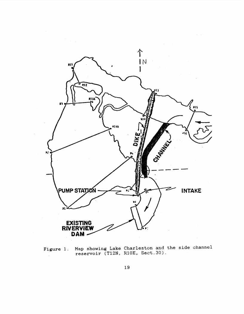
out from the mucus. Lake Charleston is located a mile and a
half south of the city of Charleston, Illinois (T12N, RlOE,
Sect.30). Lake Charleston is a highly eutrophic, man-made,
side channel reservoir which dates to 1947 and the
construction of the Riverview dam. The dam blodked off the
It:mbarrass River with the flooded area behind the dam being
used as .the water supply for Charleston. In 1978, as a
result of heavy siltation, the lake had an average depth of
0.46-0.61 m and provided a 74-day supply of water for the
<:lt..y of Charlt"'ston. In the event of a drought, however, the
water supply would have been severely threatened. With this
in mind a new dike and side channel were constructed in
1981. The depth of Lake Charleston was subsequently
increased to.2.44 m and the city's water supply increased to
174 days. The lake depth is kept more or less constant by a
station which pumps filtered water from the side channel
i.nto the reservoir (Figure 1). The lake can be successfully
stocked with fish as the fish population cannot move from
the lake.
2
The .fish being s1:lmpled, the gizzard shad, does not
<><~cur naturally in Lake Charleston. It Waf:S stocked in the
luke a~• a forage f i.rsh for pisc:i vores, especially the large
mouth bass (~ salmoides Lacepede). There is
evidence that large mouth bass only eat young gizzard shad
and do not bother older fish (Smith, 1979).
LITERATURE REVIEW
GIZZARD SHAD NATURAL HISTORY
The gizzard shad <Dot.:osoma cepedianum) is a member of
the class Osteichthyes, order Clupeiformes, family
Clupeidae. Originally, its range extended from Minnesota
~astward to the St. Lawrence River and from New Jersey
southward to the Gulf of Mexico. Today it is found in the
southwestern states where it has been introduced as a forage
fish. Although the gizzard shad is generally considered to
be a fresh water fish, it has also been recovered in
estuari~s and brackish waters. The gizzard shad is
recognized by i.ts white or silvery body, dorsal fin in which
the last ray extends into a long filament, and an inferior
or subterminal mouth. In addition, it has a very acute
sense of hearing because its swimbladder is connected to the
inner ear by a narrow diverticulum of the bladder (Moyle and
Cech~ 1988). Although similar in appearance to the threadfin
shad (Dorosoma petenense Gunther), the gizzard shad differs
3
in its mouth position, smaller scales, and higher anal fin
ray count (Smith, 1979).
The gizzard shad is found in greatest abundance in
oxbow lakes and reservoirs where it may comprise the
greatest percentage of the fish biomass. As a result of its
economic i~portance, research to date on the gizzard shad
has been centered on natural history and physiology. No
previous studies have been published on fungal spores
present in the mucus of the gizzard shad.
Spawning occurs in April, May and June. Females lay
several thousand adhesive eggs which attach to almost any
available substrate. The eggs hatch in about three days at
25° C (Smith, 1979). The shad larvae are cylindrical in
shape and exhibit a terminal mouth that contains teeth on
both jaws. The growth rate in the first growing season is
quite rapid with an average length of 100 mm attained by the
end of this period. Maturity is reached in the second year
of life with the average total life expectancy being 6-7
years (Smith, 1979).
Young gizzard shad feed on surf ace and subsurface
zooplankton until they reach 25 mm in length. As they age,
their digestive system undergoes developmental changes to
allow benthic feeding (Heinrichs, 1982). First, the mouth
changes from a supraterminal position to a subterminal
position to facilitate benthic feeding. The pharynx is then
modified for straining microscopic particles. Next,
4
pharyng.-,1d or.u;1rn~3 with goblet cells and t.ante bude develop,
becomfrn muscular to function as a crushing organ.
Intestinal length increases, most likely to accommodate the
shift in diet from zooplankton to a herbivorous diet on
benthic material (Heinrichs, 1982).
MUCUS LAYER
In fresh and salt water fish, the mucus layer is the
outermost covering. It covers the scales and epidermis and
provides an extra barrier which potential parasites must
penetrate. Since it is slick, the mucus layer may also aid
the fish in evading predators. Research completed by
Fletcher and Grant (1969) suggests that teleost fish possess
the ability to produce antibodies in the external mucus, I
aithough the fish must be alive for this inhibitory
morphogen to be present~ If the mucus is removed from the
fish or if a dea~ fish is exposed to pathogenic spores, the
external mucus is readily colonized (Wood~ .§..l., 1988). In
healthy fish; the mucus is constantly being replaced
through continuous secretions by goblet cells ln the dermis.
The old mucus is sloughed off and spores are lost with it.
FUNGI ASSOCIATED WITH FISH
Many types of aquatic fungi can attack fish. In a study
performed by Pickering and Willoughby (1977), five genera
5
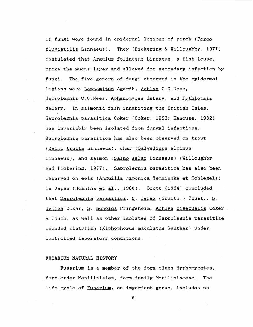
of fungi were found in epidermal lesions of perch (Perea
~~Linnaeus). They (Pickering & Willoughby, 1977)
postulated that ArguJus foliaceus Linnaeus, a fish louse,
broke the mucus layer and allowed for secondary infection by
fungi. The fiv~ genera of fungi observed in the epidermal
legions were Leptomitus Agardh, Achlya C.G.Nees,
Saprolegnia C.G.Nees, AphanomYces deBary, and Pythiopsis
deBary. In salmonid fish inhabiting the British Isles,
Saprolegnia parasitica Coker (Coker, 1923; Kanouse, 1932)
has invariably been isolated from fungal infections.
Saprolegnia parasitica has also been observed on trout
(Salmo trutta Linnaeus), char (Salyelinus alpinus
Linnaeus), and salmon (Salmo salar Linnaeus) (Willoughby
and Pickering, 1977). Saprolegn.1.Jl parasitiQ.A has also been
observed on eels (Anguilla Japonica Temmincke n Schlegels)
in Japan (Hoshina ~Al., 1960). Scott (1964) concluded
that Saprolegoia Parasitica, s. ~AX. (Gruith.) Thuet., s. deliQS\ Coker, S. ID.Qnoica Pringsheim, Achlya 'bisexualis Coker
& Couch, as well as other isolates of Saptolegnla parasitize
wounded platyfish (Xiphophorus maculatus Gunther) under
controlled laboratory conditions.
FUSABIUM NATURAL HISTORY
Fusarium is a member of the form-class Hyphomycetes,
form o~der·Moniliniales, form family Moniliniaceae. The
life cycle of Fusarium, an imperfect genus, includes no
6
known sexual phase. The mycelium is extensive and cottony
in culture, often with some tinge of pink, p~rple or yellow
i~ the my9elium or medium. The conidiophores are variable,
slender and simple or stout, short, branched irregularly or
bearing a whorl of phialides, single or grouped into
sporodochia. The conidia (phialospores) are hyaline,
variable, principally of two kinds, often held in small
moist heads: 1) macroconidia: several-celled, slightly
curved or bent at the pointed ends, typically canoe-shaped:
2) microconidia: 1-celled, ovoid or oblong, borne singly or
in chains. In additi.on, some conidia are intermediate to
these two· types and are two or three-celled, oblong or
slightly curved. Fusarium is typically parasitic on higher
plants or saprobic.bn decaying plant material (Barnett and
Hunter, 1972) ..
£YTHIUM NATURAL HISTORY
hthi.Y..m .is a member of the class Oomycetes, order
Peronosporales, family Pythiaceae (Bold li Al.., 1980).
f.£.t.b.i.wn has a we11·-developed mycelium consisting of highly
branched hyphae. The zoosporangia are either filamentous
and undifferentiated or well-defined spheroidal structures,
each .with an emission tube of variable length. In addition,
the zoosporangia may exhibit internal proliferation.
Biflagellate, reniform zoospores are expelled i.nto a vesicle
at the tip of the discharge tube where cleavage and
7
maturation occur. Pvthium thalli are monoecious. Oogonia
are spherical or ellipsoidal and may be either terminal or
iritercalary. Each oogonium produces a single egg· which
becomes an oospore after fertilization. Antheridia are of
various shapes and may be either stalked or sessile. One to
several antheridia are associated with each oogonium and
form distinct fertilization tubes. Members of the genus
Pythium are saprobic or parasitic on both plant and animal
matter in water and soil (Sparrow, 1960).
SAPROLEGNIA FERAX NATURAL HISTORY
Saprole.sn..i.g ferax (Gruith.) Thuet. is a member of the
class Oomycetes, ·order Saprolegniales, family
Saprolegn.iaceae (Bold il Jil.., 1980). The mycelium of .S..
ferax consists of stout, irregularly branched hyphae.
Sporangia, which frequently exhibit lateral proliferation
from the base, are cylindrical or slightly tapered in
appearance. Gemmae when present, are pyriform and jointed.
The thallus of .S.. ferax is monoecious, producing numerous
oogonia and antheridia. Spherical or oval oogonia ar either
terminal or intercalary, but are never arranged in chains.
Each oogonium contains a single row of up to 20 eggs.
Short, tuberous ~ntheridia are associated with each oogonium
and fertilization tubes are suppressed. Saprolegnia ferax
is found in fresh water and soil (Sparrow, 1960) and is
often a fish pathogen of economic importance (Scott, 1964).
8
ALLOMYCES ARsuecoLA NATURAL HISTORY
All.QJilJ!.Q.il 1.u.:.b.JJ..w;;i.Y.l.A Butler is a member of the class
Chytridiomycetes, order Blastocladiales, family
Blastocladiaceae (Bold At. Al.., 1980). The vegetative
thallus of Allomyces arbuscula closely resembles that of
AllomYces macrogvnus. The two species can be separated
using gametangial size. The female gametangia of Allomvces
macrogynu§ are at least twice the size of the male
. gametangia, while the male and female gametangia of
A. arbuscula are equal in size. Allomvces arbuscula
produces two or rarely more gametangia terminally on its
branches.
The resting spores of Allomvces are able to survive in
the dry state for months or years. Species included in this
genus are more often found in the warmer parts of the world,
such as the Southern United States, Mexico, Central and
South America, the West Indies, Southern Asia, the East
Indies, Africa and Southern Europe (Bessey, 1965). ·Members
of the genus Allomyces are found in both soil and water
where they are saprobic on plant and animal remains
(Sparrow, 1960) .
9
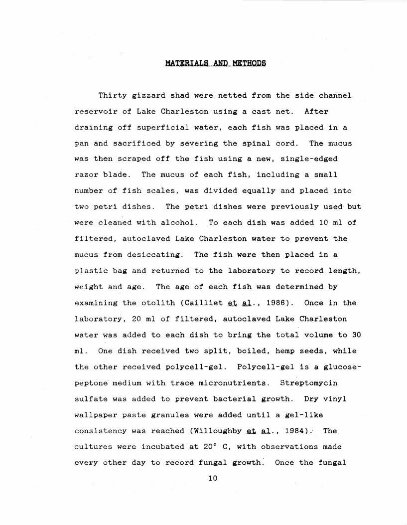
MATKBIALS AND MJtTHODS
Thirty gizzard shad were netted from the side channel
reservoir of Lake Charleston using a cast net. After
draining off superficial water, each fish was placed in a
pan and sacrificed by severing the spinal cord. The mucus
was then scraped off the fish using a new, single-edged
razor blade. The mucus of each fish, including a small
number of fish scales, was divided equally and placed into
two petri dishes. The petri dishes were previously used but
were cleaned with alcohol. To each dish was added 10 ml of
filtered, autoclaved Lake Charleston water to prevent the
mucus from desiccating. The fish were then placed in a
plastic bag and returned to the laboratory to record length,
weight and age. The age of each fish was determined by
examining the otolith (Cailliet ~ .§.l., 1986). Once in the
laboratory, 20 ml of filtered, autoclaved Lake Charleston
water was added to each dish to bring the total volume to 30
ml. One dish received two split, boiled, hemp seeds, while
the other received polycell-gel. Polycell-gel is a glucose
peptone medium with trace micronutrients. Streptomycin
sulf ate was added to prevent bacterial growth. Dry vinyl
wallpaper paste granules were added until a gel-like
consistency was reached (Willoughby il .al., 1984) .. The
cultures were incubated at 20° C, with observations made
every other day to record fungal growth. Once the fungal
10
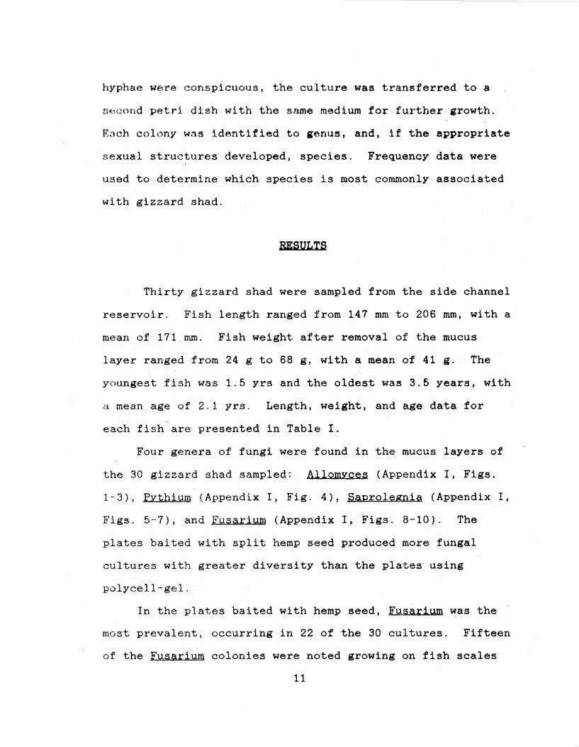
hyphae were conspicuous, the culture was transferred to a
.fit1cond petri di.sh with the same medium for fux·ther growth.
Each colony was identified to genus, and, if the appropriate
sexual structures developed, species. Frequency data were
used to determine which species is most commonly associated
with gizzard shad.
RESULTS
Thirty gizzard shad were sampled from the side channel
reservoir. Fish length ranged from 147 mm to 206 mm, with a
mean of 171 mm. Fish weight after removal of the mucus
layer ranged from 24 g to 68 g, with a mean of 41 g. The
youngest fish was 1.5 yrs and the oldest was 3.5 years, with
a mean age of 2.1 yrs. Length, weight, and age data for
each fish are presented in Table I.
Four genera of fungi were found in the mucus layers of
the 30 gizzard shad sampled: Allomvces (Appendix I, Figs.
1-3), PYthium (Appendix I, Fig. 4), Saprolegni~ (Appendix I,
Figs. 5-7), and FusariQm (Appendix I, Figs. 8-10). The
plates baited with split hemp seed produced more fungal
cultures with greater diversity than the plates using
polycell-gel.
In the plates baited with hemp seed, Fusarium was the
most prevalent, occurring in 22 of the 30 cultures. Fifteen
of the Fus~rium colonies were noted growing on fish scales
11
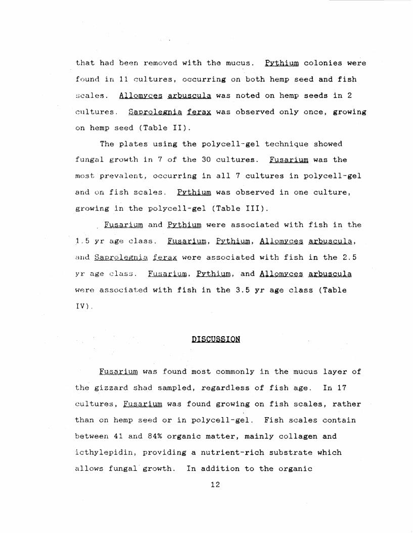
that had been removed with the mucus. fvthium colonies were
found in 11 cultures, occurring on both hemp seed and fish
~•cales. A.il.Q.m2:.Q.eJ;I. arbusculsi was noted on hemp seeds in 2
cultures. Saprolegnia ferax was observed only once, growing
on hemp seed (Table II).
The plates using the polycell-gel technique showed
fungal growth in 7 of the 30 cultures. Fusarium was the
most prevalent, occurring in all 7 cultures in polycell-gel
and on fish scales. Pythium was observed in one culture,
growing in the polycell-gel (Table III) .
. Fusarium and Pythium were associated with fish in the
1.5 yr age class. Fusarium, PYthium, Al.lomvce.,a arbµscula,
and Sap:r.olegrd,a i~~ were associated with fish in the 2. 5
yr age class. ~.d.J.un, Pvthium, and~ arbuscula
were associated with fish in the 3.5 yr age class (Table
IV).
DISCUSS I OH
,[usarium was found most commonly· in the mucus layer of
the gizzard shad iampled~ regardless of fish age. In 17
cultures, fusarium was found growing on fish scales, rather
than on hemp seed or in polycell-gel. Fish scales contain
between 41 and 84% organic matter, mainly collagen and
icthylepidin, providing a nutrient-rich substrate which
allows fungal· growth. In addition to the organic
12
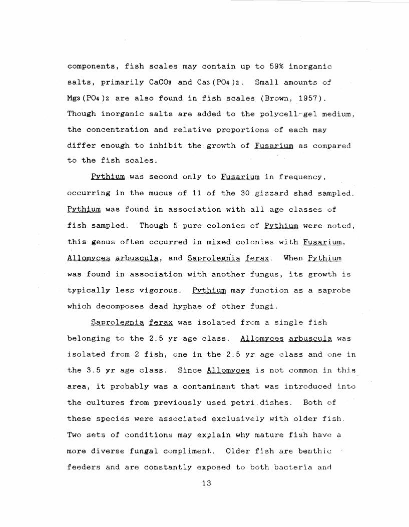
components, fish scales may contain up to 59% inorganic
salts, primarily CaC03 and Cas(P04)2. Small amounts of
Mgs(P04)2 are also found in fish scales (Brown, 1957).
Though inorganic salts are added to the polycell-gel medium,
the concentration and relative proportions of each may
differ enough to inhibit the growth of .FusaLiY.fil as compared
to the fish scales.
PYthium was second only to Fusarium in frequency,
occurring in the mucus of 11 of the 30 gizzard shad sampled.
Pythium was found in association with all age classes of
fish sampled. Though 5 pure colonies of .h:tl1i..llin were noted,
this genus often occurred in mixed colonies with Fu~arj,_11m,
Al lomyc§§ .ru;:,Puscula, and SJam;:.Q.legni?;! ;f erax. When .EY:t ... bium
was found in association with another fungus, its growth is
typically less vigorous. Pvthium may function as a saprobe
which decomposes dead hyphae of other fungi.
SaProlegnia ferax was isolated from a single fish
belonging to the 2.5 yr age class. Allomycea arbusculA was
isolated from 2 fish, one in the 2.5 yr age class and one in
the 3.5 yr age class. Since Allomyces is not common in this
area, it probably was a contaminant that was introduced into
the cultures from previously used petri dishes. Both of
these species were associated exclusively with older fish.
Two sets of conditions may explain why mature fish have a
more diverse fungal compliment. Older fish are benthic
feeders and are constantly exposed to both bacteria anrl
13
fungi on the lake bottom. The chance of encountering fungal
spores is thereby increased. Secondly, older fish have
reached reproductive age and are subjected to reproductive
stress, which may temporarily weaken their resistance to
fungal infection. When the fish were collected from the
side channel reservoir, spawning was in progress. Energy
typically allocated to life processes, such as continual
replacement of the mucus layer and the production of
associated antibodies, was used instead for reproductive
activities.
14
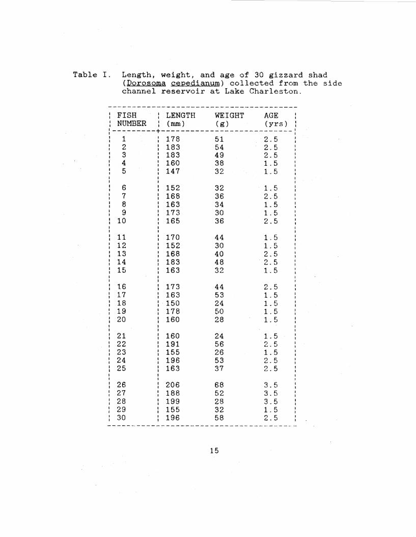
Table I. Length, weight, and age of 30 gizzard shad (Oorosoma cepedianum) collected from the side channel reservoir at Lake Charleston.
---------------------------------------FISH I LENGTH WEIGHT AGE I
NUMBER I (mm) ( g) (yrs) I I I
---------+---------------------------: 1 178 51 2.5 2 183 54 2.5 3 183 49 2.5 4 160 38 1.5 5 147 32 1. 5
6 152 32 1. 5 7 168 36 2.5 8 163 34 1. 5 9 173 30 1.5
10 165 36 2.5
11 170 44 1. 5 12 152 30 1. 5 13 168 40 2.5 14 183 48 2.5 15 163 32 1. 5
16 173 44 2.5 17 163 53 1. 5 18 150 24 1. 5 19 178 50 1.5 20 160 28 1. 5
21 160 24 1. 5 22 I 191 56 2.5 23 155 26 1. 5 24 196 53 2.5 25 163 37 2.5
26 206 68 3.5 27 188 52 3.5 28 199 28 3.5 29 155 32 1. 5 30 196 58 2.5
---------------------------------------
15
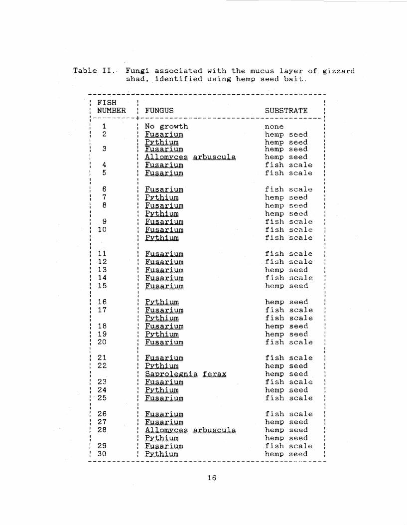
Table II. Fungi associated with the mucus layer of gizzard shad, identified using hemp seed bait.
FISH NUMBER
I I
: FUNGUS SUBSTRATE I I I I
------~--+--------------------------------------: 1 · : No growth _none 2 : Fusarium hemp seed
3
4 5
6 7 8
9 10
11 12 13 14 15
16 17
18 19 20
21 22
23 24 25
26 27 28
29 30
: Pythium hemp : Fusarium hemp : Allomvces arbuscula hemp
Fusarium fish Fusarium fish
El.A~ su: iYm fish .rzt.bi.wn hemp [u~ar1.JJm hemp fv:tbi !.lm hemp JlU1591.:ilJ.m fish [JJfHU:'. i Y.m fish fYthium fish
E~.uuu.: il.lm fish EYJi a. ;r;: i um fish Ji'.Y.!iisu:iY.m hemp Jry,i;ia..tiY.m fish
I [ysarium hemp
fY:thiY.m hemp [u§g,rj,um fish fy:thiY.m fish Ji'.Y.~Hu::i um hemp fv:thiY.m hemp Fusarium fish
[l.J.~Hl.J.:iY.m fish fy:tb1Ym hemp ~g,12;r;:Ql~snie f erax hemp Fu§a;riY.m fish fv:tbil.1m hemp Fusarium fish
EY.§s:riY.m fish Ji'.1.Uigl:iYm hemp Allomy~~§ e.;i;:lnHg;mle hemp fl!:tbiY.m hemp Ei.urn.r iYm fish fy:tbil.lm hemp
16
seed seed seed scale scale
scale seed se~'d seed scale scale s.cale
scale scale seed scale seed
seed scale scale seed seed scale
scale seed seed scale seed scale
scale seed seed seed scale seed
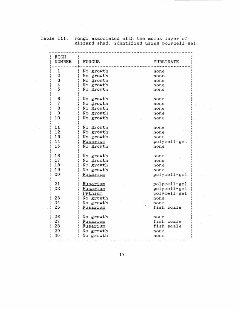
Table III.
: FISH : NUMBER
FQngi associated with the mucus layer of gizzard shad, identified using polycell-gel.
I I
: FUNGUS SUBSTRATE I I
:---------+---------------------------------------: : 1 : No growth none : : 2 : No growth none : : 3 No growth none : : 4 No growth none : : 5 No growth none : I I I I
6 7 8 9
10
11 12 13 14 15
16 17 18 19 20
21 22
23 24 25
26 27 28 29 30
No growth No growth No growth No growth No growth
No growth No growth No growth Fusarium No growth
No growth No growth No growth No growth Fusari..wn
Fusarium Fusarium PYthium No growth No growth Fusarium
No growth Fusarium Fusar1um No growth No growth
none none none none none I
I.
I I
non<:i none none polycell gel none
none none none none polycell-gel
polycell-gel polycell-gel polycell-gel none none fish scale
none fish scale fish scale none none
17
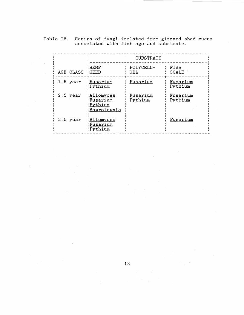
Table IV. Genera of fungi isolated from gizzard shad mucus associated with fish age and substrate.
: SUBSTRATE : '-----------------------------------------' I I
:HEMP : POLYCELL- : FISH 1
AGE CLASS : SEED : GEL : SCALE , -----------+------------+-------------+--------------: 1.5 year :Fusarium Fusarium Fusarium
:Pvthium Pythium I I
2.5 year :Allomyces :Fusarium :Pvthium :saprolegnia
3.5 year I I
:Allomyces :Fusarium :Pvthium
18
Fusarium fythium
Fusarium P;vthill.ID
Fu sari um
EXISTING RIVERVIEW·
DAM
t IN I
INTAKE
Figure 1. Map showing Lake Charleston and the side channel reservoir (T12N, R10E, Sect.30).
19
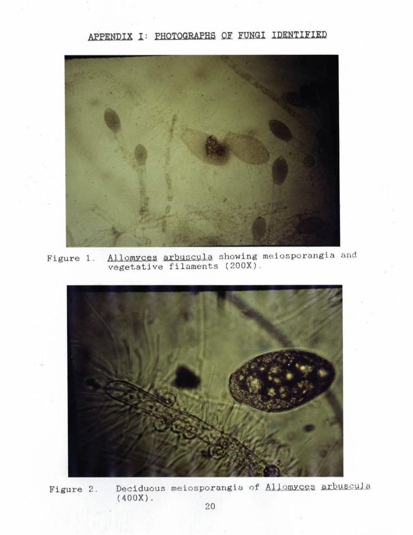
AFFENDIX I : FHOTOGRAPHS QE FUNGI IDENTIFIED
Figure 1. AllomYces a r bqsc!Jil showing meiosporangia and vegetative filaments (200X) .
Figure 2. Deciduous meiosporangio. of Allomy_®.~ il.12lL~ill...2 ( 400X).
20
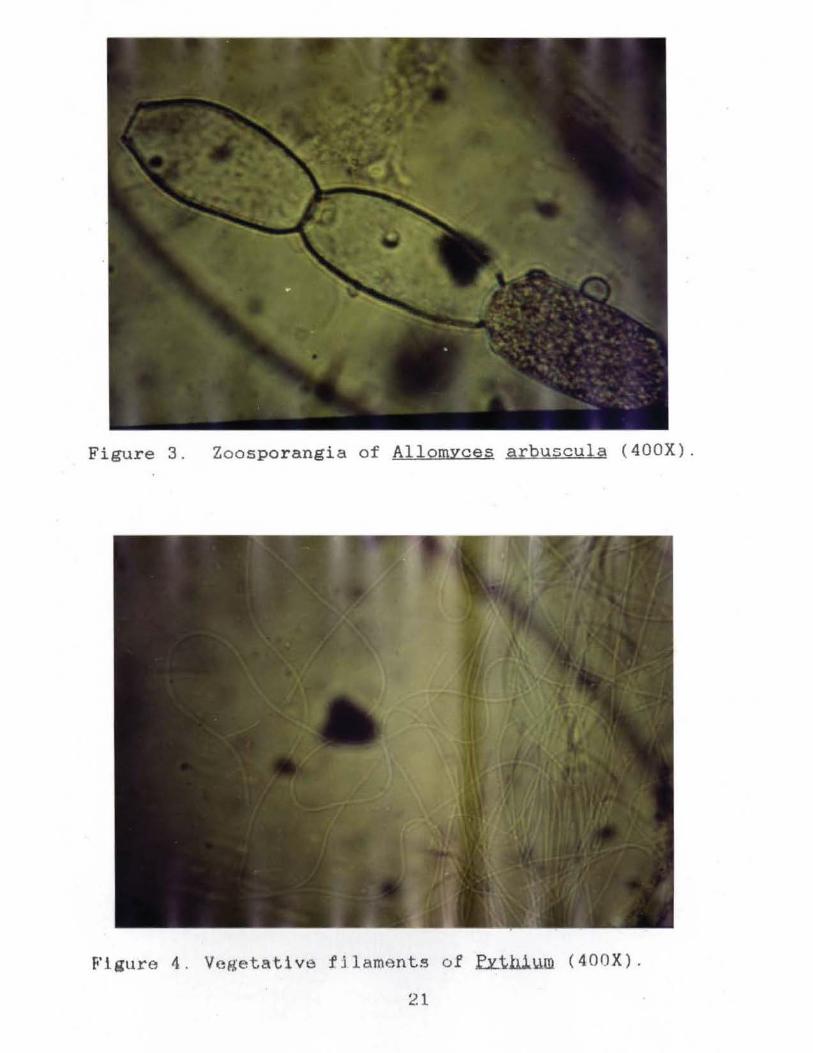
Figure 3. Zoosporangia of AllomYces arbuscula (400X).
Figure 4 . Vegetative fJl aments of ~thi~m (400X) .
21
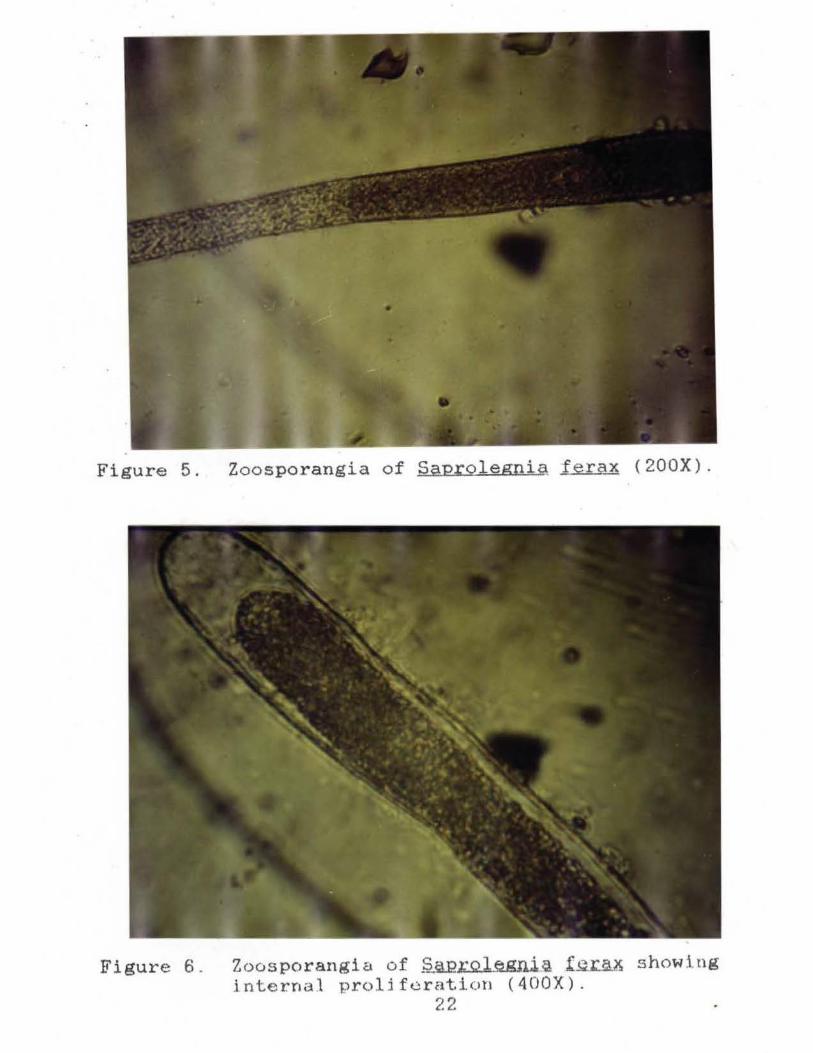
Figure 5. Zoosporangia of Saprolegnia ferax (200X).
Figure 6 . Zoosporangia of ~L:.Q.l_e.gni.~ ferax showing j nterna.l proli f eratlon ( 400X).
22
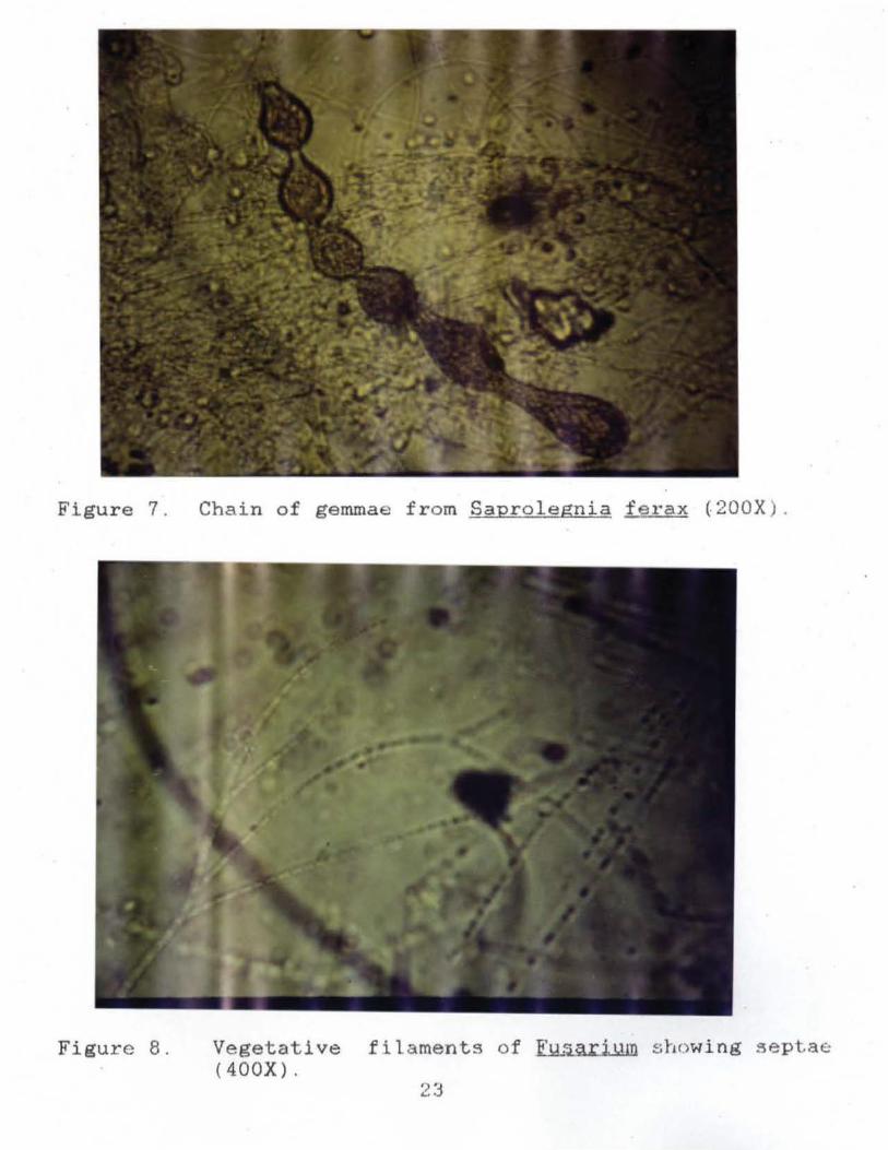
Figure 7. Chain of gemmae from Saprolegnia ferax (200X) .
Figure 8. Vegetative filaments of ~ showing sept.a«:: (400X).
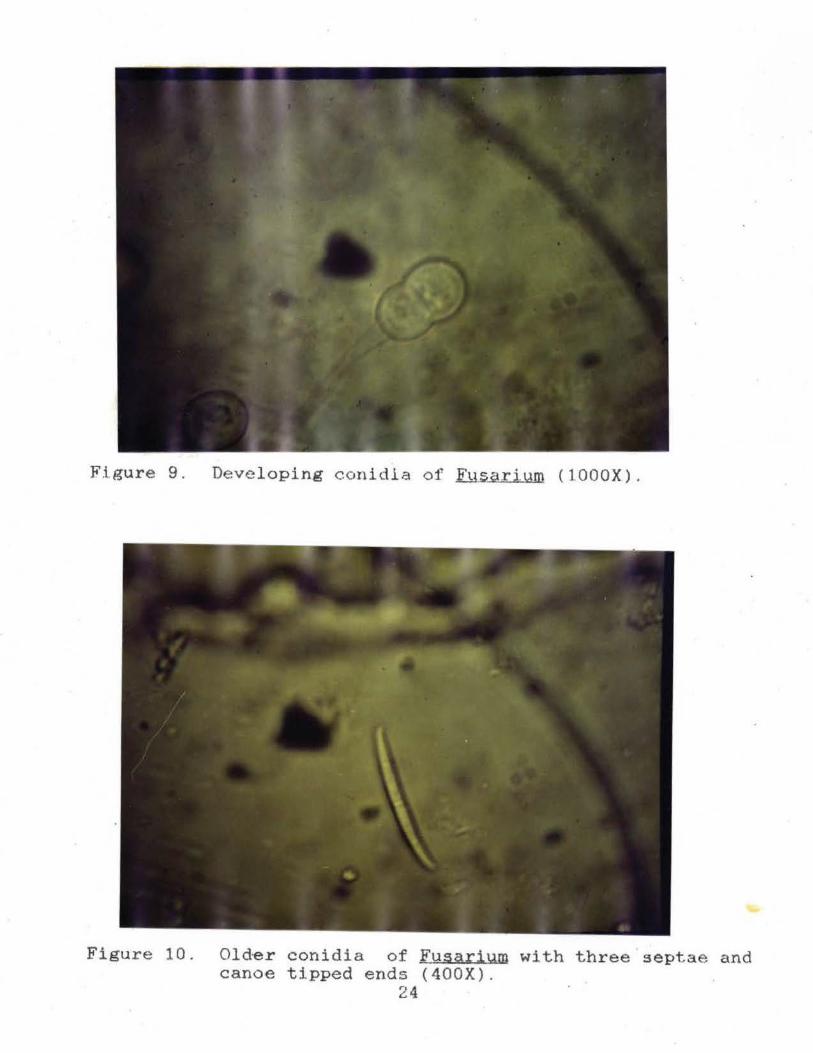
Figure 9. Deve loping conidia of Fysarium (lOOOX) .
Figure 10. Old~r conidia of Fusarium with three septae and canoe tipped ends (400X).
24
REFERENCES
Agersborg, H.P.K. 1933. Salient problems in the artificial rearing of salmonoid fisheries, with special reference to intestinal fungistosis and the cause of white-spot disease. Trans. Amer. Fish. Soc. 63: 240-250.
Barnett, H.L. and Hunter, B.B. 1972. Illustrated Genera Qf. Imperfect Fungi. Burgess Publishing Company. Minneapolis. pp 126-127.
Bessey, E.A. 1950. Morphology .fill,Q Taxonomy Q.f Fungi. ·Hafner Publishing Co., Inc., New York~ pp 84-86.
Bold, H.C., C.J. Alexopoulos, and T. Delevoryas. 1980. Morphologv Q.f. Plants .filMi Fungi. Harper & Row, Publishers, Inc., New York. 819 p.
Brown, M.E. 1957. IM Phvsiologv Qi Fishes. Academic Press, Inc., New York. 447 p.
Cailliet, G.M., M.S. Love, and A.W. Ebeling. 1986. ~.h~= A Field llll.d Laboratory M~nual 2n .T.h.~.iJ;: Structyre, Identif.kll.i.9.n, ~ N.a.tJ.U:lli History. Wadsworth Publishing Company, Belmont. 146 p.
Coker, W.C. 1923. ~ Saprolegniaceae, ~Notes Qil Other Watermolds. University of North Carolina Press, Chapel Hill.
Fletcher, T.C. and P.T.Grant. 1969. Immunoglobulins in the serum and mucus of the plaice (Pleuronectes platessa). Biochem. J. 115(5): 65.
Heinrichs, S.M. 1982. Ontogenic changes in the digestive · tract of larval gizzard shad, QQ.r.osoma cepedianum.
Trans. Amer. Microsc. Soc. 101(3): 262-275.
Hoshina, T., Sano, T. and M. Sunayama. 1960. Studies on the saprolegniasis of eel. J. Tokyo Univ. Fish. 47: 59-79.
Kanouse, B.B. 1932. A physiological and morphological study of Saprolegnia ..Qarasiti.c.a. Mycologia 24: 431-· 452.
Moyle, P. B. and J. J. Cech. 1988. El~s;:J: .An ln.t..t·9..Q.yc_tl<,..~r1 t:Q. IchthyolQ.gy. Prentice--Hall, Inc., l!-:nglewood Cliffs 221 p.·
25
Pickering, A.D. and L.G. Willoughby. lesions and fungal infection on fluviatilis L., in Windermere. 354.
1977. Epidermal the perch, . Perea J. Fish Bio. 11: 349-
Scott, W.W. 1964. Fungi associated with fish diseases. Devel. Indus. Microbio. 5: 109-123.
Smith, P.W. 1979. The Fishes of Illinois. University of Illinois Press, Urbana, pp 31-33.
Sparrow, F.K., Jr. 1960. Aouatic Phycomvcetes. The University of Michigan Press, Ann Arbor. 1187 p.
Willoughby, L.G. and A.D. Pickering. 1977. Viable saprolegniaceae spores on the epidermis of the salmonid fish~ trutt~ and Salvelinu,s .~tlp.inu~. Trans. Brit. Myc. Soc. 68(1): 91-95.
Willoughby, L.G., Pickering, A.D. and H.G. Johnson. 1984. · Polycell-gel assay of water for spores of
saprolegniaceae (fungi), especially those of the ~Qlegnia pathogen of fish. Hydrobiologia. 114: 237-248. .
Wood, S.E., Willoughby, L.G. and G.W. Beakes. 1988. Experimental studies on uptake and interaction of spores of the Saprolegnia diclina-parasitica complex with external mucus of brown trout (Salmo trutta). Trans. Brit. Myc. Soc. 90(1): 63-73.
26