Embed Size (px)
Citation preview
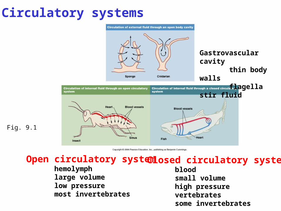
Fig. 9.1
Circulatory systems
Open circulatory systemhemolymphlarge volumelow pressuremost invertebrates
Closed circulatory systembloodsmall volumehigh pressurevertebratessome invertebrates
Gastrovascular cavity thin body walls flagella stir fluid
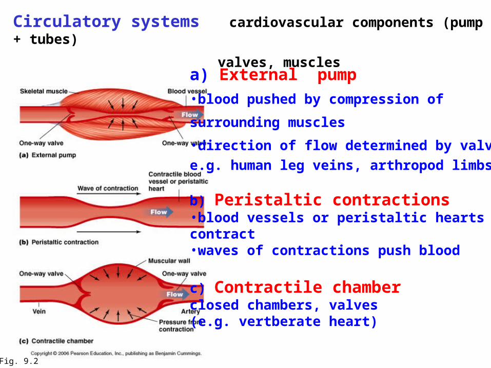
Circulatory systems cardiovascular components (pump + tubes)
valves, muscles
Fig. 9.2
a) External pump
•blood pushed by compression of
surrounding muscles
•direction of flow determined by valves
e.g. human leg veins, arthropod limbs
b) Peristaltic contractions•blood vessels or peristaltic heartscontract•waves of contractions push blood
c) Contractile chamberclosed chambers, valves(e.g. vertberate heart)

Types of circulatory systems
1.OPEN CIRCULATORY SYSTEM(most invertebrates) Low pressure (< 1.5 kPa)
High volume (30% body vol.)Slow velocity
Fig. « 9.7 »
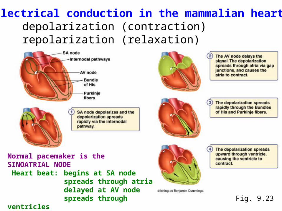
Electrical conduction in the mammalian heartdepolarization (contraction) repolarization (relaxation)
Fig. 9.23
Normal pacemaker is the SINOATRIAL NODE Heart beat: begins at SA node
spreads through atriadelayed at AV nodespreads through ventricles
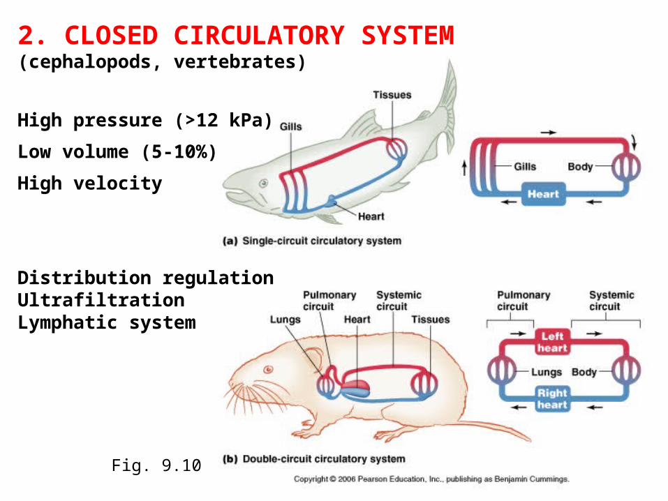
Fig. 9.10
2. CLOSED CIRCULATORY SYSTEM (cephalopods, vertebrates)
High pressure (>12 kPa)
Low volume (5-10%)
High velocity
Distribution regulationUltrafiltrationLymphatic system
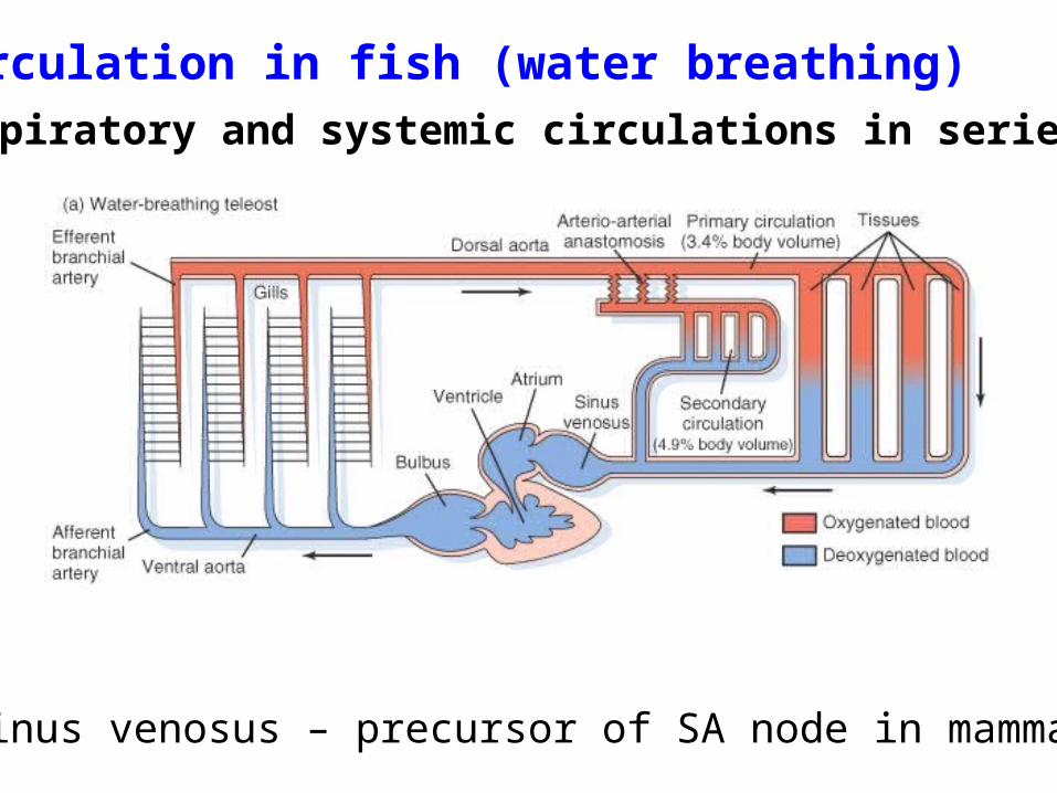
Circulation in fish (water breathing)respiratory and systemic circulations in series
sinus venosus – precursor of SA node in mammals
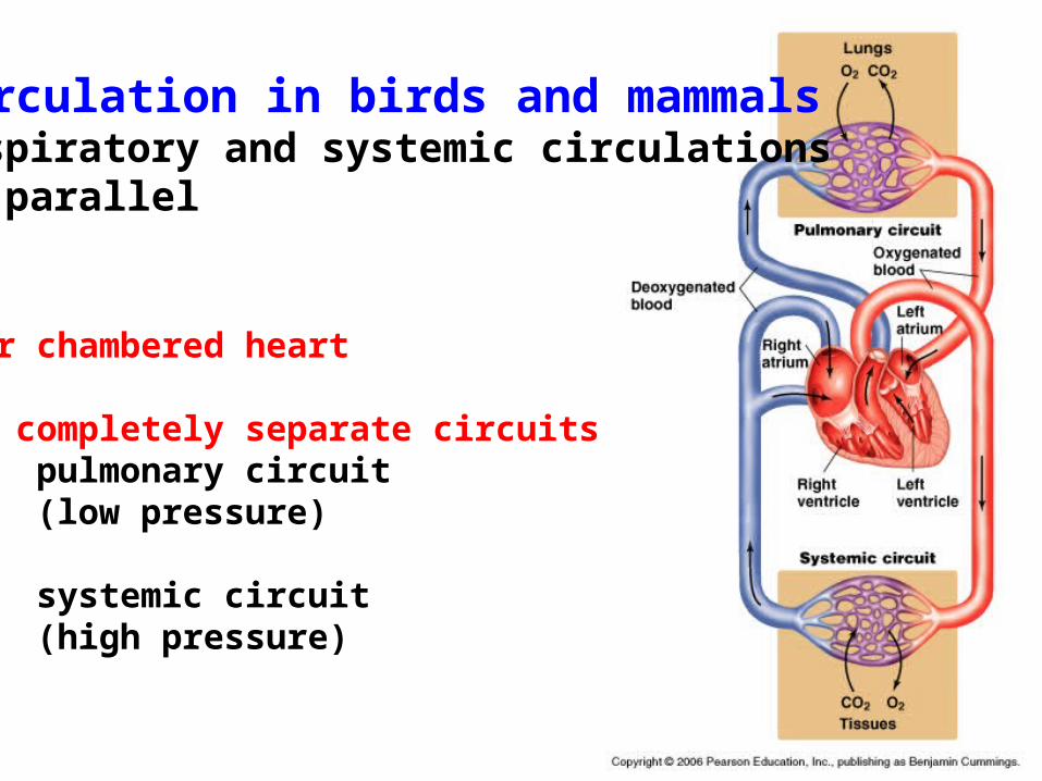
Circulation in birds and mammalsrespiratory and systemic circulations in parallel
Four chambered heart
Two completely separate circuitspulmonary circuit(low pressure)
systemic circuit(high pressure)

Anatomy of the mammalian heart
adult heartfour chamberscomplete separation of left and right heart
fetal heartforamen ovaleductus arteriosuspulmonary circuit not
functional
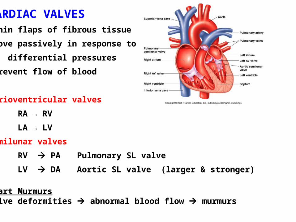
CARDIAC VALVES •thin flaps of fibrous tissue
•move passively in response to
differential pressures
•prevent flow of blood
Atrioventricular valves
RA → RV
LA → LV
Semilunar valves
RV PA Pulmonary SL valve
LV DA Aortic SL valve (larger & stronger)
Heart MurmursValve deformities abnormal blood flow murmurs
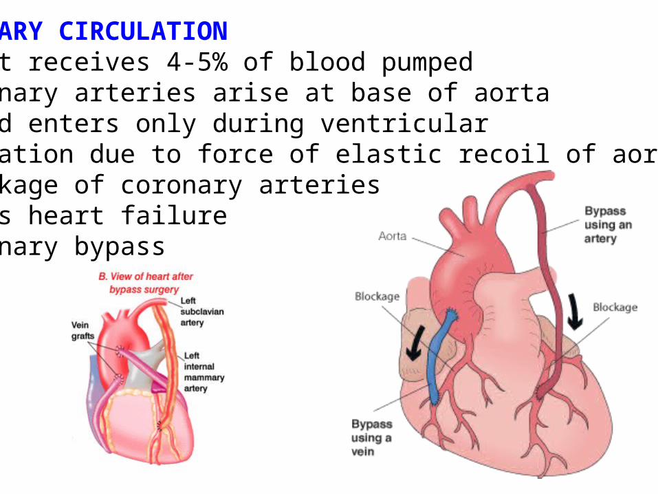
CORONARY CIRCULATION•heart receives 4-5% of blood pumped•coronary arteries arise at base of aorta•blood enters only during ventricular relaxation due to force of elastic recoil of aorta•blockage of coronary arteries causes heart failure •coronary bypass

Electrical conduction in the mammalian heartdepolarization (contraction) repolarization (relaxation)
Fig. 9.23
Normal pacemaker is the SINOATRIAL NODE Heart beat: begins at SA node
spreads through atriadelayed at AV nodespreads through ventricles

Fig. 4.2 The structure of gap junctions
GAP JUNCTIONScells coupled metabolically and electrically via hydrophilic channels
Passage of:- inorganic ions- small water-soluble molecules:
amino acidssugarsnucleotides
- electrical signals
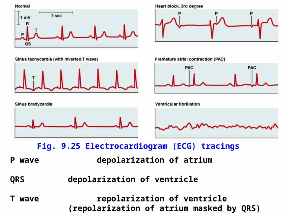
P wave depolarization of atrium
QRS depolarization of ventricle
T wave repolarization of ventricle(repolarization of atrium masked by QRS)
Fig. 9.25 Electrocardiogram (ECG) tracings
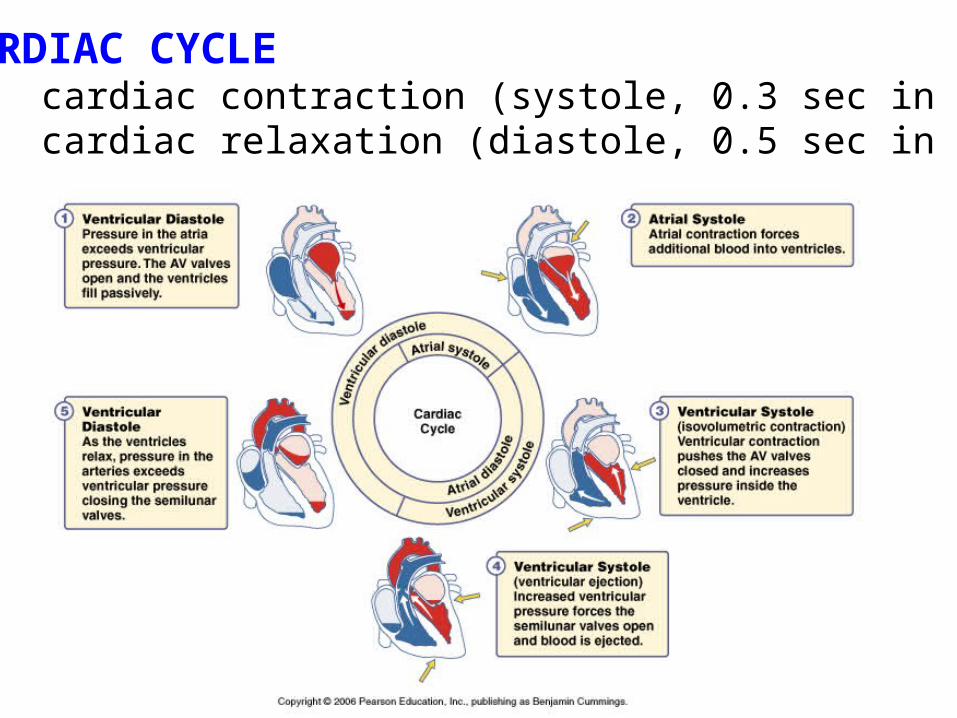
CARDIAC CYCLEcardiac contraction (systole, 0.3 sec in human)cardiac relaxation (diastole, 0.5 sec in human)
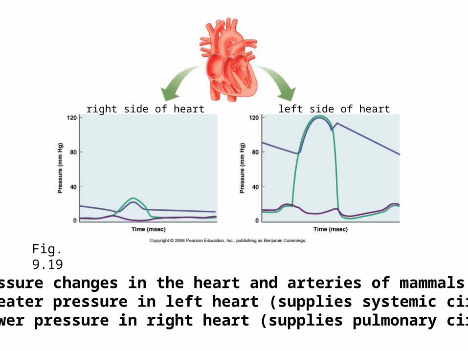
Fig. 9.19
Pressure changes in the heart and arteries of mammals•greater pressure in left heart (supplies systemic circuit)•lower pressure in right heart (supplies pulmonary circuit)
right side of heart left side of heart

Fig. 9.26
N.B. same volume changes at different pressures

STROKE VOLUME (SV)= (end diastolic vol. - end systolic vol.) in healthy humans at rest
stroke volume ~ (140 - 60) ml = 80 ml
both ventricles eject same volume of blood (total blood volume in humans 5-6 L)
SV regulated by: end diastolic volume SV EDV
mean arterial pressure SV 1/MAP
contractility SV C
End-diastolic volume determined by:
venous filling pressurevenoconstriction, skeletal muscle pumpatrial pressureventricular distensibility
filling time
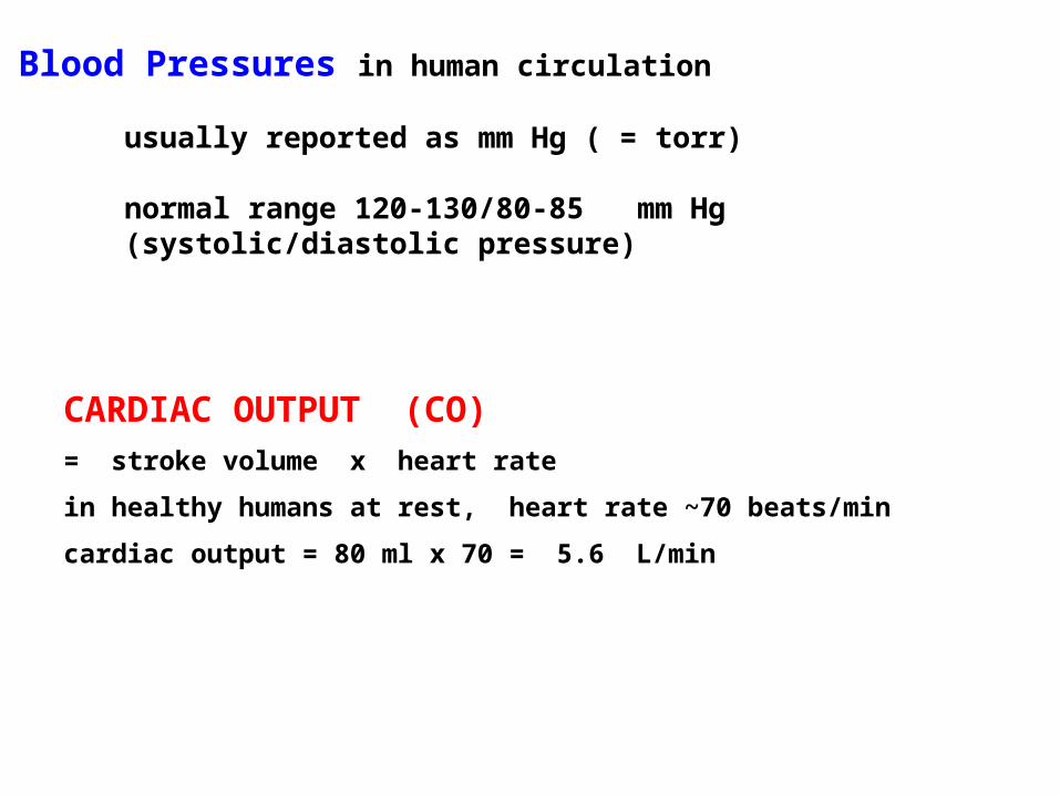
Blood Pressures in human circulation
usually reported as mm Hg ( = torr)
normal range 120-130/80-85 mm Hg (systolic/diastolic pressure)
CARDIAC OUTPUT (CO)= stroke volume x heart rate
in healthy humans at rest, heart rate ~70 beats/min
cardiac output = 80 ml x 70 = 5.6 L/min
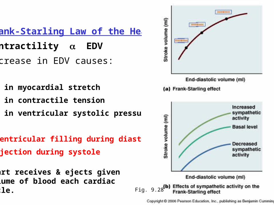
Frank-Starling Law of the Heart
Contractility EDV
Increase in EDV causes:
• in myocardial stretch
• in contractile tension
• in ventricular systolic pressure
ventricular filling during diastole,
ejection during systole
heart receives & ejects given volume of blood each cardiac cycle. Fig. 9.28
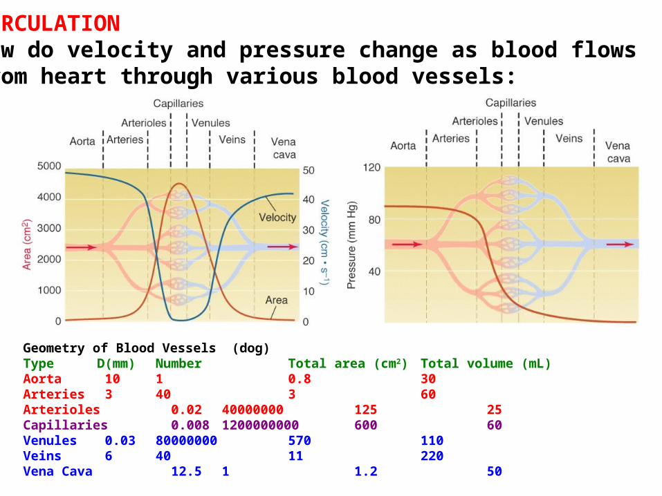
CIRCULATION How do velocity and pressure change as blood flows from heart through various blood vessels:
Geometry of Blood Vessels (dog)Type D(mm) Number Total area (cm2) Total volume (mL)Aorta 10 1 0.8 30Arteries 3 40 3 60Arterioles 0.02 40000000 125 25Capillaries 0.008 1200000000600 60Venules 0.03 80000000 570 110Veins 6 40 11 220Vena Cava 12.5 1 1.2 50
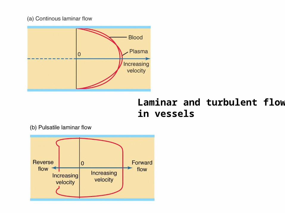
Laminar and turbulent flowin vessels
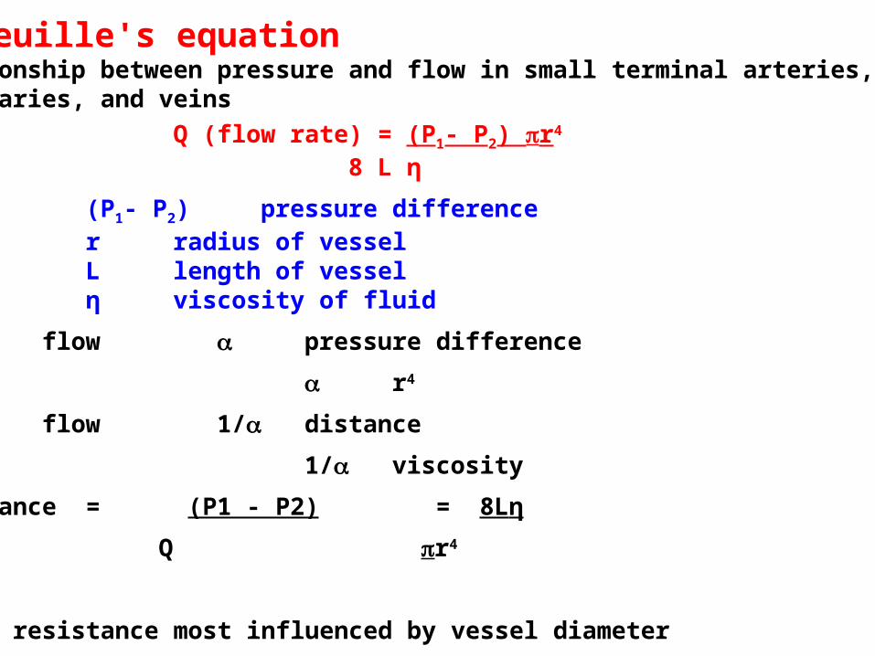
Poiseuille's equationrelationship between pressure and flow in small terminal arteries, capillaries, and veins
Q (flow rate) = (P1- P2) r4
8 L η
(P1- P2) pressure differencer radius of vesselL length of vesselη viscosity of fluid
flow pressure difference
r4
flow 1/ distance
1/ viscosity
resistance = (P1 - P2) = 8Lη
Q r4
Flow & resistance most influenced by vessel diameter
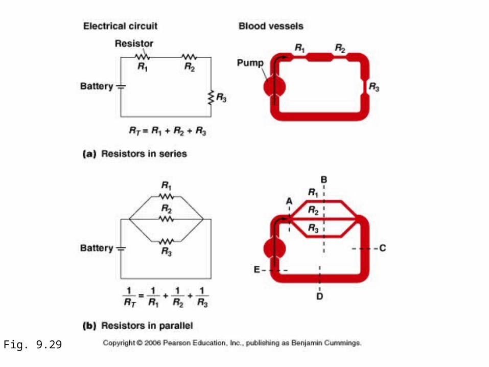
Fig. 9.29

COMPLIANCEincreased P stretch increased volumeincreased r decreased resistance increased flow
Compliance = volume/pressure
Venous system:very compliant volume reservoirlarge volume changes result in small pressure changes
Arterial system:less compliant pressure reservoirmaintain capillary flow
Compliance of veins 24x greater than arteries:
(except elastic aortae which dampen pressure oscillations)

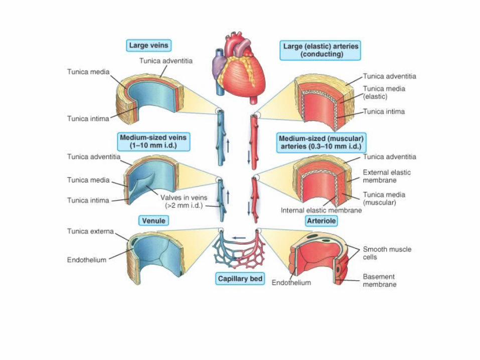
Fig.9.31
tunica intima:endothelium
tunica media:smooth muscleelastin
tunica externa:collagen

Blood flow in vertebrate circulatory systems
•high and variable pressure in ventricle
•low pressure, steady flow in capillaries, venules, veins
•velocity increase in venoussystem
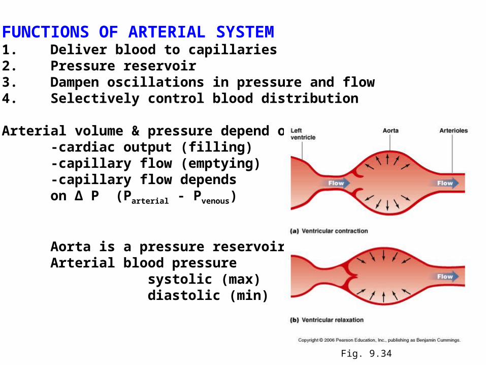
FUNCTIONS OF ARTERIAL SYSTEM1. Deliver blood to capillaries2. Pressure reservoir3. Dampen oscillations in pressure and flow4. Selectively control blood distribution
Arterial volume & pressure depend on:-cardiac output (filling)-capillary flow (emptying)-capillary flow depends on ∆ P (Parterial - Pvenous)
Aorta is a pressure reservoirArterial blood pressure
systolic (max)diastolic (min)
Fig. 9.34
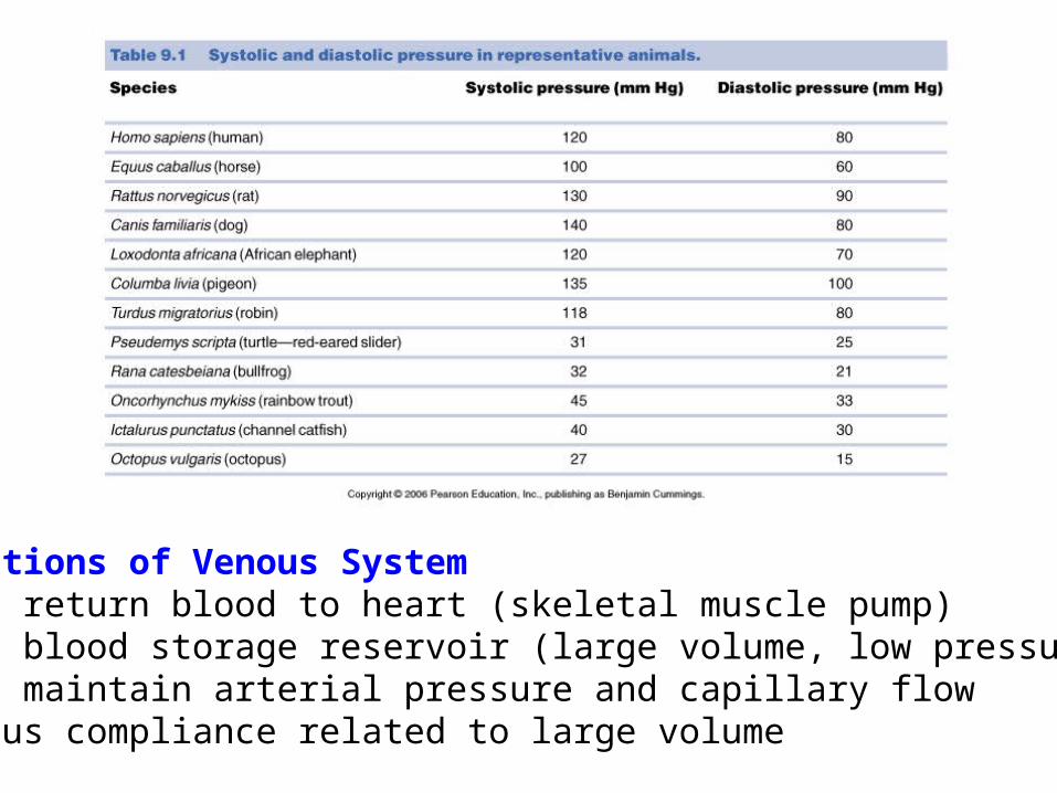
Functions of Venous System1. return blood to heart (skeletal muscle pump)2. blood storage reservoir (large volume, low pressure)3. maintain arterial pressure and capillary flowVenous compliance related to large volume
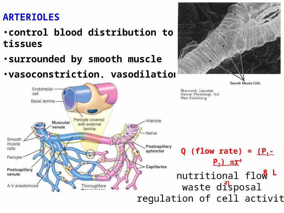
ARTERIOLES
•control blood distribution to tissues
•surrounded by smooth muscle
•vasoconstriction, vasodilation
nutritional flowwaste disposal
regulation of cell activities
Q (flow rate) = (P1- P2) r4
8 L
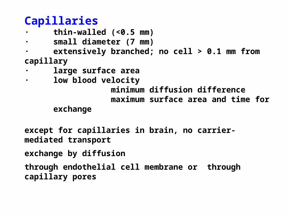
Capillaries· thin-walled (<0.5 mm)· small diameter (7 mm)· extensively branched; no cell > 0.1 mm from capillary· large surface area· low blood velocity
minimum diffusion differencemaximum surface area and time for
exchange
except for capillaries in brain, no carrier-mediated transport
exchange by diffusion
through endothelial cell membrane or through capillary pores
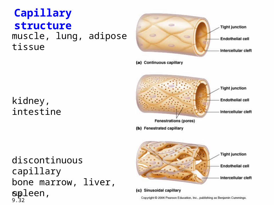
Fig. 9.32
Capillary structure
discontinuous capillarybone marrow, liver, spleen,
kidney, intestine
muscle, lung, adipose tissue
Regulation of circulation
Priorities: maintain continuous perfusion of brain & heart
then supply other organs as needed
maintain ECF volume & composition
hyperemia increased capillary flow
ischemia cessation of capillary flow
tissue metabolism vessel dilation flow
brain and heart continuously perfused, last to be deprived of capillary flow
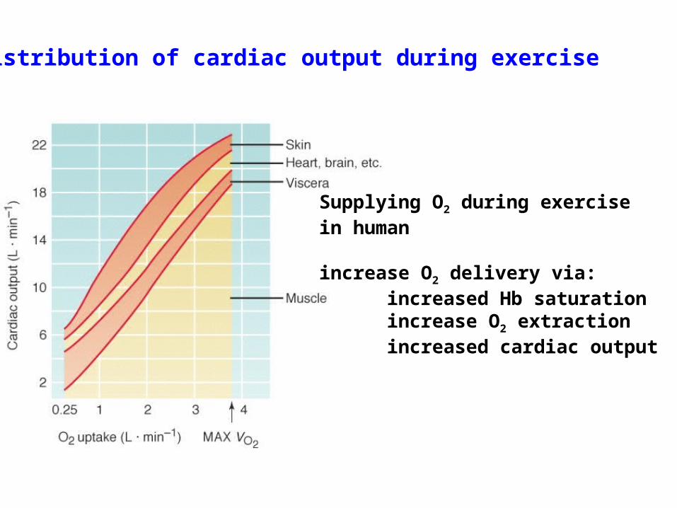
Supplying O2 during exercise in human
increase O2 delivery via:increased Hb saturationincrease O2 extractionincreased cardiac output
Distribution of cardiac output during exercise

Medullary cardiovascular center
receives inputs from
Baroreceptorscarotid sinusaortic archsubclaviancommon carotidpulmonary artery
Mechanoreceptorsatrialventricular
Chemoreceptorsarterialventricular
Skeletal muscle afferent fibres
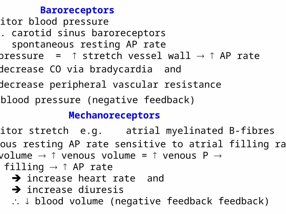
BaroreceptorsMonitor blood pressuree.g. carotid sinus baroreceptors
spontaneous resting AP rate blood pressure = stretch vessel wall AP rate
decrease CO via bradycardia and
decrease peripheral vascular resistance
blood pressure (negative feedback)
Mechanoreceptors
Monitor stretch e.g. atrial myelinated B-fibres
spontaneous resting AP rate sensitive to atrial filling rate&volume blood volume venous volume = venous P atrial filling AP rate
increase heart rate and increase diuresis blood volume (negative feedback feedback)
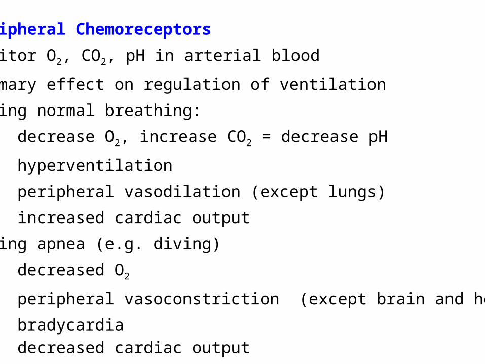
Peripheral Chemoreceptors
Monitor O2, CO2, pH in arterial blood
primary effect on regulation of ventilation
During normal breathing:
decrease O2, increase CO2 = decrease pH
hyperventilation
peripheral vasodilation (except lungs)
increased cardiac output
During apnea (e.g. diving)
decreased O2
peripheral vasoconstriction (except brain and heart)
bradycardia
decreased cardiac output
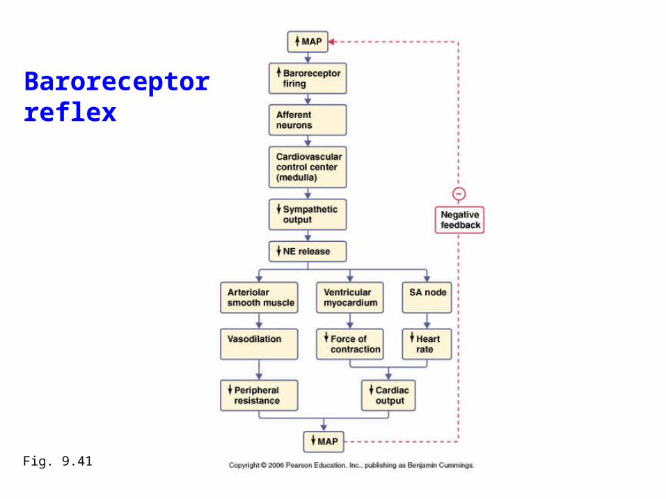
Fig. 9.41
Baroreceptorreflex
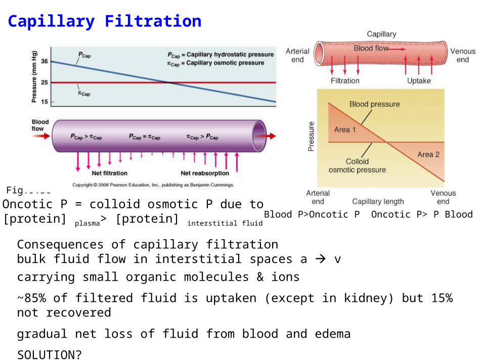
Capillary Filtration
Fig.9.36
Blood P>Oncotic P Oncotic P> P BloodOncotic P = colloid osmotic P due to [protein] plasma> [protein] interstitial fluid
Consequences of capillary filtrationbulk fluid flow in interstitial spaces a v
carrying small organic molecules & ions
~85% of filtered fluid is uptaken (except in kidney) but 15% not recovered
gradual net loss of fluid from blood and edema
SOLUTION?
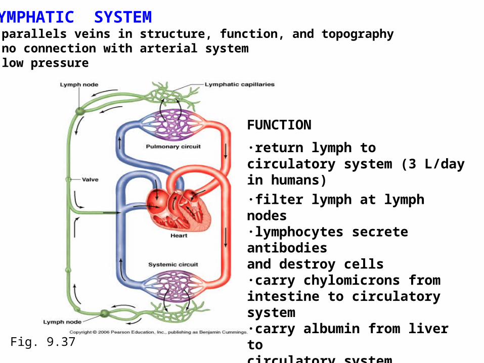
LYMPHATIC SYSTEM- parallels veins in structure, function, and topography- no connection with arterial system- low pressure
Fig. 9.37
FUNCTION
·return lymph to circulatory system (3 L/day in humans)
·filter lymph at lymph nodes·lymphocytes secrete antibodies and destroy cells·carry chylomicrons from intestine to circulatory system·carry albumin from liver to circulatory system
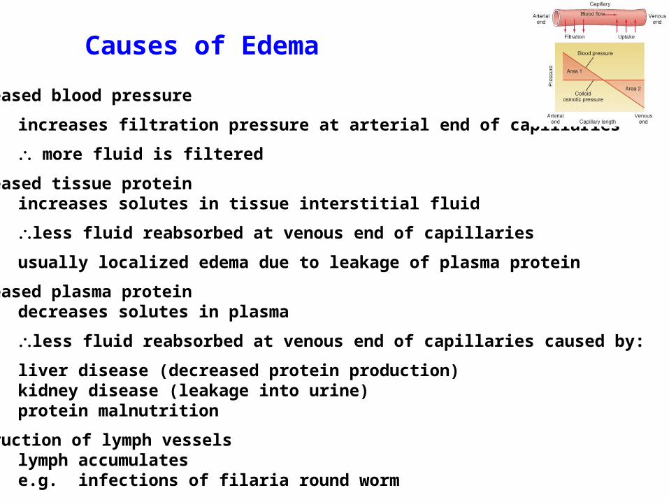
Causes of Edema
Increased blood pressure
increases filtration pressure at arterial end of capillaries
more fluid is filtered
Increased tissue proteinincreases solutes in tissue interstitial fluid
less fluid reabsorbed at venous end of capillaries
usually localized edema due to leakage of plasma protein
Decreased plasma proteindecreases solutes in plasma
less fluid reabsorbed at venous end of capillaries caused by:
liver disease (decreased protein production)kidney disease (leakage into urine)protein malnutrition
Obstruction of lymph vesselslymph accumulatese.g. infections of filaria round worm

Hb Saturation
arterial (fully saturated) ~97%
venous at rest 75%mild exercise 58%heavy exercise 27%
Increased cardiac output (= stroke vol x heart rate)
via: stroke volume
heart rate
In human:
CO (L) SV (ml) HR (bpm)
at rest 5.6 80 70heavy exercise 18 80 220trained athlete 5 100 50