Embed Size (px)
Citation preview

Fig. 19-1
0.5 µm
Chapter 19Chapter 19
1.The simplest form of life
2.Exist on the borderline between the living and the inanimate, non-biological world
3. Reveal more complex life
4. Human disease
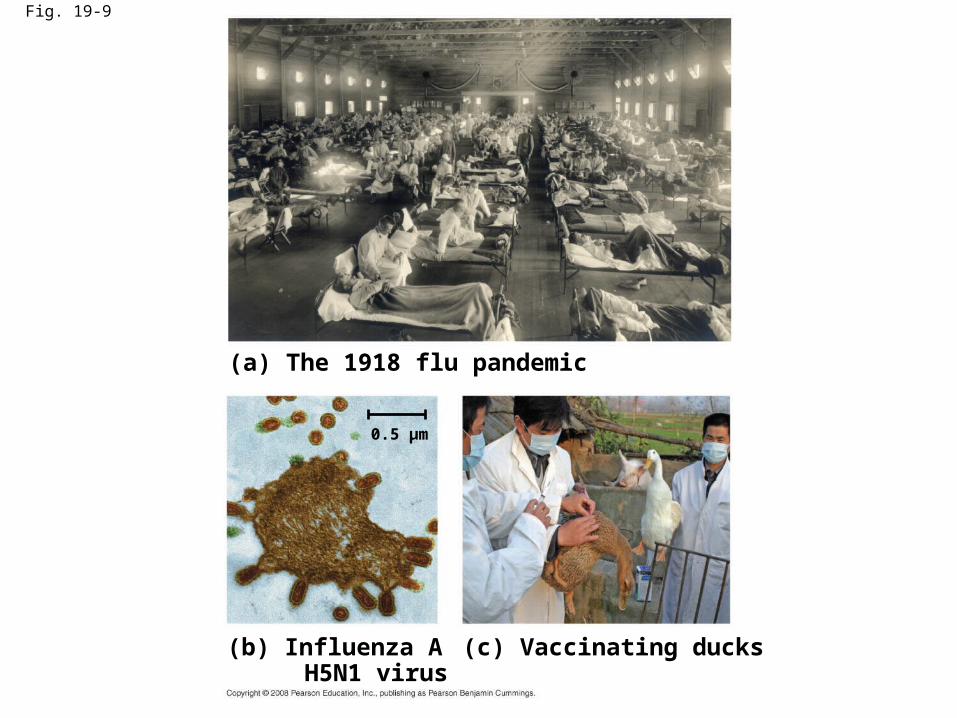
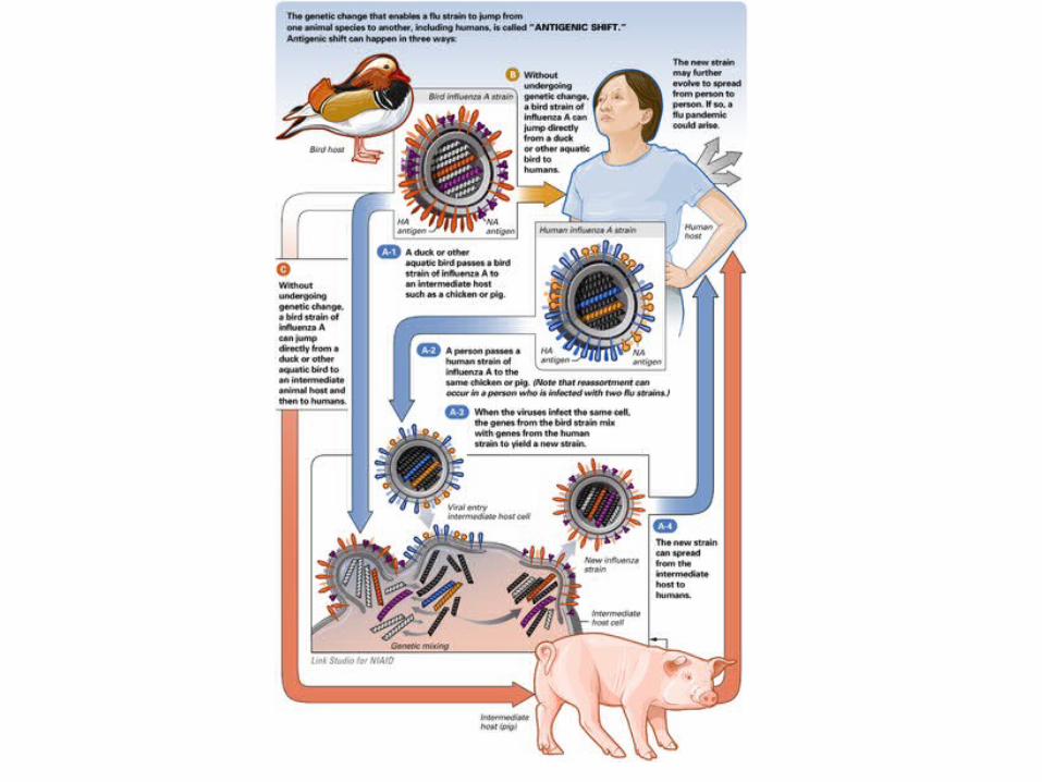
Fig. 19-9
(a) The 1918 flu pandemic
(b) Influenza A H5N1 virus
(c) Vaccinating ducks
0.5 µm

Fig. 19-2RESULTS
1 2 3Extracted sapfrom tobaccoplant withtobaccomosaic disease
Passed sapthrough aporcelain filter knownto trapbacteria
Rubbed filteredsap on healthytobacco plants
4 Healthy plantsbecame infected
•In 1935, Wendell Stanley confirmed this hypothesis by crystallizing the infectious particle, now known as tobacco mosaic virus (TMV)

• Some evolutionary biologist: make more copies of their genome
• Viruses : nucleic acid + a protein coat (capsid), protection and entrance
• Viral genomes may consist of either– Double- or single-stranded DNA, or– Double- or single-stranded RNA
• Depending on its type of nucleic acid, a virus is called a DNA virus or an RNA virus
• NA+ protein coat= nucleocapsid, in animal virus +a membranous envelope (the lipid membrane)
• A good source of pure DNA before the advent of gene cloning– eg: SV40 virus (a ds DNA virus , 5 genes
• Obligate parasite (either as degenerate cells or renegade cellular genes)
• Rapid genetic change has obscured or erased ant relationships
Structure of Viruses
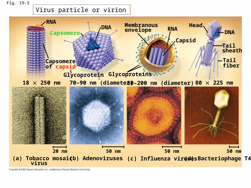
Fig. 19-3
RNA
Capsomere
Capsomereof capsid
DNA
Glycoprotein
18 250 nm 70–90 nm (diameter)
Glycoproteins
80–200 nm (diameter) 80 225 nm
Membranousenvelope RNA
Capsid
HeadDNA
Tailsheath
Tailfiber
50 nm50 nm50 nm20 nm
(a) Tobacco mosaic virus
(b) Adenoviruses (c) Influenza viruses (d) Bacteriophage T4
Virus particle or virion

viral envelopes
• Some viruses have membranous envelopes that help them infect hosts – (membrane-scavenging strategy)
• These viral envelopes surround the capsids of influenza viruses and many other viruses found in animals – (encephalitis virus, smallpox, rabies, herpes virus and the HIV)
• Viral envelopes, which are derived from the host cell’s membrane, contain a combination of viral (virus-encoded proteins) and host cell molecules – (N-terminal protrude outwrd, C-terminal ofter contact the nucleocapsid)
– Easily dissolved by detergents but gastrointestinal viruses (including poliovirus) have purely protein

Bacteriophages
• Bacteriophages, also called phages, are viruses that infect bacteria
• They have the most complex capsids found among viruses
• Phages have an elongated capsid head that encloses their DNA
• A protein tail piece attaches the phage to the host and injects the phage DNA inside
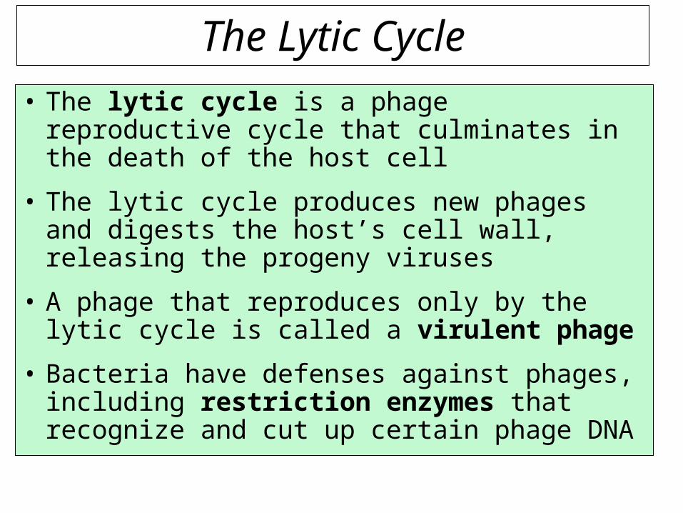
The Lytic Cycle• The lytic cycle is a phage reproductive cycle that culminates in the death of the host cell
• The lytic cycle produces new phages and digests the host’s cell wall, releasing the progeny viruses
• A phage that reproduces only by the lytic cycle is called a virulent phage
• Bacteria have defenses against phages, including restriction enzymes that recognize and cut up certain phage DNA

The lysogenic cycle• The lysogenic cycle replicates the phage genome without destroying the host
• The viral DNA molecule is incorporated into the host cell’s chromosome
• This integrated viral DNA is known as a prophage
• Every time the host divides, it copies the phage DNA and passes the copies to daughter cells

Temperate phages
• An environmental signal can trigger the virus genome to exit the bacterial chromosome and switch to the lytic mode
• Phages that use both the lytic and lysogenic cycles are called temperate phages

Transcriptionand manufactureof capsid proteins
Self-assembly of new virus particles and their exit from the cell
Entry anduncoating
Fig. 19-4VIRUS1
2
3
DNA
Capsid
4
Replication
HOST CELL
Viral DNA
mRNA
Capsidproteins
Viral DNA
1. Adsorption penetration
22 cytoplasm (smallpox and RNA virus) or
3. nucleus (DNA virus)

Fig. 19-UN1
PhageDNA
Bacterialchromosome
The phage attaches to ahost cell and injects its DNA
Prophage
Lysogenic cycle• Temperate phage only• Genome integrates into bacterial chromosome as prophage, which (1) is replicated and passed on to daughter cells and (2) can be induced to leave the chromosome and initiate a lytic cycle
Lytic cycle• Virulent or temperate phage• Destruction of host DNA• Production of new phages• Lysis of host cell causes release of progeny phages

Fig. 19-6
PhageDNA
Phage
The phage injects its DNA.
Bacterialchromosome
Phage DNAcircularizes.
Daughter cellwith prophage
Occasionally, a prophageexits the bacterialchromosome,initiating a lytic cycle.
Cell divisionsproducepopulation ofbacteria infectedwith the prophage.
The cell lyses, releasing phages.
Lytic cycle
Lytic cycleis induced or Lysogenic cycle
is entered
Lysogenic cycle
Prophage
The bacterium reproduces,copying the prophage andtransmitting it to daughter cells.
Phage DNA integrates intothe bacterial chromosome,becoming a prophage.
New phage DNA and proteinsare synthesized andassembled into phages.

Reproductive Cycles of Animal Viruses
• There are two key variables used to classify viruses that infect animals:– DNA or RNA?– Single-stranded or double-stranded?
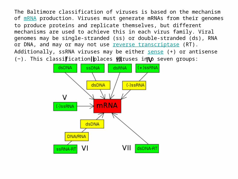
The Baltimore classification of viruses is based on the mechanism of mRNA production. Viruses must generate mRNAs from their genomes to produce proteins and replicate themselves, but different mechanisms are used to achieve this in each virus family. Viral genomes may be single-stranded (ss) or double-stranded (ds), RNA or DNA, and may or may not use reverse transcriptase (RT). Additionally, ssRNA viruses may be either sense (+) or antisense (−). This classification places viruses into seven groups:

Table 19-1
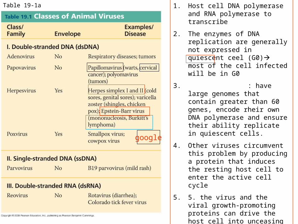
Table 19-1a 1. Host cell DNA polymerase and RNA polymerase to transcribe
2. The enzymes of DNA replication are generally not expressed in quiescent ceel (G0) most of the cell infected will be in G0
3. : have large genomes that contain greater than 60 genes, encode rheir own DNA polymerase and ensure their ability replicate in quiescent cells.
4. Other viruses circumvent this problem by producing a protein that induces the resting host cell to enter the active cell cycle
5. 5. the virus and the viral growth-promoting proteins can drive the host cell into unceasing growth and cell division (oncogene)
6. HPV: cervical carcinoma
EB: mononucleosis
Burkitt’s lymphoma (a childhood tumor) in central Africa
Nasopharyngeal carcinoma in Southeast Asia

Table 19-1b
德國麻疹
馬象皮病毒
SS DNA
1. A simple genome
2. Have a simple genome—one gene for a viral nucleocapside protein and another gene for a DNA replication enzyme
3. only template for transcription ds DNA, 3’ as a primer
(+) ssRNA+ an mRNA
1. pico—small
2. mRNA make viral proteins that can replicate the ssRNA genome as well as the proteins needed for the capsid.
Fig 19-7
+
-

Fig. 19-7
Capsid
RNA
Envelope (withglycoproteins)
Capsid and viral genomeenter the cell
HOST CELL
Viral genome (RNA)
Template
mRNA
ER
Glyco-proteins
Capsidproteins Copy of
genome (RNA)
New virus
The reproductive cycle of an
enveloped RNA virus (Class V)
--- Template for mRNA synthesis

2009 flu pandemic

Influenza hemagglutinin (HA) or haemagglutinin (British English)
1. is a type of hemagglutinin found on the surface of the influenza viruses.
2. 2. It is an antigenic glycoprotein. It is responsible for binding the virus to the cell that is being infected.
3. The name "hemagglutinin" comes from the protein's ability to cause red blood cells (erythrocytes) to clump together ("agglutinate") in vitro

• Viral neuraminidase is an enzyme on the surface of influenza viruses that enables the virus to be released from the host cell.
• Drugs that inhibit neuraminidase, known as neuraminidase inhibitors, are used to treat influenza.
• When influenza virus reproduces, it attaches to the cell surface using hemagglutinin, a molecule found on the surface of the virus which binds to sialic acid groups.
• Sialic acids are found on various glycoproteins at the host cell surface,
• and the virus exploits these groups to bind the host cell. In order for the virus to be released from the cell, neuraminidase must enzymatically cleave the sialic acid groups from host glycoproteins.
• In some viruses, a hemagglutinin-neuraminidase protein combines the neuraminidase and hemagglutinin functions in a single protein.

Influenza virus replication

De Clercq Nature Reviews Drug Discovery 5, 1015–1025 (December 2006) | doi:10.1038/nrd2175

RNA as Viral Genetic Material
• The broadest variety of RNA genomes is found in viruses that infect animals
• Retroviruses use reverse transcriptase to copy their RNA genome into DNA
• HIV (human immunodeficiency virus) is the retrovirus that causes AIDS (acquired immunodeficiency syndrome)

Fig. 19-8Glycoprotein Viral envelope
Capsid
RNA (twoidenticalstrands)Reverse
transcriptase HIV
HIVMembrane ofwhite blood cell
HIV entering a cell
0.25 µm
Viral RNA
RNA-DNAhybrid
HOST CELL
Reversetranscriptase
DNA
NUCLEUS
Provirus
ChromosomalDNA
RNA genomefor the next viralgeneration
mRNA
New virusNew HIV leaving a cell

Fig. 19-8aGlycoprotein
Reversetranscriptase HIV
RNA (twoidenticalstrands)
Capsid
Viral envelope
HOST CELL
Reversetranscriptase
Viral RNA
RNA-DNAhybrid
DNA
NUCLEUS
Provirus
ChromosomalDNA
RNA genomefor thenext viralgeneration
mRNA
New virus

Fig. 19-8b
HIVMembrane ofwhite blood cell
HIV entering a cell
0.25 µm
New HIV leaving a cell

Emerging Viruses
• Emerging viruses are those that appear suddenly or suddenly come to the attention of scientists
• Severe acute respiratory syndrome (SARS) recently appeared in China
• Outbreaks of “new” viral diseases in humans are usually caused by existing viruses that expand their host territory

Structure of the Virus

Evolution of viruses
• Hypothesis: naked bits of cellular nucleic acisds that moved from one cell to another
• Mobile genetic elements– Plasmid– transposons
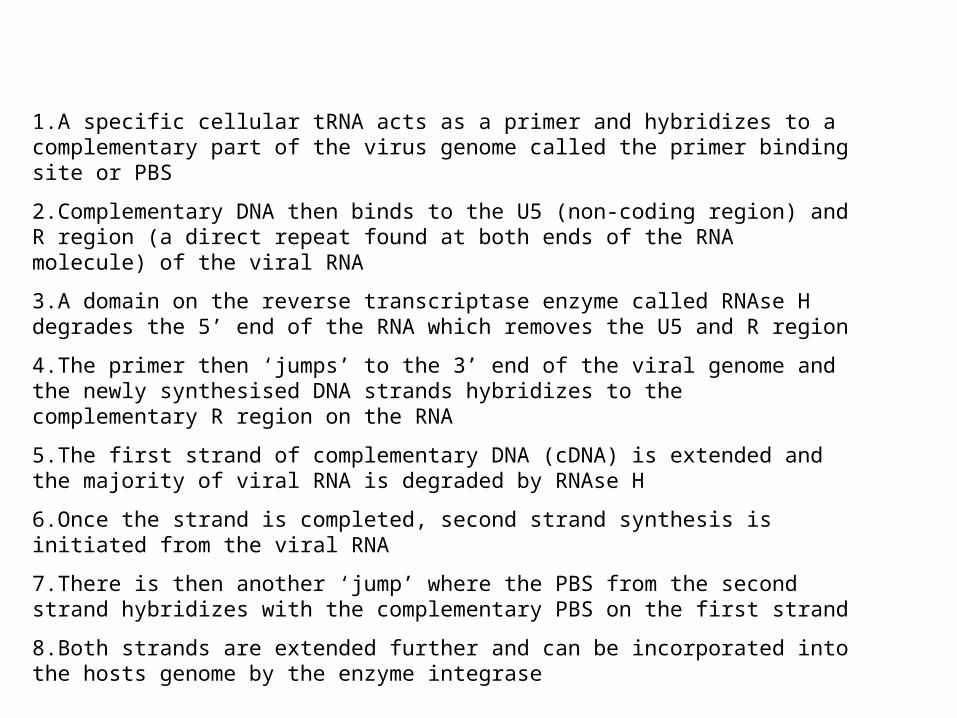
1.A specific cellular tRNA acts as a primer and hybridizes to a complementary part of the virus genome called the primer binding site or PBS
2.Complementary DNA then binds to the U5 (non-coding region) and R region (a direct repeat found at both ends of the RNA molecule) of the viral RNA
3.A domain on the reverse transcriptase enzyme called RNAse H degrades the 5’ end of the RNA which removes the U5 and R region
4.The primer then ‘jumps’ to the 3’ end of the viral genome and the newly synthesised DNA strands hybridizes to the complementary R region on the RNA
5.The first strand of complementary DNA (cDNA) is extended and the majority of viral RNA is degraded by RNAse H
6.Once the strand is completed, second strand synthesis is initiated from the viral RNA
7.There is then another ‘jump’ where the PBS from the second strand hybridizes with the complementary PBS on the first strand
8.Both strands are extended further and can be incorporated into the hosts genome by the enzyme integrase

2. RNA-dependent RNAP
1.RNase H
1.RNase H
3. DNA-dependent DNAP
Reverse transcriptase Specific tRNA as primer

Fig. 19-10
Viral infection of plants
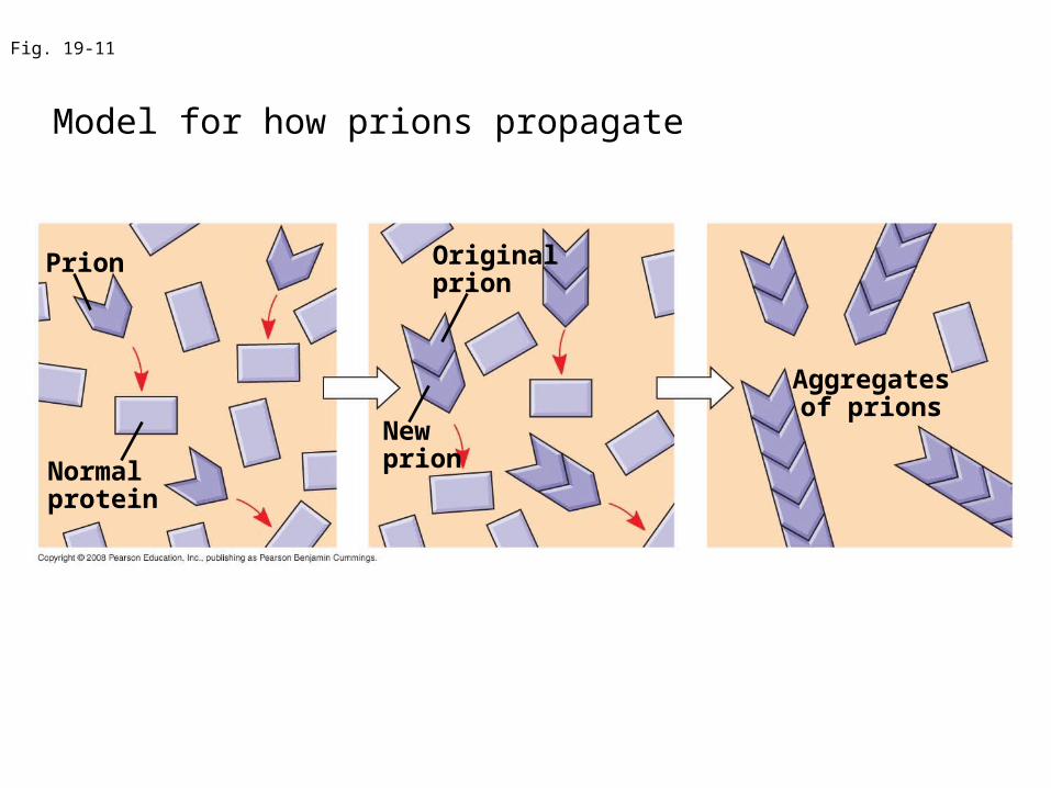
Fig. 19-11
Prion
Normalprotein
Originalprion
Newprion
Aggregatesof prions
Model for how prions propagate

Fig. 19-UN3