Embed Size (px)
Citation preview
FIELD DEPLOYMENT OF A PORTABLE XRD/XRF INSTRUMENT ON MARS ANALOG TERRAIN
P. Sarrazin1, D. Blake2, S. Feldman2, S. Chipera3, D. Vaniman3, and D. Bish4
1 inXitu, PO Box 730, Mountain View, CA 94042 USA 2 NASA Ames Research Center, MS 239-4, Moffett Field, CA 94035
3 Los Alamos National Laboratory, MS D469, Los Alamos, NM 87545 4 Indiana University, 1001 E 10th St., Bloomington IN 47405
ABSTRACT
CheMin is a miniature X-ray diffraction/X-ray fluorescence (XRD/XRF) instrument that is included in the payload of the Mars 2009 Mars Science Laboratory mission. A portable CheMin prototype was built to test the capability of the instrument for remote in-situ mineralogical characterization of geological materials. The instrument was successfully deployed at a variety of Mars analog sites in Death Valley, California, in May 2004.
INTRODUCTION
The principal goals of NASA’s Mars exploration program [1] are to determine whether conditions existed on the planet that could have supported life, and if so, if life developed. The search for evidence of life, prebiotic chemistry or volatiles supportive of life on Mars will require the identification of rock types that could have preserved these. Anything older than a few million years (>99.9% of Mars history) will either be a rock, or will only be interpretable in the context of the rocks that contain it. The key role that definitive mineralogy plays in this search is a consequence of the fact that minerals are thermodynamic phases, having known and specific ranges of temperature, pressure and composition within which they are stable. More than simple compositional analysis, definitive mineralogical analysis can provide information about pressure/temperature conditions of formation, past climate, water activity and the like. Definitive mineralogical analyses are necessary to establish the origin or provenance of a sample. The search for evidence of extant or extinct life on Mars will initially be a search for evidence of present or past conditions supportive of life (e.g., evidence of water), not for life itself.
The CheMin XRD/XRF instrument performs definitive mineralogical analysis of crushed or powdered samples through the combined application of X-ray diffraction (mineral structure analysis) and X-ray fluorescence (elemental compositional analysis). The two techniques are extraordinarily powerful and complementary; they constitute the preferred methods for mineralogical analysis of unknowns in terrestrial laboratories. Other techniques such as Mössbauer spectroscopy, infrared absorption spectroscopy (IR), Raman spectroscopy, differential thermal analysis or thermogravimetric analysis, while able to identify features attributable to specific minerals or to generally identify classes of minerals, are seldom definitive or quantitative and cannot unravel the complex mineral assemblages found in nature.
Copyright ©JCPDS - International Centre for Diffraction Data 2005, Advances in X-ray Analysis, Volume 48. 194

In the past, XRD was not seriously considered for robotic spacecraft missions because powder sample preparation was difficult and X-ray diffractometers typically required massive power supplies, high-power X-ray tubes and complex mechanical motions of the sample, detector or both (a review of XRD and XRF instruments intended for or deployed to solar system targets is presented in Blake [2]). The CheMin XRD/XRF, which has been proposed for robotic spacecraft missions to Venus, the Earth’s moon, Mars, Europa, and a comet nucleus, is a compact instrument with no moving parts that performs simultaneous XRD and XRF. CheMin utilizes a highly collimated miniature low-power X-ray source, a transmission sample geometry and a two-dimensional charge-coupled device (CCD) detector capable of both spatial and energy resolution of individual X-ray photons. Early CheMin prototypes built and tested at NASA Ames Research Center in 1992-1997 demonstrated the capabilities of the concept but offered limited performance [3-6]. Recent improvements in X-ray CCD detectors and small X-ray tube technology were implemented in a third-generation CheMin prototype (CheMin III). A novel sample vibration system [7] incorporated into CheMin III allows the analysis of coarse-grained samples that have simply been crushed and sieved, eliminating the requirement for fine-grained powder samples. A field-deployable CheMin III instrument was built and tested at a variety of Mars analog sites in Death Valley, California.
PRINCIPLE OF OPERATION
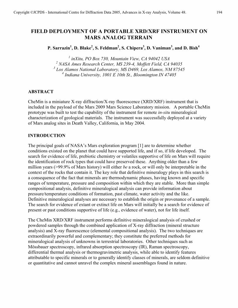
In operation, a collimated X-ray beam from an X-ray tube source is directed through powdered or crushed sample material. An X-ray sensitive CCD imager is positioned on the opposite side of the sample from the source and directly detects X-rays diffracted or fluoresced by the sample [Figure 1(a)]. The CCD detector is operated in single-photon counting mode (the detector is read out at a frequency that ensures that the vast majority of pixels contain charge from either zero or one photon). The CCD detector is exposed to the X-ray flux, read out and erased a large number of times (100-1000 exposures). When operated in this manner, the CCD can be used to measure the charge generated by each photon (and hence its energy). Diffracted primary beam characteristic X-rays strike the detector and are identified by their energy producing a two-dimensional image that constitutes the diffraction pattern. At incremental radii this pattern is summed circumferentially about the central undiffracted beam to yield a one- dimensional 2θ plot comparable to conventional diffractometer data [Figure 1(b)]. All of the X-rays detected by the CCD are summed into a histogram of number of photons vs. photon energy that constitutes an XRF analysis of the sample [Figure 1(c)]. Quantitative mineralogical results are obtained from XRD data by Rietveld refinement and other full-pattern fitting techniques [8-9]. Both crystalline and amorphous materials can be analyzed in this way. Quantitative elemental compositions can be obtained from XRF data by fundamental parameters (FP) calculations [10].
CHEMIN III
The CheMin III instrument can be separated into three groups of components (see Figure 2): the CCD camera, the sample holder, and the X-ray source and collimator. The CCD camera is a Be-window sealed, thermoelectrically cooled AndorTM X-ray camera, having a front-illuminated deep-depleted E2V 5530 CCD (1251x1151 array of 22.5 µm pixels) that is directly exposed to the X-ray flux. The sample cell consists of a 5 mm circular opening, fitted with two parallel Mylar windows held at a distance of 175 µm from each other. A piezoelectric actuator shakes
Copyright ©JCPDS - International Centre for Diffraction Data 2005, Advances in X-ray Analysis, Volume 48. 195
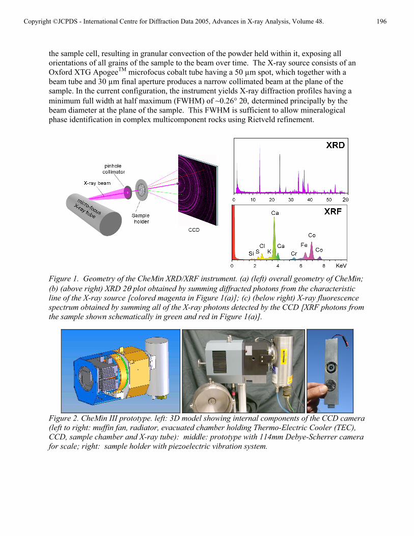
the sample cell, resulting in granular convection of the powder held within it, exposing all orientations of all grains of the sample to the beam over time. The X-ray source consists of an Oxford XTG ApogeeTM microfocus cobalt tube having a 50 µm spot, which together with a beam tube and 30 µm final aperture produces a narrow collimated beam at the plane of the sample. In the current configuration, the instrument yields X-ray diffraction profiles having a minimum full width at half maximum (FWHM) of ~0.26° 2θ, determined principally by the beam diameter at the plane of the sample. This FWHM is sufficient to allow mineralogical phase identification in complex multicomponent rocks using Rietveld refinement.
Figure 1. Geometry of the CheMin XRD/XRF instrument. (a) (left) overall geometry of CheMin; (b) (above right) XRD 2θ plot obtained by summing diffracted photons from the characteristic line of the X-ray source [colored magenta in Figure 1(a)]; (c) (below right) X-ray fluorescence spectrum obtained by summing all of the X-ray photons detected by the CCD [XRF photons from the sample shown schematically in green and red in Figure 1(a)].
Figure 2. CheMin III prototype. left: 3D model showing internal components of the CCD camera (left to right: muffin fan, radiator, evacuated chamber holding Thermo-Electric Cooler (TEC), CCD, sample chamber and X-ray tube): middle: prototype with 114mm Debye-Scherrer camera for scale; right: sample holder with piezoelectric vibration system.
Copyright ©JCPDS - International Centre for Diffraction Data 2005, Advances in X-ray Analysis, Volume 48. 196

Figure 3. XRD images and corresponding 1D patterns of a starkeite powder collected at various integration times: left 5 min, middle 50 min, right 500 min; search-match allows automatic identification of the phase in all three cases.
Figure 3 shows performance characteristics of the instrument with a cobalt X-ray tube operated at 40 kV and 250 mA (10 W total power). Integration times as short as 5 minutes are sufficient to identify a complex single mineral such as starkeite (MgSO4•4H2O). Figure 4 shows the low-2θ performance of the instrument. Hydrous minerals such as clays are important indicators of water activity and habitability. The ability to identify and characterize the large d-values (low 2θ angles) characteristic of clay minerals and zeolites is essential in a mineralogical instrument intended for Mars exploration. The characterization of complex mineral assemblages having several minerals in varying amounts will require longer periods of integration. The rock type (e.g., basalt, andesite, sedimentary) and the type of mineralogic information required will dictate the duration of analysis. For example, in a basalt, one might want to know the identity and amount of all major and minor minerals, as well as refined unit-cell parameters for some of the phases. These data, in addition to the overall XRF analysis of the sample, can then be used to fully characterize the structure and composition of each phase. Figure 5 shows results obtained from a mineralogically complex rock (an andesite) containing seven major minerals. The results of a Rietveld refinement using the data are shown as an inset to the figure.
In order to minimize CCD background noise during analyses, the TEC is normally used to cool the CCD to –60° C or below. In the laboratory, chilled water is used to remove the waste heat from the TEC. However, in the field or ultimately during remote deployment on Mars, water cooling will not be used. We performed tests in which the CCD was warmed in 10° C increments to study the effect of thermal noise on the X-ray signal. We found that although the increase in background degraded the energy-resolving capability of the CCD relatively rapidly (above about –40° C, XRF analysis became impossible), it was possible to perform acceptable XRD analysis at CCD temperatures as high as +10° C.
Copyright ©JCPDS - International Centre for Diffraction Data 2005, Advances in X-ray Analysis, Volume 48. 197

Figure 4. low-2θ detection limit of the CheMin III Prototype; left: XRD pattern of non-purified silver behenate CH3(CH2)20COO-Ag with the first ring at d001=58.38Å, 1.75° 2θ Co Kα, right: XRD pattern of a smectite clay (SWa-1) with trace of quartz.
Figure 5. CheMin III data collected from a crushed sample of andesite (LANL #P52, sieved to <150 µm); left: 2D XRD image; right: 1D pattern obtained by integration of the diffraction image and results of the Rietveld refinement.
FIELD DEPLOYABLE INSTRUMENT
A portable version of the CheMin III instrument was developed for field deployment on Mars analog terrain. The design considerations for the portable instrument were: The instrument and all peripherals must be transportable in a small car and hand-carried to a geological site of interest; the instrument must be battery powered and controlled by a laptop computer; all instrument control, data collection and data analysis functions must be performed on-site; and sample preparation must be simple and minimal.
In the field-deployable version, the CheMin III instrument was mounted inside a sturdy aluminum framework which enclosed the camera controller, thermoelectric cooler controller, X-ray tube power supply and sample vibration system (Figure 6). The portable CheMin III instrument (which weighs ~15 kg) was connected by umbilical to a laptop computer that was used for both instrument operation and data reduction. The same umbilical connected a battery pack to the instrument. Sample preparation was performed manually, but the normally exhaustive process of powdering the material with a mortar and pestle was eliminated through
Mineral computed wt%
albite 13.3(8) labradorite 50.6(6) enstatite 13.9(10) augite 13.4(7) cristobalite 7.2(3) magnetite 1.0(2) forsterite 0.7(5)
Copyright ©JCPDS - International Centre for Diffraction Data 2005, Advances in X-ray Analysis, Volume 48. 198

the use of the vibrating sample holder [7]. Rocks or consolidated sediments were simply smashed with a hammer and sieved to <150 µm prior to inserting them into the sample holder.
Figure 6. Portable version of the CheMin III prototype with geologist’s hammer for scale.
FIELD TEST IN DEATH VALLEY
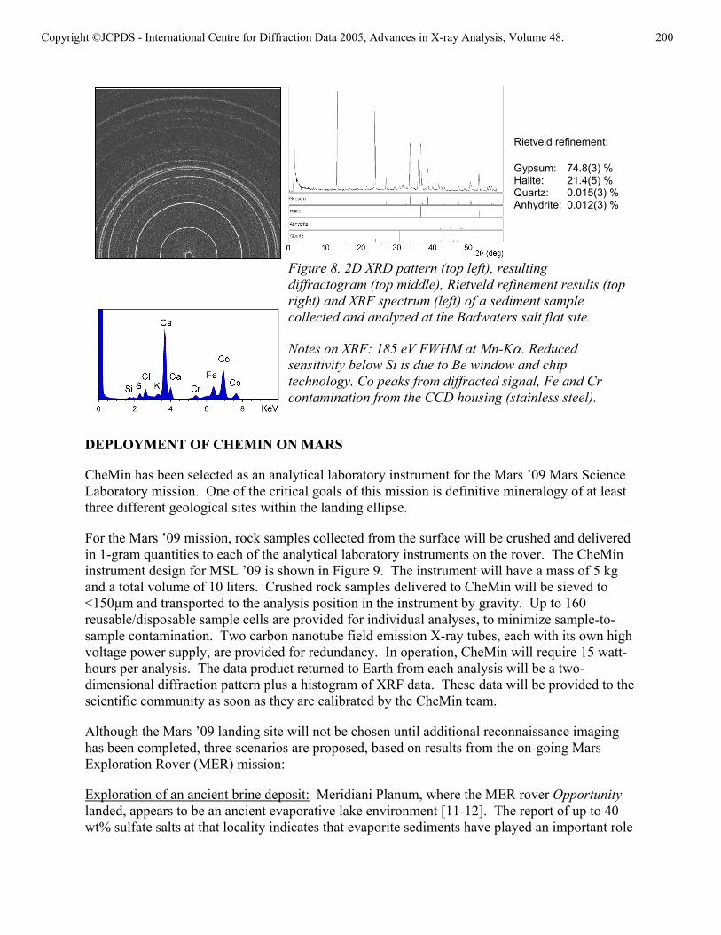
The CheMin III instrument was field-tested in Death Valley, California, in May, 2004. Death Valley National Monument has within its boundaries a large number of Mars analog sites including evaporites and playa lake deposits, igneous terrains, dune fields and sedimentary deposits. Many of these sites are appropriate analogs for Mars surface terrains. Figure 7 shows the CheMin instrument in situ at Badwaters Flat, an evaporative environment containing halite and calcium sulfate hydrates. The TEC was able to maintain the CCD at –60° C without water cooling during the early morning hours. Figure 8 shows a 2-D diffraction pattern from material prepared and analyzed on-site, as well the resulting diffractogram and search-match results obtained from a commercial phase identification program run from the laptop. Results of a Rietveld refinement of these data are shown in the table.
Figure 7. deployment of CheMin III in Badwaters Salt Flat, Death Valley, California.
TEC cooled camera with deep depleted CCD
X-ray tube power supply
Microfocus X-ray tube (Co, 10W)
Sample handling and collimation stage
TEC controller
Function generator and amplifier for sample vibrator
Umbilical to battery packs and laptop computer
Copyright ©JCPDS - International Centre for Diffraction Data 2005, Advances in X-ray Analysis, Volume 48. 199
DEPLOYMENT OF CHEMIN ON MARS
CheMin has been selected as an analytical laboratory instrument for the Mars ’09 Mars Science Laboratory mission. One of the critical goals of this mission is definitive mineralogy of at least three different geological sites within the landing ellipse.
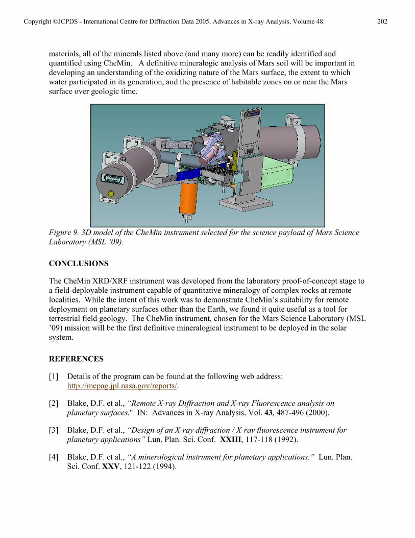
For the Mars ’09 mission, rock samples collected from the surface will be crushed and delivered in 1-gram quantities to each of the analytical laboratory instruments on the rover. The CheMin instrument design for MSL ’09 is shown in Figure 9. The instrument will have a mass of 5 kg and a total volume of 10 liters. Crushed rock samples delivered to CheMin will be sieved to <150µm and transported to the analysis position in the instrument by gravity. Up to 160 reusable/disposable sample cells are provided for individual analyses, to minimize sample-to-sample contamination. Two carbon nanotube field emission X-ray tubes, each with its own high voltage power supply, are provided for redundancy. In operation, CheMin will require 15 watt-hours per analysis. The data product returned to Earth from each analysis will be a two-dimensional diffraction pattern plus a histogram of XRF data. These data will be provided to the scientific community as soon as they are calibrated by the CheMin team.
Although the Mars ’09 landing site will not be chosen until additional reconnaissance imaging has been completed, three scenarios are proposed, based on results from the on-going Mars Exploration Rover (MER) mission:
Exploration of an ancient brine deposit: Meridiani Planum, where the MER rover Opportunity landed, appears to be an ancient evaporative lake environment [11-12]. The report of up to 40 wt% sulfate salts at that locality indicates that evaporite sediments have played an important role
Figure 8. 2D XRD pattern (top left), resulting diffractogram (top middle), Rietveld refinement results (top right) and XRF spectrum (left) of a sediment sample collected and analyzed at the Badwaters salt flat site.
Notes on XRF: 185 eV FWHM at Mn-Kα. Reduced sensitivity below Si is due to Be window and chip technology. Co peaks from diffracted signal, Fe and Cr contamination from the CCD housing (stainless steel).
Rietveld refinement:
Gypsum: 74.8(3) % Halite: 21.4(5) % Quartz: 0.015(3) %Anhydrite: 0.012(3) %
Copyright ©JCPDS - International Centre for Diffraction Data 2005, Advances in X-ray Analysis, Volume 48. 200

in the hydrogeologic history of Mars. Data available to date support the presence of the mineral jarosite (a hydrous Fe-sulfate), Mg-sulfate, and lesser amounts of salts containing Cl and Br. These data imply that several sulfates, mixed with halogen salts, combine to form a complex salt assemblage. One of the most exciting features of the Meridiani sediments is the possibility that the salts may be hydrated. Water abundances in hydrated salts on Earth can be considerably greater than water abundances in hydrous silicates such as clays and zeolites. Water storage in minerals may be a significant source of the elevated hydrogen abundances seen in some of the equatorial regions by the Odyssey spacecraft, with abundances up to 8-9 wt% water equivalent present in areas where water ice should not be stable [13]. Salt hydrates in evaporite sediments might account for some equatorial water. Could such a water-rich system harbor life at depth, or at least preserve evidence of brine-dwelling organisms? The ability to quantify hydrated mineral assemblages such as those found at Meridiani Planum will be important for reconstructing brine evolution and for determining the nature of interactions between brine minerals and detrital mineralogy. CheMin XRD/XRF data will be highly useful in interpreting brine chemistry and the extent and nature of the ancient habitable zone that existed on early Mars.
Exploration of a basaltic terrain: The present-day surface environment of Mars appears to be inhospitable to life. The most likely habitable zones in the present or remote past would have existed in the sub-surface [14]. Should life have evolved in the subsurface, chemosynthesis (the utilization of chemical energy from the environment by primitive organisms) must have been the principal energy-harvesting strategy. If one is searching for evidence of subsurface chemosynthetic life, exploration of a basaltic terrain is an important component of this search. Ferrous iron contained in the mineral olivine (as well as in basaltic glass, pyroxene and some oxide minerals) could provide a source of redox energy for chemosynthetic life). CheMin can determine the amount and composition of the major rock-forming minerals in basaltic rocks. In addition, refined unit-cell parameters of phases such as olivine, (Fe,Mg)2SiO4 allow the determination of the amounts of Fe++ available for reaction with water. Once primary mineralogy is determined, one must search for and quantify secondary mineral assemblages that can indicate water-rock interactions. Even the passage of small amounts of water through the rock system can leave signatures in the form of hydrated phases such as clays, zeolites, chlorite, serpentine and a host of other H2O- or OH-bearing minerals as well as non-hydrous alteration minerals. Such assemblages, if found on Mars, would suggest the presence of a habitable zone having both liquid water and an energy source.
Mineralogy and Chemistry of the Global Soil Component: The mineralogic compositions of Martian soils, which include globally distributed components, are still poorly constrained. Initial modeling of Viking XRF results suggested a mixture of clays, kieserite, calcite and rutile [15]. The minerals of detrital basalt plus a Cl-bearing phase must also be present. In a summary of the Pathfinder soils, Bell [16] suggests that IR spectral data are consistent with the presence of poorly crystalline or nanophase ferric oxide(s), sometimes mixed with small but varying amounts of well-crystallized ferric and ferrous phases. An important objective of soil analysis will be to determine the mineralogy and hydration state of the Martian soil “duricrust” which contains a significant amount of sulfate. Widespread distribution of sulfate salts as cementing agents in soil might be due to recently active pedogenic processes, possibly associated with acidic weathering, that do not require surface water or groundwater [17]. The extent to which water participated in the generation of the Mars soil(s) is important in determining the presence/absence of surface habitable zones over Mars history. With the possible exception of some of the nanophase
Copyright ©JCPDS - International Centre for Diffraction Data 2005, Advances in X-ray Analysis, Volume 48. 201
materials, all of the minerals listed above (and many more) can be readily identified and quantified using CheMin. A definitive mineralogic analysis of Mars soil will be important in developing an understanding of the oxidizing nature of the Mars surface, the extent to which water participated in its generation, and the presence of habitable zones on or near the Mars surface over geologic time.
Figure 9. 3D model of the CheMin instrument selected for the science payload of Mars Science Laboratory (MSL ‘09).
CONCLUSIONS
The CheMin XRD/XRF instrument was developed from the laboratory proof-of-concept stage to a field-deployable instrument capable of quantitative mineralogy of complex rocks at remote localities. While the intent of this work was to demonstrate CheMin’s suitability for remote deployment on planetary surfaces other than the Earth, we found it quite useful as a tool for terrestrial field geology. The CheMin instrument, chosen for the Mars Science Laboratory (MSL ’09) mission will be the first definitive mineralogical instrument to be deployed in the solar system.
REFERENCES
[1] Details of the program can be found at the following web address: http://mepag.jpl.nasa.gov/reports/.
[2] Blake, D.F. et al., “Remote X-ray Diffraction and X-ray Fluorescence analysis on planetary surfaces." IN: Advances in X-ray Analysis, Vol. 43, 487-496 (2000).
[3] Blake, D.F. et al., “Design of an X-ray diffraction / X-ray fluorescence instrument for planetary applications” Lun. Plan. Sci. Conf. XXIII, 117-118 (1992).
[4] Blake, D.F. et al., “A mineralogical instrument for planetary applications.” Lun. Plan. Sci. Conf. XXV, 121-122 (1994).
Copyright ©JCPDS - International Centre for Diffraction Data 2005, Advances in X-ray Analysis, Volume 48. 202

[5] Sarrazin, P., et al., “A miniature XRD/XRF Instrument for in situ characterization of Martian soils and rocks.” J. Phys. IV France 8: 465-470 (1998).
[6] Vaniman, D. et al., “Landed XRD/XRF analysis of prime targets in the search for past or present Martian life.” J. Geophys. Res. 103(E13):31,477-31,489 (1998).
[7] Sarrazin et al., “Vibrating sample holder for XRD analysis with minimal sample preparation.” IN: Advances in X-ray Analysis Vol. 48 (this proceedings) (2004).
[8] D. Bish and J. Post, “Quantitative mineralogical analysis using the Rietveld full-pattern fitting method.” Amer. Min., 78, 932-942 (1993).
[9] Chipera, S.J., and D.L. Bish, “FULLPAT: A full-pattern quantitative analysis program for X-ray powder diffraction using measured and calculated patterns.” J. Applied Crystallography, 35, 744-749 (2002).
[10] Sherman, J., “Theoretical derivation of fluorescent X-ray intensities form mixtures.” Spectrochim. Acta 7, 283-306 (1955).
[11] MER Rover web site: http://www.jpl.nasa.gov/missions/mer/.
[12.] Kerr, R.A., “A wet early Mares seen in salty deposits.” Science 303, 1450 (2004).
[13] Feldman, W.C. et al., “The global distribution of near-surface hydrogen on Mars.” Sixth Intl. Conf. On Mars, abstract #3218, Lunar and Planetary Institute, Houston (CD-ROM) (2003).
[14] Fisk, M.R. and S.J. Giovannoni, “Sources of nutrients and energy for a deep biosphere on Mars.” J. Geophys. Res. 104(E5), 11,805-11,815 (1999).
[15] Baird, A.K. et al., “Mineralogic and petrologic implications of Viking geochemical results from Mars: Interim report.” Science 194, 1288-1293 (1976).
[16] Bell, J.F., et al., “Mineralogic and compositional properties of the Martian soil and dust: Results from Mars Pathfinder.” JGR-Planets 105, 1721-1755 (2000).
[17] Banin et al., “Acidic volatiles and the Mars soil.” J. Geophys. Res. 102, 13,341-13,356 (1997).
Copyright ©JCPDS - International Centre for Diffraction Data 2005, Advances in X-ray Analysis, Volume 48. 203