Embed Size (px)
Citation preview
Postgrad. med. J. (April 1969) 45, 266-271.
Fibula autograft survival following resection ofosteoclastoma of radius
M. L. H. LEE J. C. SANDEMANM.A., M.B., F.R.C.S. M.B., M.Ch.Orth., F.R.C.S.E., F.C.S.S.A., F.R.C.S.
Consultant Orthopaedic Surgeon, Consultant Orthopaedic Surgeon andBournemouth and East Dorset Research Fellow, University of Liverpool
Hospital Group
GIANT-CELL tumours of bone may be malignant fromthe onset or malignancy may develop during treat-ment. They are, therefore, best treated by completeexcision where this is surgically possible, with bonegrafting of the resultant defect. A patient presentedwith an osteoclastoma of the lower end of the radius,which was completely excised and replaced by anautogenous fibula graft. The successful completeincorporation and function of this whole bonereplacement, with satisfactory joint function, wasfollowed by an injury and a fracture. The lack ofradiological change within the grafted bone shaft,the normal appearance at subsequent operation, anddelayed union following the fracture are of interest,and valuable when the incorporation of freshautogenous bone grafts is compared with thosewhose viability has been destroyed.
Case historyIn September 1965 a 22-year-old builder's labourer
complained of a swelling over the palmar surface ofhis left wrist. It had been present for 7 weeks andvaried in size.
Examination revealed a firm thickening of thedistal end of the radius, most marked on the palmaraspect. There was slight restriction of all movementsof the wrist. X-ray revealed a cystic lesion of theradius (Fig. 1) which was diagnosed as an osteo-clastoma. X-ray of his chest and haematologicalinvestigations were normal.An open biopsy 1 week later revealed a cellular
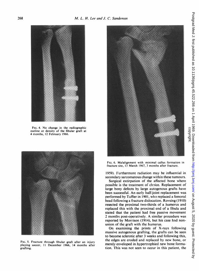
tumour (Fig. 2) composed of multinucleated giantcells and stromal cells, considered to be an osteo-clastoma. The distal end of the radius was subse-quently excised by extra-periosteal resection. Thetumour was noted to lie immediately beneath thearticular cartilage of the radius, and a furtherhistological report revealed that tumour tissue hadextended through fibrous and muscle tissue into avessel.
Address for reprints: J. C. Sandeman, Department ofOrthopaedic Surgery, University of Liverpool, Liverpool 3.
··i·
i··ii:·i. ii·' Iii·:i··'·:;ds:.' :
I··rjii..iI·i:if:::' ·Ifil·
9.2ii....I. :: :i:.I
lil.ii,l:;.·:··i:····i·
'I·: ii.ii
s:I:
i·::::: i:i: '''I: r .:..i;.li.liii:I.;.i:;i:.i:. i:;.I i:.....:::
r·:···:·.·.'" lhr3slEB.B...ff.B.&..Ci;iil;iiii'iir
'':. :di:i ·········;i:i:iii::':'':
FIG. 1. Osteoclastoma lower end of the radius, 9 September1965.
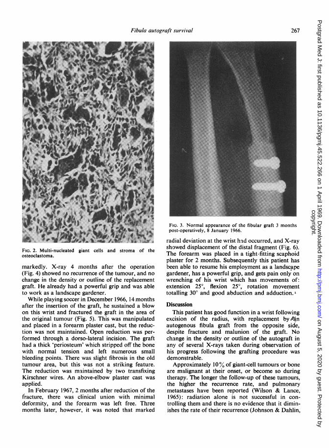
At operation, the proximal end of the right fibulawas removed by sub-periosteal resection, step cutsmade in the appropriate ends of the radius andfibula, and the fibula fixed as a forearm graft by twovitallium screws. The soft tissues of the wrist werenot reconstructed, but the graft was transfixed tothe carpus by a Kirschner wire. The graft wasimmobilized in an above-elbow plaster cast whichwas worn for 3 months.On removal of the plaster, forearm and wrist
alignment appeared normal, and X-ray in January1966 (Fig. 3) showed incorporation of the graft whichappeared of normal density and outline throughoutits length.
After mobilization of the right arm for 1 weekhis elbow movements were 50-135°, and wristextension 20° and flexion 20°, but rotation waspractically nil and with time this only achieved atotal of 30° whereas all other movements improved
copyright. on A
ugust 5, 2020 by guest. Protected by
http://pmj.bm
j.com/
Postgrad M
ed J: first published as 10.1136/pgmj.45.522.266 on 1 A
pril 1969. Dow
nloaded from
Fibula autograft survival 267
'I;
i,...'..''..1..'_5 " :;fi c6._,§.... _ !:"
;·- .3ll' m .: "._
O',,di 1." .'......
......b. -: ~i t~~~~ _j,7r .... :::::;....:.L.:.. ·..'....... ........· ::.l:. .... _w^._[#s 8.s Xt..s* { = a _.
= ^ ^ * ^ s s - f ~ ~ ~~~~~~~~~~~~......:...,:..].:..~.................e.g
s _ ^t j ~~~~.. :.did /~ '? :'''i.i-..., '.. _ t - w 2 i b .-Ij # w Wi*A s#-
-i_ N
*b: _i javp _
iCJ4ste F ftAL: iiJ_ t* }s* iZ FA _ EFIG. 2. Multi-nucleated giant cells and stroma of theosteoclastoma.
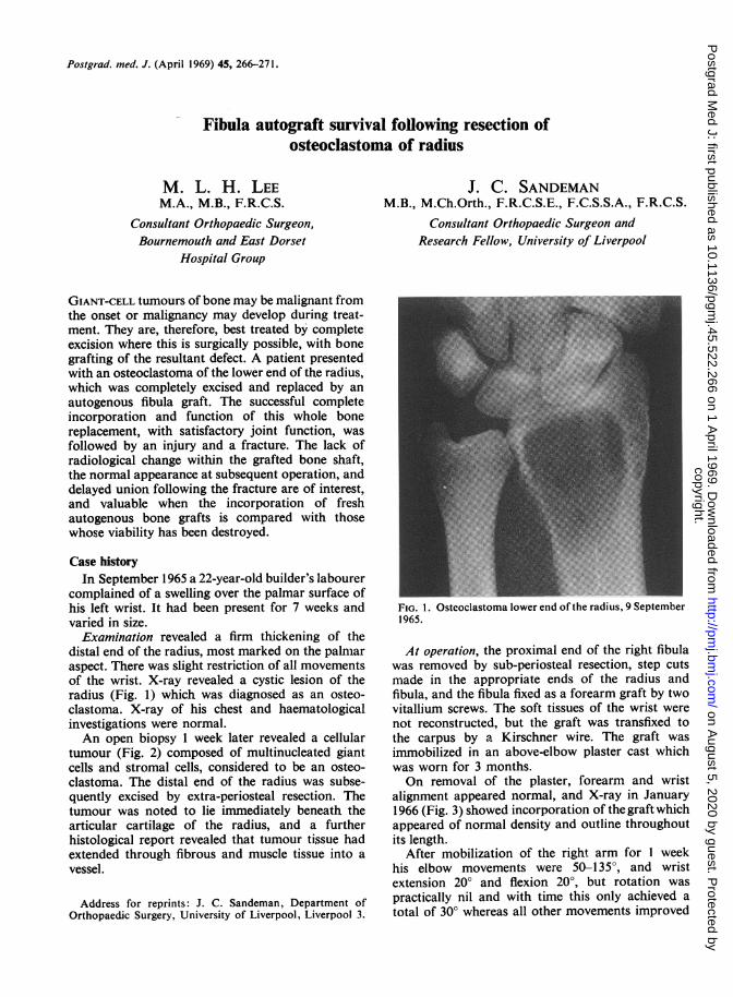
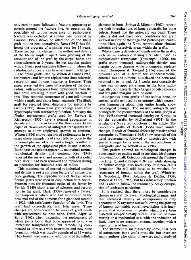
markedly. X-ray 4 months after the operation(Fig. 4) showed no recurrence of the tumour, and nochange in the density or outline of the replacementgraft. He already had a powerful grip and was ableto work as a landscape gardener.While playing soccer in December 1966, 14 months
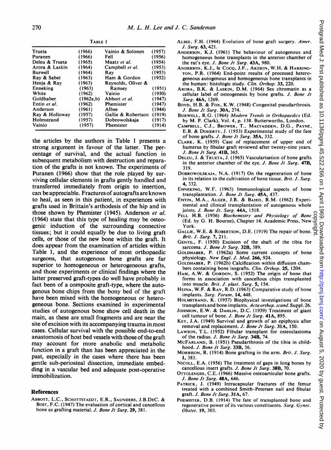
after the insertion of the graft, he sustained a blowon this wrist and fractured the graft in the area ofthe original tumour (Fig. 5). This was manipulatedand placed in a forearm plaster cast, but the reduc-tion was not maintained. Open reduction was per-formed through a dorso-lateral incision. The grafthad a thick 'periosteum' which stripped off the bonewith normal tension and left numerous smallbleeding points. There was slight fibrosis in the oldtumour area, but this was not a striking feature.The reduction was maintained by two transfixingKirschner wires. An above-elbow plaster cast wasapplied.
In February 1967, 2 months after reduction of thefracture, there was clinical union with minimaldeformity, and the forearm was left free. Threemonths later, however, it was noted that marked
ii?lii.ia.,ilii
:·:·:,:::
liir:::::;:··:: ::i.i.i.ii''li:i:!:·;:····r::i··:r·······;·':
;·:'I:!i%ifli:gl."i:K:i··l·;·:i;iisilgli:iSi.i i··l' li.'· ···il, :··.n
··:'I
Pl.iii.::·I:s:
::.:::r:i:H:: :;!:
··'l''c::$iXilf:i
·. ·'·i·IB.;··l..:·sns:·..···
i..i:i.ii.i.i(·,:li.
·i:.
:i
iiii:
i: '.BB3.aa.P.B%B.f·iii; .:...B.i.a.s.lqigpCsss...:li·. · ·':if
;.i. ::.t.&.gE.:.B..!.ii:·ii··i'
irilli
:*:·
;:· .I:.searre%8n.8lrrs...ec·:.·
FIG. 3. Normal appearance of the fibular graft 3 monthspost-operatively, 8 January 1966.
radial deviation at the wrist had occurred, and X-rayshowed displacement of the distal fragment (Fig. 6).The forearm was placed in a tight-fitting scaphoidplaster for 2 months. Subsequently this patient hasbeen able to resume his employment as a landscapegardener, has a powerful grip, and gets pain only onwrenching of his wrist which has movements of:extension 25°, flexion 25°, rotation movementtotalling 30° and good abduction and adduction.
DiscussionThis patient has good function in a wrist following
excision of the radius, with replacement by.anautogenous fibula graft from the opposite side,despite fracture and malunion of the graft. Nochange in the density or outline of the autograft inany of several X-rays taken during observation ofhis progress following the grafting procedure wasdemonstrable.
Approximately 10% of giant-cell tumours or boneare malignant at their onset, or become so duringtherapy. The longer the follow-up of these tumours,the higher the recurrence rate, and pulmonarymetastases have been reported (Wilson & Lance,1965): radiation alone is not successful in con-trolling them and there is no evidence that it dimin-ishes the rate of their recurrence (Johnson & Dahlin,
copyright. on A
ugust 5, 2020 by guest. Protected by
http://pmj.bm
j.com/
Postgrad M
ed J: first published as 10.1136/pgmj.45.522.266 on 1 A
pril 1969. Dow
nloaded from
268 M. L. H. Lee and J. C. Sandeman
l~~~~~~~~~~~~~~~~~~~::: .i:·M:|~~~~~~~~~~~~~· ·ll|f.F.:..;~~~~~~~~~~~~~~~~:·::
FIG. 4. No change in the radiographicoutline or density of the fibular graft at4 months, 12 February 1966.
:::·· ·:·":191·
·.i·l,ilB·'"· ''':
ifi'·l::r.::.::: ..i.i··.·..
ir;.i::.l.ii:ii.r'·.········ .I: .· .· '.a·:-dr: i:·:··i/:.l'f·:·:.. ·:··:·I:···
·i·. ····;·······::·.··
·: ·:· ::·::·:·:i i:··:·i::.::I:;::· I·.: ::::::...i... ··.:::·.·::::.,:;..:..·· :·t· i: ·:
:·'··:···i· :::; 'd::i:····· ·i·:'·'
"·':'
:·::'
FIG. 5. Fracture through fibular graft after an injuryplaying soccer, 11 December 1966, 14 months aftergrafting.
*:·i···:.. \w; .:··
_E: 0.,,* q;. ,>-, . . :,,.:. i:' ::..
: .·: ··:: :.· ;·
i~~.....,::i.·· ,\ .': ·i ·::.... Y;·.:
:.:.::. X..t·i:: ·:; ·.·.. :.: :: :.....'
·;
,: ..-· ....::
- L ;.: .La·i:· :::·
FIG. 6. Malalignment with minimal callus formation infracture site, 13 March 1967, 3 months after fracture.
1959). Furthermore radiation may be influential insecondary sarcomatous change within these tumours.
Surgical extirpation of the affected bone wherepossible is the treatment of choice. Replacement oflarge bony defects by large autogenous grafts havebeen successful. An early half-joint replacement wasperformed by Tuffier in 1901, who replaced a femoralhead following a fracture dislocation. Rovsing (1910)resected the proximal two-thirds of a humerus andreplaced this with the proximal end of a fibula andstated that the patient had free passive movement2 months post-operatively. A similar procedure wasreported by Morrison (1914), but his case had non-union of the graft with the humerus.On examining the prints of X-rays following
massive autogenous grafting, the grafts can be seento become sclerotic after 3 weeks and following this,the edges are eroded and replaced by new bone, ormerely enveloped in hypertrophied new bone forma-tion. This was not seen to occur in this patient, the
copyright. on A
ugust 5, 2020 by guest. Protected by
http://pmj.bm
j.com/
Postgrad M
ed J: first published as 10.1136/pgmj.45.522.266 on 1 A
pril 1969. Dow
nloaded from
Fibula autograft survival
only erosion seen, followed a fracture, appearing asporosis around the fracture line. At operation thepossibility of tumour recurrence or pathologicalfracture was excluded. A similar case reported byLawson (1952) shows no alteration of the fibulagraft outline post-operatively. Ross (1953) has fol-lowed the progress of a similar case for 15 years.There has been no change in the outline and densityof the fibular implant apart from grooving of thearticular end of the graft by the carpal bones andsome sclerosis at 9 years. He has another patientwith a 5-year post-operative follow-up in whom theradiological appearances have remained unchanged.The fibula grafts used by Wilson & Lance (1965)
for humeral and femoral replacement show sclerosis,resorption and in one instance, a fracture. Theirpaper presented five cases of resection of the distalradius, with autogenous bone replacement from theiliac crest, resulting in cure with good function intwo. They reported recurrence of giant-cell tumourwithin a graft, and also a lung metastasis. The fibulagraft for resected tibial diaphysis for sarcoma byGentil (1950), showed no graft change apart fromhypertrophy with satisfactory function. Similarly thefibular replacement grafts used by Stewart &Richardson (1952) have a normal appearance indensity and outline in two of the three cases. In hispaper of eleven cases of autograft transplants withattempt to allow epiphyseal growth to continue,Wilson (1966) shows reprints of radiographs in twocases where transplants of short tubular bone to theproximal phalanx of a toe and a finger, resulted inthe growth of the epiphyseal plate in one instance.Both these transplants apparently maintained normalradiological density and contour. Key (1949)reported the survival and normal growth of a radialhead after it had been removed and replaced duringan operation for fractured neck of radius.
This maintenance of normal radiological outlineand density is not a common feature of autogenousbone grafting. The reproductions of X-rays, wherefibular grafts were used in conjunction with Smith-Petersen pins for fractured necks of the femur byPatrick (1949) show areas of sclerosis and resorp-tion in the graft. Clark (1959) reported a 29-yearfollow-up on a patient who had an excision of theproximal end of the humerus for a giant-cell tumourin 1929, with satisfactory function of the limb. Thisgraft had osteosclerotic and lytic areas. Thesechanges are considered to be areas of graft deathwith replacement by host bone. Entin, Alger &Baird (1962) when discussing the replacement ofwhole joints found that bone appeared to surviveimmediate transplantation, but remodelling com-menced at 15 weeks with resorption and new boneformation which was usually completed at 35 weeks.They found there was survival of some of the cellular
elements in bone. Stringa & Mignani (1967), report-ing their investigations of large autografts for bonedefects, found that the autograft was dead. Thesepatients did not have ideal conditions for graftsurvival in view of their age and pathological condi-tions. Here again the X-ray reproductions showedsclerosis and osteolytic areas within the grafts.Where there is definite cell-death within the grafts,
such as in cadaveric hgmografts when used asosteoarticular transplants (Ottolenghi, 1966), thegrafts show increased radiographic density andmargin outline changes while undergoing replace-ment. Thompson & Streggal (1956) resected theproximal end of a femur for chondrosarcoma,curetted out the tumour, autoclaved the bone andreturned it to its bed. At 2 weeks post-operativelythere was no apparent change in the bone radio-logically, but thereafter the changes of osteosclerosisand irregular margins were obvious.Autogenous bone grafts of cancellous bone, or
cortical grafts removed by osteotomy which necessi-tates hammering along their entire length, showradiological changes. The massive autografts andhomografts for pseudarthrosis of the tibia (Boyd &Fox, 1948) showed increased density on X-rays, asdo the autografts by McFarland (1951) in thesatisfactory X-ray reproductions of his article. Thecancellous grafts of Nicoll (1956) have similarchanges. Repair of femoral defects by massive tibialautografts by Phemister (1945) show sclerosis of thetibial replacements, followed by hypertrophy, withsimilar changes being seen in the reproductions ofautografts used by Abbott et al. (1947).
This patient showed no radiological changes inbone density or outline until he sustained his fracturefollowing football. Osteoporosis around the fractureline (Fig. 5), and subsequent X-rays, while showingno further change, also reveal very little new callusformation. He will still have to be watched forrecurrence of tumour within the graft (Windeyer& Woodyatt, 1949; Johnson & Dahlin, 1959;Wilson & Lance, 1965), but has satisfactory functionand is able to follow the reasonably heavy occupa-tion of landscape gardening.
It is realized that there must be considerablechange in a graft to reveal radiological changes, andthat increased density or osteoporosis is onlyapparent on X-ray some weeks following the graftingof known non-viable bone; nevertheless it is inter-esting that sometimes in grafts which have beendissected sub-periosteally without the use of ham-mering or a mechanical saw with the induction ofheat, there is no apparent change, together with arapid return of function.The statement is maintained by some, that cells
of autogenous bone grafts must die, but there aremany authors who claim otherwise: and a study of
269
copyright. on A
ugust 5, 2020 by guest. Protected by
http://pmj.bm
j.com/
Postgrad M
ed J: first published as 10.1136/pgmj.45.522.266 on 1 A
pril 1969. Dow
nloaded from
270 M. L. H. Lee and J. C. Sandeman
TABLE 1
Trueta (1966) Vainio & Solonen (1957)Puranen (1966) Fell (1956)Deleu & Trueta (1965) Maatz et al. (1954)Arora & Laskin (1964) Campbell et al. (1953)Burwell (1964) Ray (1953)Ray & Sabet (1963) Ham & Gordon (1952)Henja & Ray (1963) Reynolds, Oliver &Enneking (1963) Ramsey (1951)White (1962) Vainio (1950)Goldhaber (1962a,b) Abbott et al. (1947)Entin et al. (1962) Phemister (1947)Anderson (1961) Albee (1944)Ray & Holloway (1957) Gallie & Robertson (1919)Holmstrand (1957) Dobrowolskaja (1917)Vainio (1957) Phemister (1914)
the articles by the authors in Table 1 presents astrong argument in favour of the latter. The per-centage of survival, and the actual function insubsequent metabolism with destruction and repara-tion of the grafts is not known. The experiments ofPuranen (1966) show that the role played by sur-viving cellular elements in grafts gently handled andtransferred immediately from origin to insertion,can be appreciable. Fractures of autografts areknownto heal, as seen in this patient, in experiences withgrafts used in Brittain's arthodesis of the hip and inthose shown by Phemister (1945). Anderson et al.(1964) state that this type of healing may be osteo-genic induction of the surrounding connectivetissues; but it could equally be due to living graftcells, or those of the new bone within the graft. Itdoes appear from the examination of articles withinTable 1, and the experience of most orthopaedicsurgeons, that autogenous bone grafts are farsuperior to homogeneous or heterogeneous grafts,and those experiments or clinical findings where thelatter preserved graft-types do well have probably infact been of a composite graft-type, where the auto-genous bone chips from the bony bed of the grafthave been mixed with the homogeneous or hetero-geneous bone. Sections examined in experimentalstudies of autogenous bone show cell death in themain, as these are small fragments and are near thesite of excision with its accompanying trauma inmostcases. Cellular survival with the possible end-to-endanastomosis ofhost bed vessels with those of the graftmay account for more anabolic and metabolicfunction in a graft than has been appreciated in thepast, especially in the cases where there has beengentle sub-periosteal dissection, immediate embed-ding in a vascular bed and adequate post-operativeimmobilization.
ReferencesABBOTT, L.C., SCHOTTSTAEDT, E.R., SAUNDERS, J.B.DEC. &
BOST, F.C. (1947) The evaluation of cortical and cancellousbone as grafting material. J. Bone Jt Surg. 29, 381.
ALBEE, F.H. (1944) Evolution of bone graft surgery. Amer.J. Surg. 63, 421.
ANDERSON, K.J. (1961) The behaviour of autogenous andhomogeneous bone transplants in the anterior chamber ofthe rat's eye. J. Bone Jt Surg. 43A, 980.
ANDERSON, K.J., le COCQ, J.F., AKESON, W.H. & HARRING-TON, P.R. (1964) End-point results of processed hetero-geneous autogenous and homogeneous bone transplants inthe human: histologic study. Clin. Orthop. 33, 220.
ARORA, B.K. & LASKIN, D.M. (1964) Sex chromatin as acellular label of osteogenesis by bone grafts. J. Bone JtSurg. 46A, 1269.
BOYD, H.B. & Fox, K.W. (1948) Congenital pseudarthrosis.J. Bone Jt Surg. 30A, 274.
BURWELL, R.G. (1964) Modern Trends in Orthopaedics (Ed.by M. P. Clark), Vol. 4, p. 138. Butterworths, London.
CAMPBELL, C.J., BROWER, T., MACFADDEN, D.G., PAYNE,E.B. & DOHERTY, J. (1953) Experimental study of the fateof bone grafts. J. Bone Jt Surg. 35A, 332.
CLARK, K. (1959) Case of replacement of upper end ofhumerus by fibular graft reviewed after twenty-nine years.J. Bone Jt Surg. 41B, 365.
DELEU, J. & TRUETA, J. (1965) Vascularisation of bone graftsin the anterior chamber of the eye. J. Bone Jt Surg. 47B,319.
DOBROWOLSKAJA, N.A. (1917) On the regeneration of bonein its relation to the cultivation of bone tissue. Brit. J. Surg.4, 332.
ENNEKING, W.F. (1963) Immunological aspects of bonetransplantation. J. Bone Jt Surg. 45A, 437.
ENTIN, M.A., ALGER, J.R. & BAIRD, R.M. (1962) Experi-mental and clinical transplantation of autogenous wholejoints. J. Bone Jt. Surg. 44A, 1518.
FELL H.B. (1956) Biochemistry and Physiology of Bone(Ed. by G. H. Bourne), Chapter 14. Academic Press, NewYork.
GALLIE, W.E. & ROBERTSON, D.E. (1919) The repair of bone.Brit. J. Surg. 7, 211.
GENTIL, F. (1950) Excision of the shaft of the tibia forsarcoma. J. Bone Jt Surg. 32B, 389.
GOLDHABER, P. (1962a) Some current concepts of bonephysiology. New Engl. J. Med. 266, 924.
GOLDHABER, P. (1962b) Calcification within diffusion cham-bers containing bone isografts. Clin. Orthop. 25, 1204.
HAM, A.W. & GORDON, S. (1952) The origin of bone thatforms in association with cancellous chips transplantedinto muscle. Brit. J. plast. Surg. 5, 154.
HEJNA, W.F. & RAY, R.D. (1963) Comparative study of boneimplants. Surg. Forum, 14, 448.
HOLMSTRAND, K. (1957) Biophysical investigations of bonetransplants and bone implants. Acta orthop. scand. Suppl. 26.
JOHNSON, E.W. & DAHLIN, D.C. (1959) Treatment of giantcell tumour of bone. J. Bone Jt Surg. 41A, 895.
KEY, J.A. (1949) Survival and growth of an epiphysis afterremoval and replacement. J. Bone Jt Surg. 31A, 150.
LAWSON, T.L. (1952) Fibular transplant for osteoclastomaof the radius. J. Bone Jt Surg. 34B, 74.
MCFARLAND, B. (1951) Pseudarthrosis of the tibia in child-hood. J. Bone Jt Surg. 33B, 36.
MORRISON, R. (1914) Bone grafting in the arm. Brit. J. Surg.1, 383.
NICOLL, E.A. (1956) The treatment of gaps in long bones bycancellous insert grafts. J. Bone Jt Surg. 38B, 70.
OTTOLENGHI, C.E. (1966) Massive osteoarticular bone grafts.J. Bone Jt Surg. 48A, 646.
PATRICK, J. (1949) Intracapsular fractures of the femurtreated with a combined Smith-Petersen nail and fibulargraft. J. Bone Jt Surg. 31A, 67.
PHEMISTER, D.B. (1914) The fate of transplanted bone andregenerative power of its various constituents. Surg. Gynec.Obstet. 19, 303.
copyright. on A
ugust 5, 2020 by guest. Protected by
http://pmj.bm
j.com/
Postgrad M
ed J: first published as 10.1136/pgmj.45.522.266 on 1 A
pril 1969. Dow
nloaded from
Fibula autograft survival 271
PHEMISTER, D.B. (1945) Rapid repair of defect of femur bymassive bone grafts after resection for tumours. Surg.Gynec. Obstet. 80, 120.
PHEMISTER, D.B. (1947) Treatment of ununited fractures byonlay bone grafts without screw or tie fixation and withoutbreaking down of the fibrous union. J. Bone Jt Surg. 29A,946.
PURANEN, J. (1966) Reorganisation of fresh and preservedbone transplants. Acta orthop. scand. Suppl. 92.
RAY, R.D. (1953) Discussion on the fate of bone grafts.J. Bone Jt Surg. 35A, 343.
RAY, R.D. & HOLLOWAY, J.A. (1957) Bone implants. Pre-liminary report of an experimental study. J. Bone Jt Surg.39A, 1119.
RAY, R.D. & SABET, T.Y. (1963) Bone grafts: cellular survivalversus induction. J. Bone Jt Surg. 45A, 337.
REYNOLDS, F.C., OLIVER, D.R. & RAMSEY, R. (1951) Clinicalevaluation of the merthiolate bone bank and homogeneousbone grafts. J. Bone Jt Surg. 33A, 873.
Ross, W.T. (1953) Osteoclastoma of radius. J. Bone Jt Surg.35B, 326.
ROVSING, T. (1910) Ein fall von freier knochentransplantationzum ersatz der zwei oberen drittel des oberarmes mit hilfeder fibula des patienten. Zbl. Chir. 37, 870.
STEWART, M.J. & RICHARDSON, T.R. (1952) Giant-celltumour of bone. J. Bone Jt Surg. 34A, 372.
STRINGA, G. & MIGNANI, G. (1967) Microradiographicinvestigation of bone grafts in man. Acta orthop. scand.Suppl. 99.
THOMPSON, V.P. & STEGALL, C.T. (1956) Chondrosarcoma ofthe proximal portion of the femur treated by resection andbone replacement. J. Bone Jt Surg. 38A, 357.
TRUETA, J. (1966) Osteogenesis: studies since Macewen'stime. Scot. med. J. 11, 33.
VAINIO, S. (1950) Observations on the regeneration of anautogenous transplant of the bone. Acta orthop. scand.100, 86.
VAINIO, S. (1957) Transplantation of bone. Ann. chir. gynec.Fenn. Suppl. 67.
VAINIO, S. & SOLONEN, K. (1957) The regenerative ability ofthe autoplastic and deep-frozen homoplastic transplant ofbone. Ann. chir. gynec. Fenn. 46, 222.
WHITE, R.G. (1962) Studies in transplantation of bone: anew approach. J. Bone Jt Surg. 44B, 3.
WILSON, J.N. (1966) Epiphyseal transplantation. J. Bone JtSurg. 48A, 245.
WILSON, P.D. & LANCE, E.M. (1965) Surgical reconstructionof the skeleton following segmental resection for bonetumours. J. Bone Jt Surg. 47A, 1629.
WINDEYER, B.W. & WOODYATT, P.B. (1949) Osteoclastoma.J. Bone Jt Surg. 31B, 225.
copyright. on A
ugust 5, 2020 by guest. Protected by
http://pmj.bm
j.com/
Postgrad M
ed J: first published as 10.1136/pgmj.45.522.266 on 1 A
pril 1969. Dow
nloaded from