Embed Size (px)
Citation preview

TUMORS OF THE UTERUS
Fahad zakwan


Classification• Benign tumors
Uterine muscles (myometrium)
Endometrial tissues
• Malignant tumors
Uterine muscle
Endometrial tissues

Benign tumors• Leiomyoma (The most common)
• Adenomyosis
• Endometrial Polyps
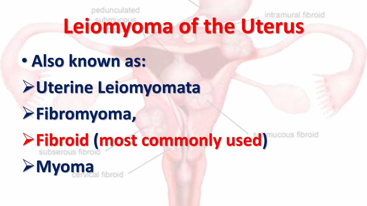
Leiomyoma of the Uterus
• Also known as:
Uterine Leiomyomata
Fibromyoma,
Fibroid (most commonly used)
Myoma
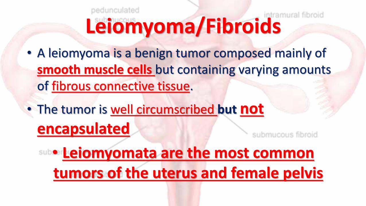
Leiomyoma/Fibroids• A leiomyoma is a benign tumor composed mainly of
smooth muscle cells but containing varying amounts of fibrous connective tissue.
• The tumor is well circumscribed but not encapsulated
• Leiomyomata are the most common tumors of the uterus and female pelvis

• It is well recognized that the incidence is much higher in black women than in white
• some studies found the incidence among black women to be three and one third times that among white women.
• There is no explanation for this racial difference.

• Leiomyomata also are larger and occur at a younger age in black women.
• However, they are uncommon in any women before 20 years of age.
• Patients with uterine leiomyomata often have a positive family history of uterine leiomyomata. This suggests the presence of a gene encoding for their development

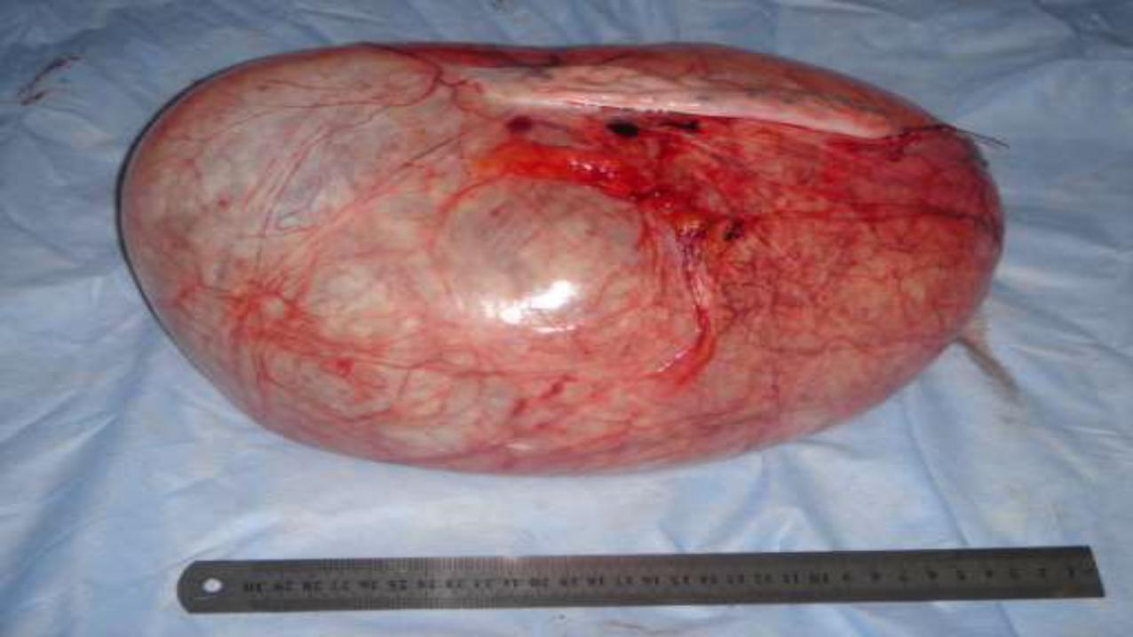
Pathology• Leiomyomas are usually multiple, discrete, and either
spherical or irregularly lobulated.
• Their pseudocapsule usually clearly demarcates them from the surrounding myometrium.
• They can be often easily and cleanly enucleated from the surrounding myometrial tissue.
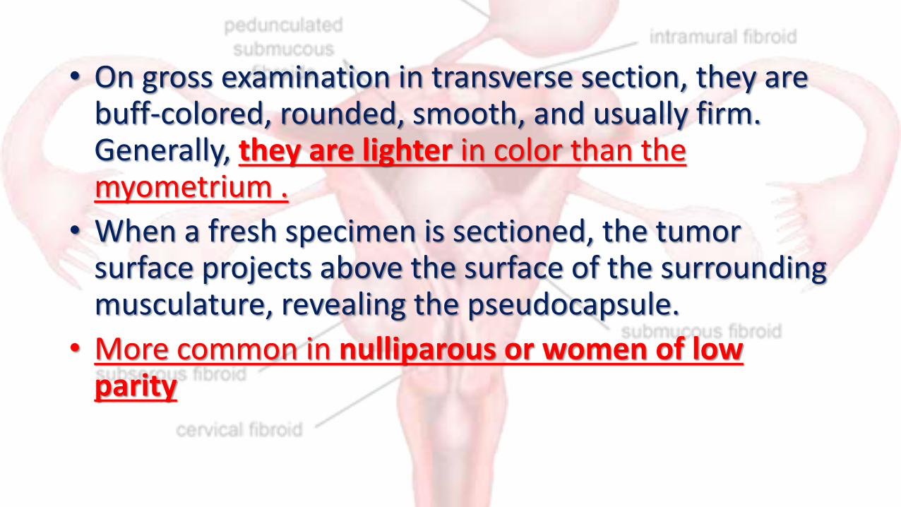
• On gross examination in transverse section, they are buff-colored, rounded, smooth, and usually firm. Generally, they are lighter in color than the myometrium .
• When a fresh specimen is sectioned, the tumor surface projects above the surface of the surrounding musculature, revealing the pseudocapsule.
• More common in nulliparous or women of low parity

Multiple myoma removed from one patient at Dodoma Hospital

Classification• Uterine leiomyomas originate in the myometrium
and are classified by anatomic location
• These anatomical locations has a tremendous effects to the clinical presentation, treatment modalities as well as future pregnancy outcomes
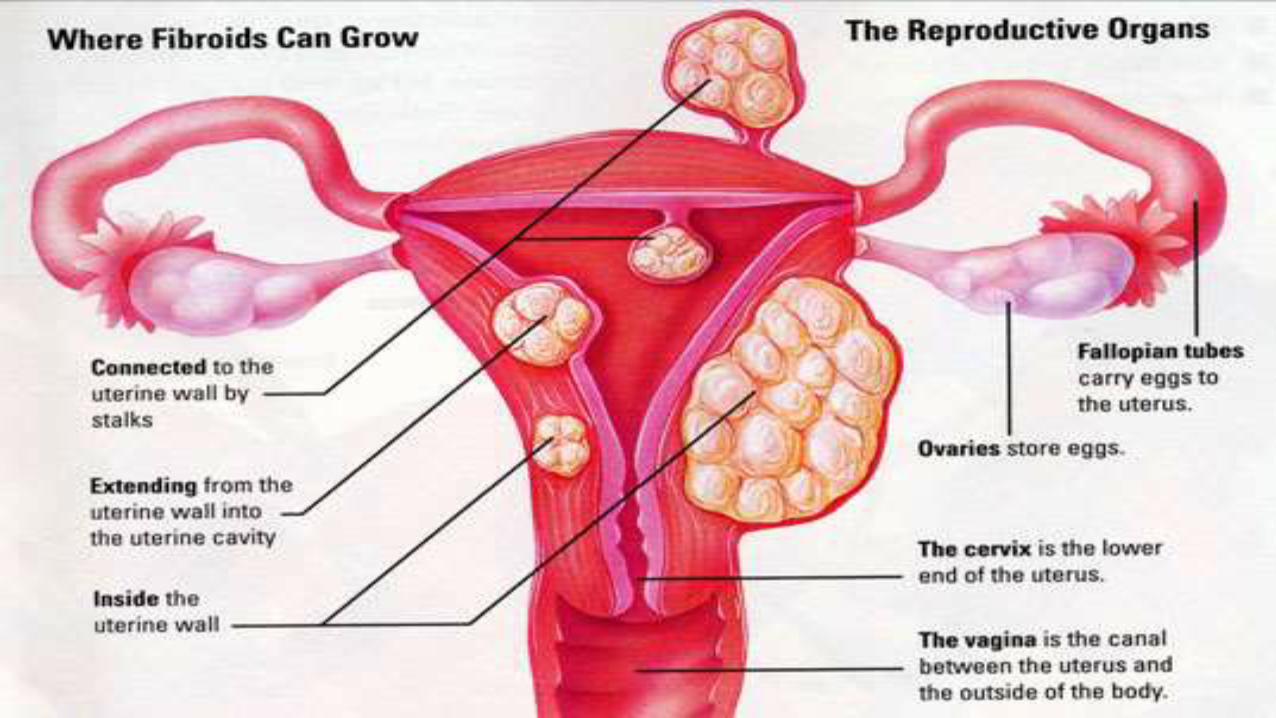
classification
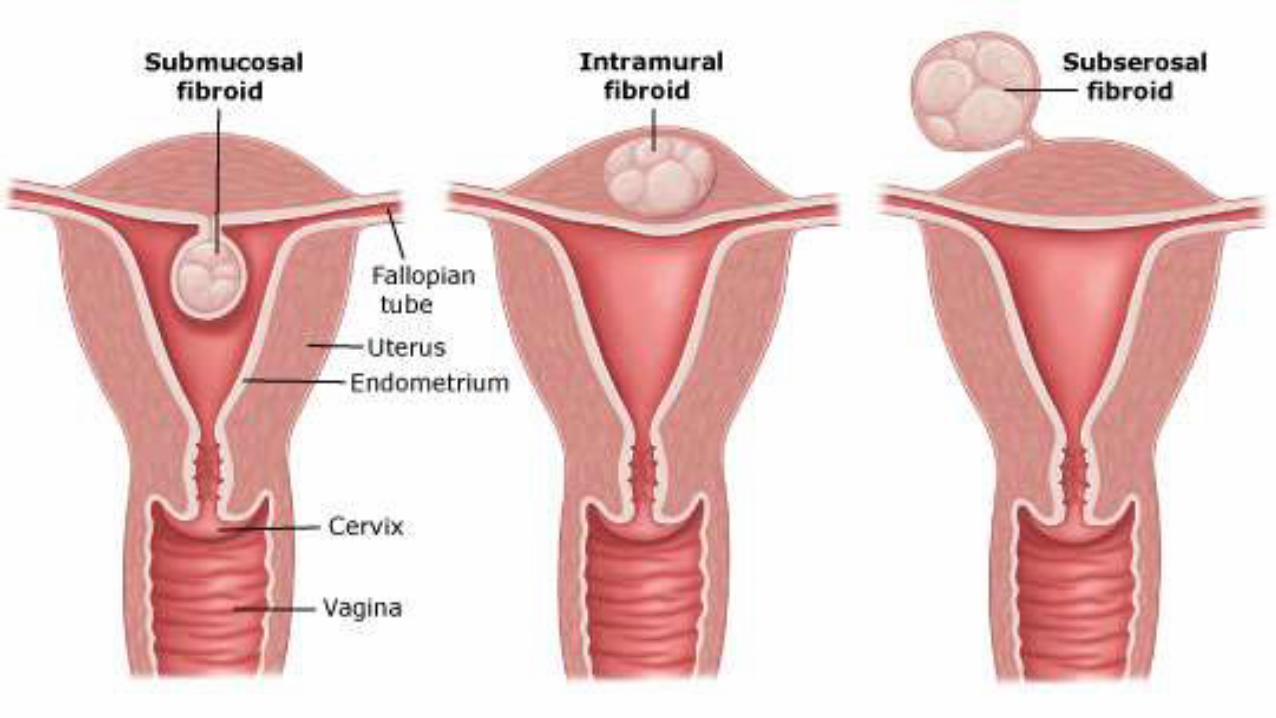

1. Submucous leiomyomas
• Submucous leiomyomas lie just beneath the endometrium and tend to compress it as they grow toward the uterine lumen.
• Their impact on the endometrium and its blood supply most often leads to irregular uterine bleeding.
• These Leiomyomata may develop pedicles and protrude fully into the uterine cavity.
• Occasionally they pass through the cervical canal while still attached within the corpus by a long stalk. When this occurs, leiomyomata are subject to torsion or infection,

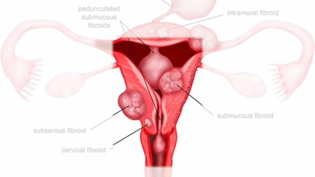

2. Intramural• Intramural or interstitial leiomyomas lie within the uterine
wall, giving it a variable consistency
These are the most common
type of leiomyomata
in women
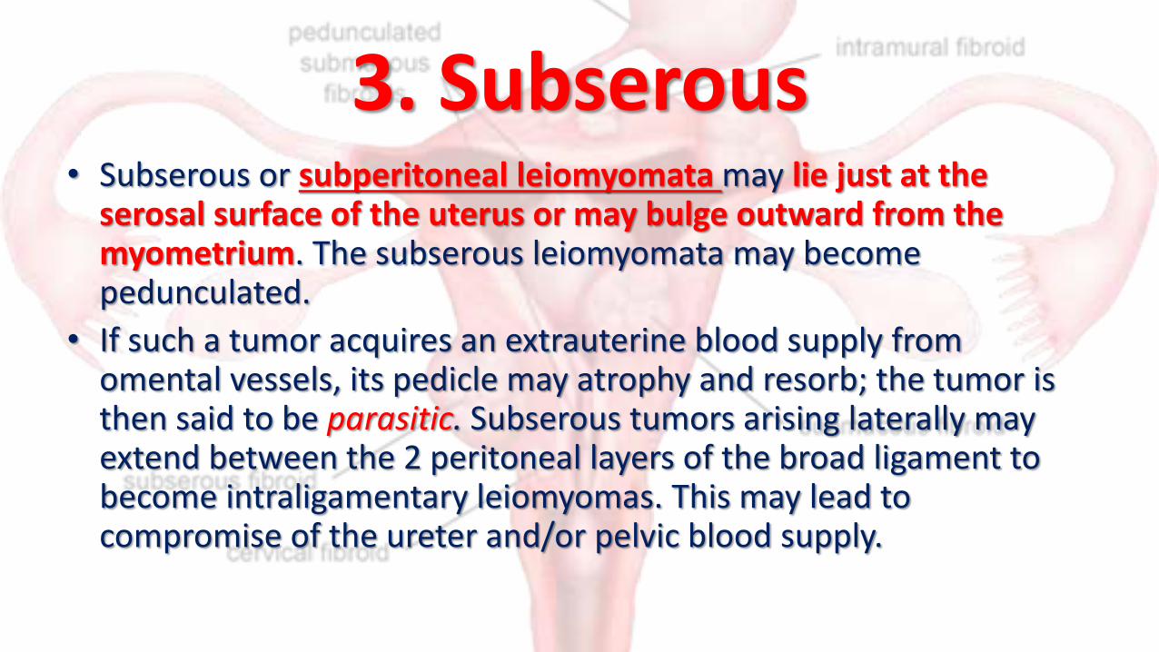
3. Subserous• Subserous or subperitoneal leiomyomata may lie just at the
serosal surface of the uterus or may bulge outward from the myometrium. The subserous leiomyomata may become pedunculated.
• If such a tumor acquires an extrauterine blood supply from omental vessels, its pedicle may atrophy and resorb; the tumor is then said to be parasitic. Subserous tumors arising laterally may extend between the 2 peritoneal layers of the broad ligament to become intraligamentary leiomyomas. This may lead to compromise of the ureter and/or pelvic blood supply.


Aetiology• Doctors don't know the cause of uterine fibroids, but research and clinical
experience point to these factors: • Genetic alterations. Many fibroids contain alterations in genes that are
different from those in normal uterine muscle cells.• Hormones. Estrogen and progesterone, two hormones that stimulate
development of the uterine lining during each menstrual cycle in preparation for pregnancy, appear to promote the growth of fibroids. Fibroids contain more estrogen and progesterone receptors than do normal uterine muscle cells.
• Other chemicals. Substances that help the body maintain tissues, such as insulin-like growth factor, may affect fibroid growth
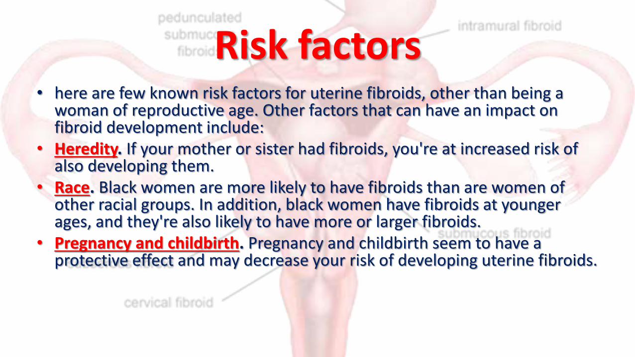
Risk factors• here are few known risk factors for uterine fibroids, other than being a
woman of reproductive age. Other factors that can have an impact on fibroid development include:
• Heredity. If your mother or sister had fibroids, you're at increased risk of also developing them.
• Race. Black women are more likely to have fibroids than are women of other racial groups. In addition, black women have fibroids at younger ages, and they're also likely to have more or larger fibroids.
• Pregnancy and childbirth. Pregnancy and childbirth seem to have a protective effect and may decrease your risk of developing uterine fibroids.
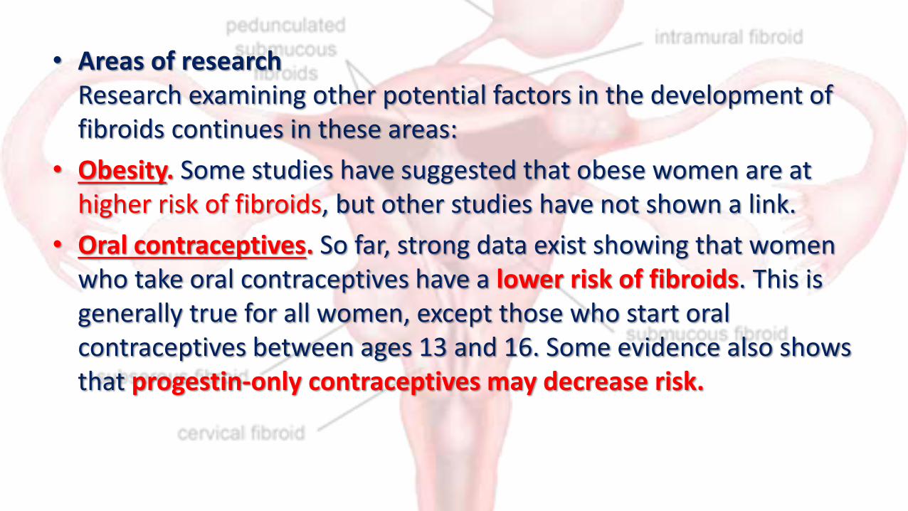
• Areas of researchResearch examining other potential factors in the development of fibroids continues in these areas:
• Obesity. Some studies have suggested that obese women are at higher risk of fibroids, but other studies have not shown a link.
• Oral contraceptives. So far, strong data exist showing that women who take oral contraceptives have a lower risk of fibroids. This is generally true for all women, except those who start oral contraceptives between ages 13 and 16. Some evidence also shows that progestin-only contraceptives may decrease risk.

Clinical Findings• Symptoms are present in only 35–50% of patients with
leiomyomas. Thus, most leiomyomata do not produce symptoms, and even very large ones may remain undetected, particularly in obese patients.
• Symptoms from leiomyomas depend on their
location,
size, state of preservation,
and whether the patient is pregnant.

Symptoms1. Abnormal Uterine Bleeding• Abnormal uterine bleeding is the most common and most
important clinical manifestation of leiomyomas, being present in up to 30% of patients.
• Most commonly, the patient has prolonged, heavy menses(menorrhagia), premenstrual spotting, or prolonged light staining following menses; however, any type of abnormal bleeding is possible.
• Minor degrees of metrorrhagia (Intermenstual bleeding) may be present.

2. Pain• Leiomyomata may cause pain when vascular compromise
occurs. Thus, pain may result from degeneration associated with vascular occlusion, infection, torsion of a pedunculated tumor, or myometrial contractions to expel a subserous myoma from the uterine cavity.
• The pain associated with infarction from torsion or red degeneration can be excruciating and produce a clinical picture consistent with acute abdomen.
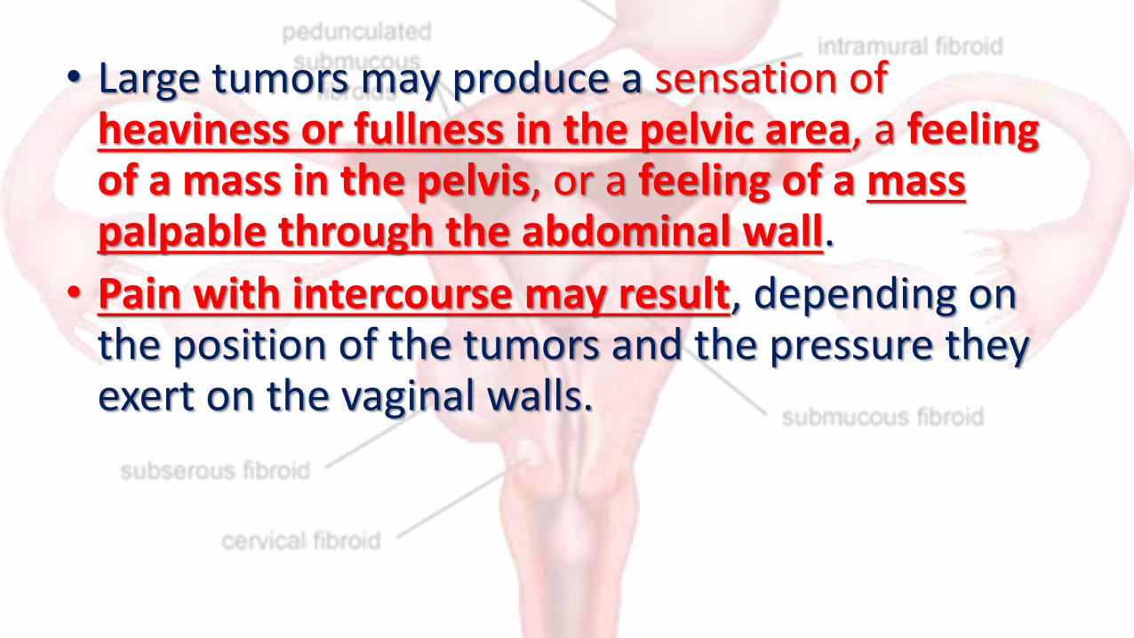
• Large tumors may produce a sensation of heaviness or fullness in the pelvic area, a feeling of a mass in the pelvis, or a feeling of a mass palpable through the abdominal wall.
• Pain with intercourse may result, depending on the position of the tumors and the pressure they exert on the vaginal walls.
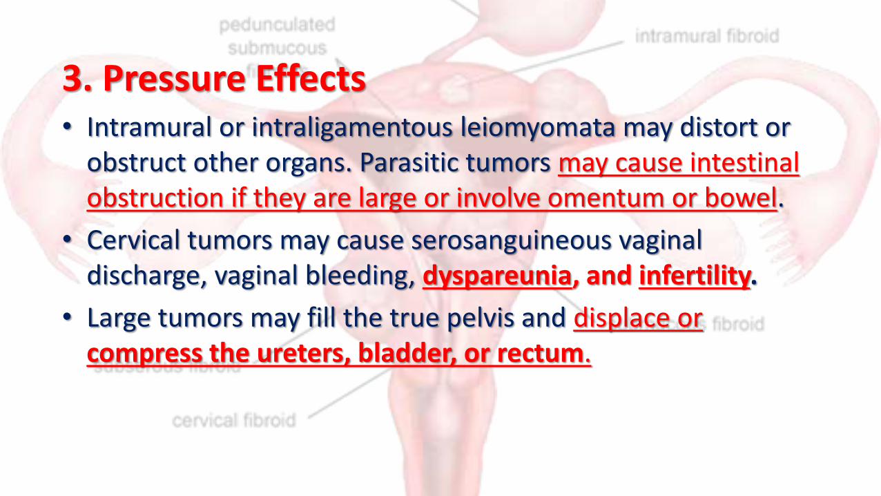
3. Pressure Effects• Intramural or intraligamentous leiomyomata may distort or
obstruct other organs. Parasitic tumors may cause intestinal obstruction if they are large or involve omentum or bowel.
• Cervical tumors may cause serosanguineous vaginal discharge, vaginal bleeding, dyspareunia, and infertility.
• Large tumors may fill the true pelvis and displace or compress the ureters, bladder, or rectum.

• Compression of surrounding structures may result in urinary symptoms or hydroureter.
• Large tumors may cause pelvic venous congestion and lower extremity edema or constipation.
• Rarely, a posterior fundal leiomyoma carries the uterus into extreme retroflexion, distorting the bladder base and causing urinary retention.
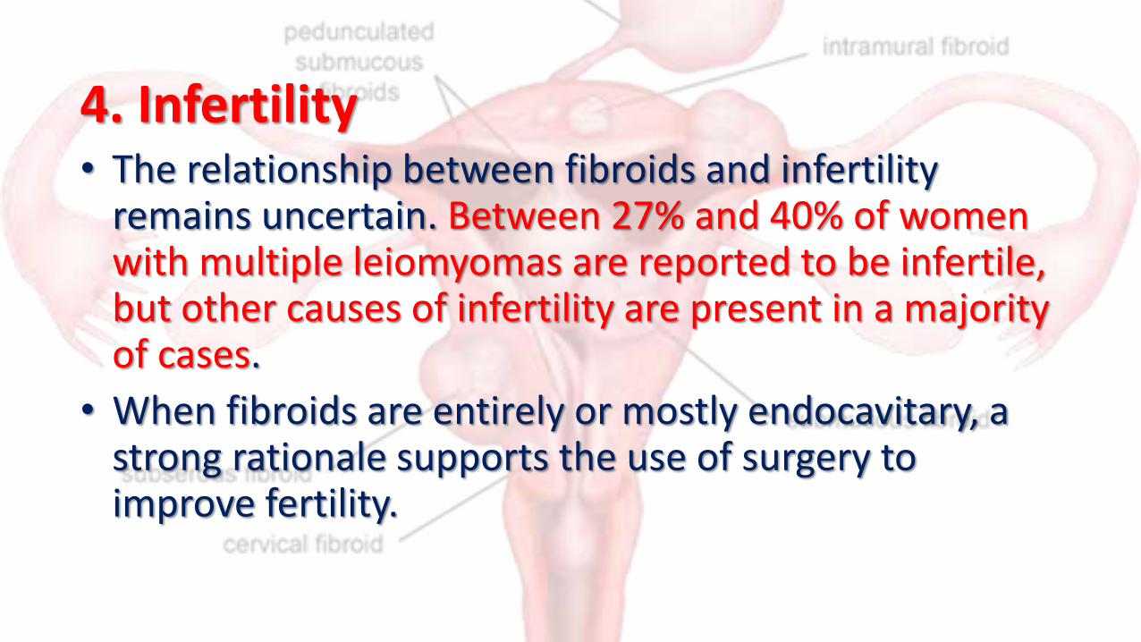
4. Infertility• The relationship between fibroids and infertility
remains uncertain. Between 27% and 40% of women with multiple leiomyomas are reported to be infertile, but other causes of infertility are present in a majority of cases.
• When fibroids are entirely or mostly endocavitary, a strong rationale supports the use of surgery to improve fertility.

5. Spontaneous Abortion• The incidence of spontaneous abortion secondary to
leiomyoma is unknown but is possibly 2 times the incidence in normal pregnant women.
• For example, the incidence of spontaneous abortion prior to myomectomy is approximately 40% and following myomectomy is approximately 20%.

Examination• Most myomas are discovered by routine bimanual
examination of the uterus or sometimes by palpation of the lower abdomen.
• The diagnosis is obvious when the normal uterine contour is distorted by 1 or more smooth, spherical, firm masses, but often it is difficult to be absolutely certain that such masses are part of the uterus

Imaging studies• A pelvic ultrasound generally assists in
establishing the diagnosis, as does excluding pregnancy as a cause of uterine enlargement. Magnetic resonance imaging (MRI) may better delineate the size and position of myomas but is not always clinically necessary.

• Pelvic ultrasound examinations are useful in confirming the diagnosis of leiomyomata. Although ultrasound should never be a substitute for a thorough pelvic examination, it can be extremely helpful in identifying leiomyomata, detailing the cause of other pelvic masses, and identifying pregnancy
• Large leiomyomata typically appear as soft tissue masses on x-ray films of the lower abdomen and pelvis; however, attention is sometimes drawn to the tumors by calcifications.

• Hysterosalpingography may be useful in detailing an intrauterine leiomyoma in the infertile patient.
• Intravenous urography may be useful in the work-up of any pelvic mass because it frequently reveals ureteral deviation or compression and identifies urinary anomalies. MRI can also be used to evaluate the urinary tract and is highly accurate in depicting the number, size, and location of leiomyomata.

Special Examinations
• Hysteroscopy may assist in identification, and may also be used for removal, of submucous leiomyomata.
• Laparoscopy is often definitive in establishing the precise origin of leiomyomata and is increasingly being used for myomectomy (see later).

Laboratory Findings• Anemia is a common consequence of leiomyomata due to
excessive uterine bleeding and depletion of iron reserves.
• However, occasional patients display erythrocytosis.
• Hematocrit levels return to normal following removal of the uterus, and elevated erythropoietin levels have been reported in such cases.

• Moreover, the recognized association of polycythemia and renal disease has led to speculation that leiomyomas compress the ureters, causing ureteral back pressure and thus inducing renal erythropoietin production.
• Leukocytosis, fever, and an elevated sedimentation rate may be present with acute degeneration or infection.

Differential Diagnosis• Any pelvic mass, including pregnancy, may be mistaken for a
leiomyoma
1. ovarian carcinoma,
2. Adenomyosis
3. tubo-ovarian abscess,
4. Endometriosis
5. Ovarian cysts or neoplasia
6. tubo-ovarian inflammatory or neoplastic masses

Treatment• Asymptomatic leiomyomas are usually managed
expectantly.
• Choice of treatment depends on the patient's symptoms, age, parity, pregnancy status, reproductive plans, and general health, as well as the size and location of the leiomyomas. Other causes of pelvic masses must be ruled out.

Watchful waiting
• Many women with uterine fibroids experience no signs or symptoms.
• Watchful waiting (expectant management) could be the best option.
• Fibroids aren't cancerous.
• They rarely interfere with pregnancy.
• They usually grow slowly — or not at all — and tend to shrink after menopause when levels of reproductive hormones drop.

Medications
• Medications for uterine fibroids target hormones that regulate your menstrual cycle, treating symptoms such as heavy menstrual bleeding and pelvic pressure. They don't eliminate fibroids, but may shrink them. Medications include:
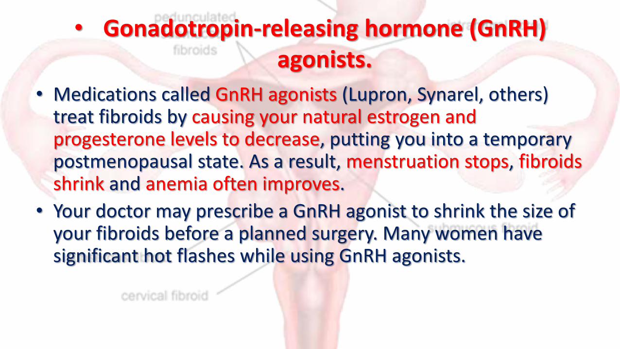
• Gonadotropin-releasing hormone (GnRH) agonists.
• Medications called GnRH agonists (Lupron, Synarel, others) treat fibroids by causing your natural estrogen and progesterone levels to decrease, putting you into a temporary postmenopausal state. As a result, menstruation stops, fibroids shrink and anemia often improves.
• Your doctor may prescribe a GnRH agonist to shrink the size of your fibroids before a planned surgery. Many women have significant hot flashes while using GnRH agonists.
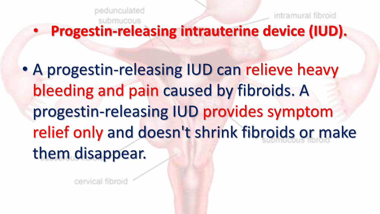
• Progestin-releasing intrauterine device (IUD).
• A progestin-releasing IUD can relieve heavy bleeding and pain caused by fibroids. A progestin-releasing IUD provides symptom relief only and doesn't shrink fibroids or make them disappear.

• Androgens.
• Danazol, a synthetic drug similar to testosterone, may effectively stop menstruation, correct anemia and even shrink fibroid tumors and reduce uterine size.
• However, this drug is rarely used to treat fibroids.
• Unpleasant side effects, such as weight gain, dysphoria (feeling depressed, anxious or uneasy), acne, headaches, unwanted hair growth and a deeper voice, make many women reluctant to take this drug

• Other medications
• Oral contraceptives or progestins can help control menstrual bleeding, but they don't reduce fibroid size.
• Nonsteroidal anti-inflammatory drugs (NSAIDs), which are not hormonal medications, may be effective in relieving pain related to fibroids, but they don't reduce bleeding caused by fibroids.

Surgical Measures
• Surgery is the mainstay of treatment of leiomyomas.
• Before definitive surgery, necessary blood volume should be replenished, and other measures such as administration of prophylactic antibiotics
• Types of surgery
1. Myomectomy
2. Hysterectomy

Myomectomy• Myomectomy is an option for the symptomatic patient who
wishes to preserve fertility or conserve the uterus.
The disadvantage is the significant risk for future leiomyomas.
Five years post myomectomy, 50–60% of patients will have new myomas detected on ultrasound, and up to 25% will require a second major surgery.
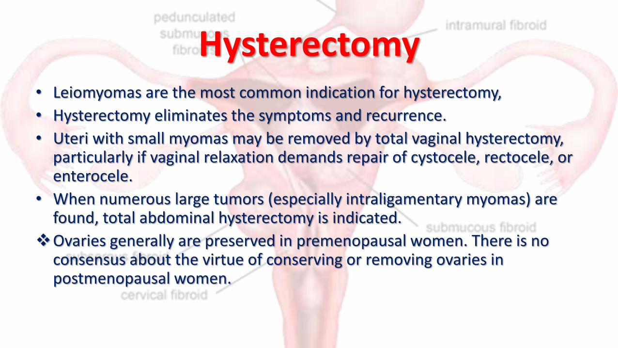
Hysterectomy• Leiomyomas are the most common indication for hysterectomy,
• Hysterectomy eliminates the symptoms and recurrence.
• Uteri with small myomas may be removed by total vaginal hysterectomy, particularly if vaginal relaxation demands repair of cystocele, rectocele, or enterocele.
• When numerous large tumors (especially intraligamentary myomas) are found, total abdominal hysterectomy is indicated.
Ovaries generally are preserved in premenopausal women. There is no consensus about the virtue of conserving or removing ovaries in postmenopausal women.

Other treatment modalities
• Uterine Fibroid Embolization
• Endometrial Ablation
• Myolysis
• Laparoscopic Uterine Artery Occlusion
• Magnetic Resonance-Guided Focused Ultrasound Surgery
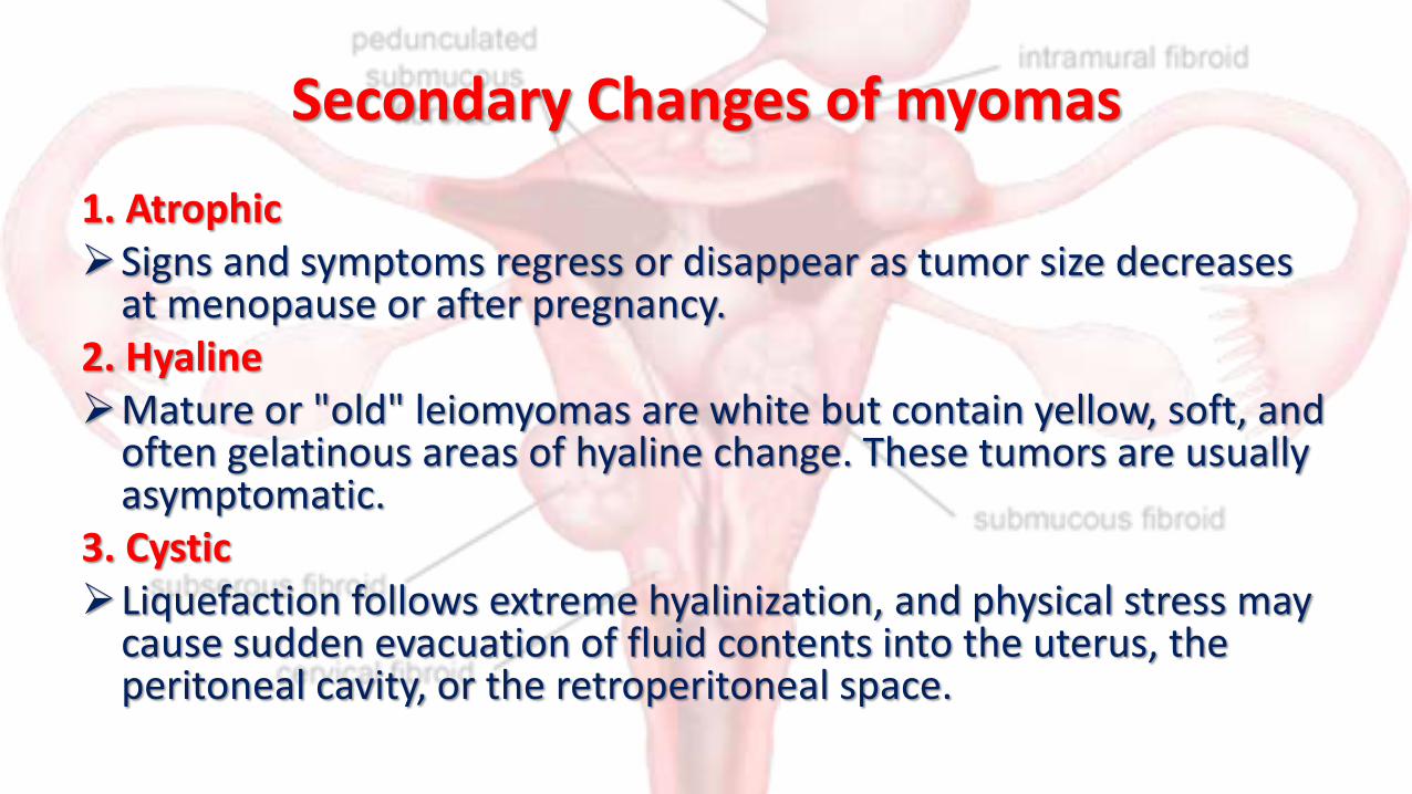
Secondary Changes of myomas
1. AtrophicSigns and symptoms regress or disappear as tumor size decreases
at menopause or after pregnancy.2. HyalineMature or "old" leiomyomas are white but contain yellow, soft, and
often gelatinous areas of hyaline change. These tumors are usually asymptomatic.
3. CysticLiquefaction follows extreme hyalinization, and physical stress may
cause sudden evacuation of fluid contents into the uterus, the peritoneal cavity, or the retroperitoneal space.

4. Calcific (Calcareous)
Subserous leiomyomata are most commonly affected by circulatory deprivation, which causes precipitation of calcium carbonate and phosphate within the tumor.
5. Septic
Circulatory inadequacy may cause necrosis of the central portion of the tumor followed by infection. Acute pain, tenderness, and fever result.

6. Carneous (Red)
Venous thrombosis and congestion with interstitial hemorrhage are responsible for the color of a leiomyoma undergoing red degeneration . During pregnancy, when carneous degeneration is most common, edema and hypertrophy of the myometrium occur.

The physiologic changes in the leiomyoma are not the same as in the myometrium; the resultant anatomic discrepancy impedes the blood supply, resulting in aseptic degeneration and infarction. The process is usually accompanied by pain but is self-limited. Potential complications of degeneration in pregnancy include preterm labor and, rarely, initiation of disseminated intravascular coagulation.
7. ?Malignant change