Embed Size (px)
Citation preview
J Cutan Pathol 2010: 37: 987–990doi: 10.1111/j.1600-0560.2009.01453.xJohn Wiley & Sons. Printed in Singapore
Copyright © 2009 John Wiley & Sons A/S
Fibrofolliculoma withancient/pseudosarcomatous features
Fibrofolliculoma is a benign skin lesion that, when multiple, can be partof the Birt-Hogg-Dube syndrome. We report on a case of solitaryfibrofolliculoma arisen on the nose of a 63-year-old woman, withpeculiar histological and immunohistochemical features. The lesion wascharacterized by the presence of bizarre multinucleated perifollicularstromal cells, positive for factor XIIIa, in a background ofCD34-positive cells, and by a peripheral population of CD34-positivespindle cells organized in fascicles haphazardly infiltrating the deepdermis, and surrounded by scattered factor XIIIa-positive dendrocytes.We consider the bizarre perifollicular cellular component as an‘ancient’ feature of fibrofolliculoma, hypothesis corroborated by theco-expression of CD34 and factor XIIIa, whereas the peripheralspindle cell fascicles represent a pseudosarcomatous proliferation ofCD34-positive cells, normally surrounding adnexal structures,stimulated by factor XIIIa-positive dendrocytes.
Cesinaro AM, Rusev BC, Kutzner H. Fibrofolliculoma withancient/pseudosarcomatous features.J Cutan Pathol 2010; 37: 987–990. © 2009 John Wiley & Sons A/S.
Anna Maria Cesinaro1, BorislavChavdarov Rusev1 andHeinz Kutzner2
1Department of Pathology, University ofModena e Reggio Emilia, Modena, Italy and2Dermatopathologische Gemeinschaftspraxis,Friedrichshafen, Germany
Anna Maria Cesinaro, MD, DipartimentoIntegrato di Servizi Diagnostici, di Laboratorio eMedicina Legale, Sezione di AnatomiaPatologica, Universita di Modena e ReggioEmilia, Policlinico, via del Pozzo 71, 41100Modena, ItalyTel: + 39 059 4224808Fax: + 39 059 4224820e-mail: [email protected]
Accepted for publication August 28, 2009
Fibrofolliculoma and its late stage version, trichodis-coma, are solitary or multiple hamartomatous cuta-neous lesions, mostly located on the face and neck ofadult patients.1 When multiple, they are part of theBirt-Hogg-Dube syndrome.2 An immunohistochem-ical study of this group of lesions has demonstrated thepositivity of perifollicular spindle cells for vimentinand CD34, but not for factor XIIIa.3,4
We describe herein a peculiar case of fibrofollicu-loma, solitary type, showing perifollicular bizarre andmultinucleated cells, positive for factor XIIIa, in abackground of CD34-positive cells, and a peripheralpopulation of spindle cells CD34-positive, infiltrat-ing the reticular dermis. Additionally, we propose anexplanation of these peculiar histologic and immuno-histochemical features.
Case reportA 63-year-old woman sought medical attentionbecause of a nodule present on the right pinna ofthe nose for several months. The lesion was solitary,1.2 × 1 cm in diameter and showed a skin-colored,
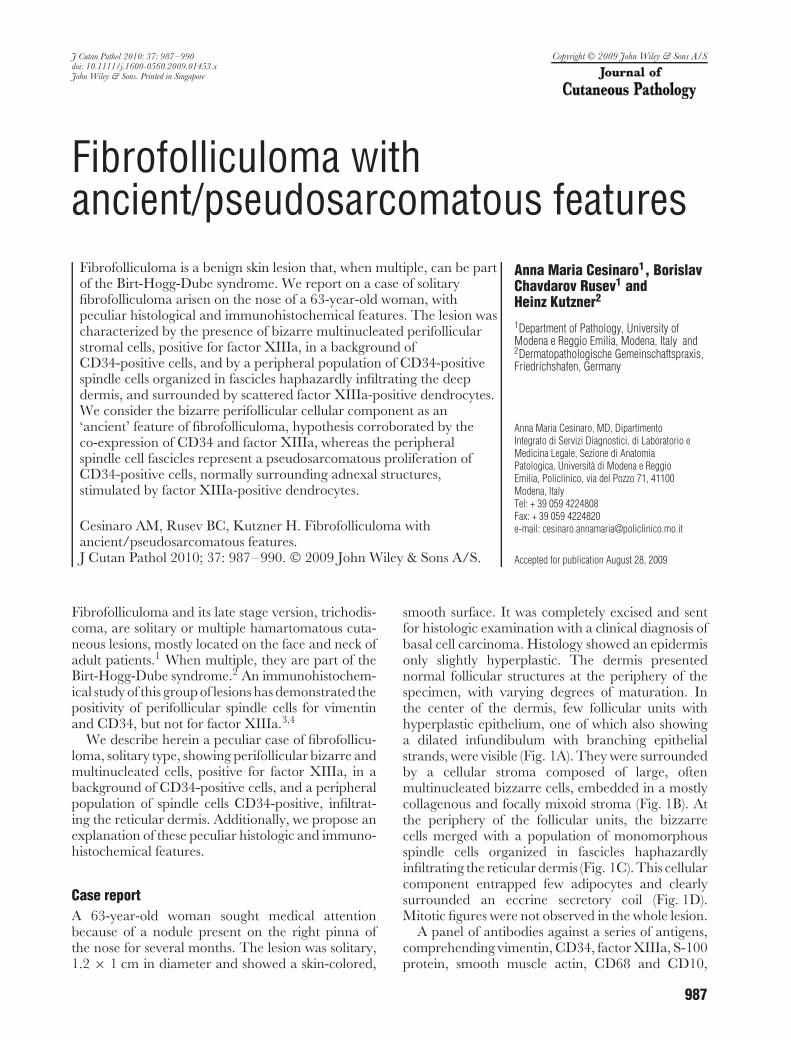
smooth surface. It was completely excised and sentfor histologic examination with a clinical diagnosis ofbasal cell carcinoma. Histology showed an epidermisonly slightly hyperplastic. The dermis presentednormal follicular structures at the periphery of thespecimen, with varying degrees of maturation. Inthe center of the dermis, few follicular units withhyperplastic epithelium, one of which also showinga dilated infundibulum with branching epithelialstrands, were visible (Fig. 1A). They were surroundedby a cellular stroma composed of large, oftenmultinucleated bizzarre cells, embedded in a mostlycollagenous and focally mixoid stroma (Fig. 1B). Atthe periphery of the follicular units, the bizzarrecells merged with a population of monomorphousspindle cells organized in fascicles haphazardlyinfiltrating the reticular dermis (Fig. 1C). This cellularcomponent entrapped few adipocytes and clearlysurrounded an eccrine secretory coil (Fig. 1D).Mitotic figures were not observed in the whole lesion.
A panel of antibodies against a series of antigens,comprehending vimentin, CD34, factor XIIIa, S-100protein, smooth muscle actin, CD68 and CD10,
987
Cesinaro et al.
Fig. 1. Overview of the lesion showing a dilated infundibulum with branching epithelial strands (A) surrounded by bizarre, often multinucleatedstromal cells (B) merging with a peripheral population of spindle cells organized in fascicles (C) infiltrating the deep dermis and surroundingan eccrine secretory coil (D).
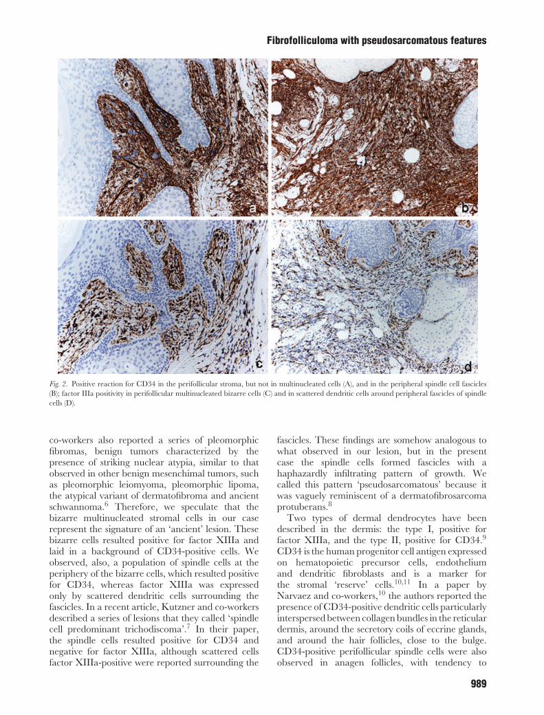
was applied to better characterize the nature ofthe lesion. Vimentin was diffusely positive in thewhole specimen. Immunohistochemical reactionsfor actin, CD68 and S-100 protein were negativein the lesion. CD10 marked spindle cells aroundnormal follicular structures and eccrine glands,whereas the bizarre cells and the spindle cellsorganized in fascicles resulted unstained. CD34strongly decorated the entire cellular componentaround the follicles, with the exception of the bizarrecells (Fig. 2A), and the spindle cells organized infascicles at the periphery (Fig. 2B). Factor XIIIaresulted expressed by the perifollicular bizarre cells(Fig. 2C), whereas it was negative in the peripheralspindle cell component, although scattered positivedendritic cells surrounding the fascicles were visible(Fig. 2D).
Because of the overall configuration, a diagnosisof fibrofolliculoma, solitary type, was rendered,and, given the presence of peculiar features,we christened the lesion as ‘fibrofolliculoma with
ancient/pseudosarcomatous features’. The patientand her family were investigated to exclude the Birt-Hogg-Dube syndrome, with negative results. Sheresulted in good health and after 6 months she hadnot developed local recurrence.
DiscussionWe report on a case of solitary fibrofolliculoma arisenon the nose of a woman, in which peculiar histologicaland immunohistochemical features were observed.The lesion was characterized by the presence of twopopulations of specialized stromal cells: the first one,with strictly perifollicular distribution, was composedof cells, several of which with multinucleation andbizarre nuclei; the second, peripherally located, wasmade up of monomorphous spindle cells organizedin fascicles, merging with the multinucleated bizarrecells. The presence of multinucleated nuclei has beenreported in trichodiscoma,5 that is considered thelate stage version of a fibrofolliculoma.1 Kamino and
988
Fibrofolliculoma with pseudosarcomatous features
Fig. 2. Positive reaction for CD34 in the perifollicular stroma, but not in multinucleated cells (A), and in the peripheral spindle cell fascicles(B); factor IIIa positivity in perifollicular multinucleated bizarre cells (C) and in scattered dendritic cells around peripheral fascicles of spindlecells (D).
co-workers also reported a series of pleomorphicfibromas, benign tumors characterized by thepresence of striking nuclear atypia, similar to thatobserved in other benign mesenchimal tumors, suchas pleomorphic leiomyoma, pleomorphic lipoma,the atypical variant of dermatofibroma and ancientschwannoma.6 Therefore, we speculate that thebizarre multinucleated stromal cells in our caserepresent the signature of an ‘ancient’ lesion. Thesebizarre cells resulted positive for factor XIIIa andlaid in a background of CD34-positive cells. Weobserved, also, a population of spindle cells at theperiphery of the bizarre cells, which resulted positivefor CD34, whereas factor XIIIa was expressedonly by scattered dendritic cells surrounding thefascicles. In a recent article, Kutzner and co-workersdescribed a series of lesions that they called ‘spindlecell predominant trichodiscoma’.7 In their paper,the spindle cells resulted positive for CD34 andnegative for factor XIIIa, although scattered cellsfactor XIIIa-positive were reported surrounding the
fascicles. These findings are somehow analogous towhat observed in our lesion, but in the presentcase the spindle cells formed fascicles with ahaphazardly infiltrating pattern of growth. Wecalled this pattern ‘pseudosarcomatous’ because itwas vaguely reminiscent of a dermatofibrosarcomaprotuberans.8
Two types of dermal dendrocytes have beendescribed in the dermis: the type I, positive forfactor XIIIa, and the type II, positive for CD34.9
CD34 is the human progenitor cell antigen expressedon hematopoietic precursor cells, endotheliumand dendritic fibroblasts and is a marker forthe stromal ‘reserve’ cells.10,11 In a paper byNarvaez and co-workers,10 the authors reported thepresence of CD34-positive dendritic cells particularlyinterspersed between collagen bundles in the reticulardermis, around the secretory coils of eccrine glands,and around the hair follicles, close to the bulge.CD34-positive perifollicular spindle cells were alsoobserved in anagen follicles, with tendency to
989
Cesinaro et al.
disappear in catagen-telogen follicles.12 Factor XIIIais expressed by dermal dendritic cells located inthe upper reticular and papillary dermis.13 Duringembryogenesis, it has been found in cells differentfrom macrophages and lacking CD68 expression,thus probably originating from primitive CD34-positive mesenchymal cells.14 This hypothesis seemsto be confirmed by the observation of tumors withco-expression of CD34 and factor XIIIa.9 In our caseof fibrofolliculoma, we found immunohistochemicalexpression of factor XIIIa in the bizarre perifollicularcells in a background of CD34-positive spindle cells,further corroborating the hypothesis of a ‘temporal’link between the two types of dendrocytes. Indeed,we think that the expression of factor XIIIa issubsequent to the ‘senescence’ of the CD34+ stromalcells.
The peripheral spindle cell component infiltratingthe deep dermis expressed CD34. We explain thesefeatures by the fact that these cells derivate from theperifollicular and the perieccrine spindle cells, knownto be CD34 positive. Moreover, the presence of factorXIIIa+ dendrocytes, interspersed among fascicles ofCD34+ spindle cells, recalls similar observationsin certain mesenchymal tumors, with dendrocyteshaving a stimulating role in the proliferation ofspindle cells.9
In conclusion, the peculiarity of our case offibrofolliculoma relies on the co-expression ofCD34 and factor XIIIa in the perifollicularspecialized stromal cells, unreported until now infibrofolliculomas, and supporting a common originfor the two types of dermal dendritic cells, or, atleast, a possible transition from one type to theother, depending on the evolution of the lesion.The second peculiar feature is the presence of apseudosarcomatous spindle cells component CD34-positive haphazardly infiltrating the deep dermis,that we considered a benign proliferation of thespecialized periappendageal stromal cells.
AcknowledgementsThe authors would like to thank Drs Hantschke, Ruetten, Mentzel,Schaerer and Paredes (Dermatopathologische Gemeinschaftspraxis,Friedrichshafen, Germany) for their contribution to the discussionof the case.
References1. Ackerman AB, Reddy VB, Soyer HP. Fibrofolliculoma and
trichodiscoma. In Ackerman AB, Reddy VB, Soyer HP, eds.Neoplasma with follicular differentiation. New York: ArdorScribendi, 2001; 221.
2. Birt AR, Hogg GR, Dube WJ. Hereditary multiple fibrofollicu-loma with trichodiscoma and acrochordons. Arch Dermatol1977; 113: 1674.
3. Collins GL, Somach S, Morgan MB. Histomorphologic andimmunophenotypic analysis of fibrofolliculomas and trichodis-comas in Birt-Hogg-Dube syndrome and sporadic disease. JCutan Pathol 2002; 29: 529.
4. Schulz T, Ebschner U, Hartschuh W. Localized Birt-Hogg-Dube syndrome with prominent perivascular fibromas. AmJ Dermatopathol 2001; 23: 149.
5. Starink TM, Kisch LS, Meijer CJLM. Familiar multipletrichodiscomas: a clinicopathologic study. Arch Dermatol 1985;121: 888.
6. Kamino H, Lee JY, Berke A. Pleomorphic fibroma of the skin:a benign neoplasm with cytologic atypia. A clinicopathologicstudy of eight cases. Am J Surg Pathol 1989; 13: 107.
7. Kutzner H, Requena L, Rutten A, Mentzel T. Spindle cellpredominant trichodiscoma: a fibrofolliculoma/trichodiscomavariant considered formerly to be a neurofollicular hamartoma.A clinicopathological and immunohistochemical analysis of 17cases. Am J Dermatopathol 2006; 28: 1.
8. Fletcher CDM, Evans BJ, Macartney JC, et al. Dermatofi-brosarcoma protuberans: a clinicopathological and immunohis-tochemical study with a review of the literature. Histopathology1985; 9: 921.
9. Silverman JS, Tamsen A. CD34 and Factor XIIIa positivemicrovascular dendritic cells and the family of fibrohistocyticmesenchimal tumors. Am J Dermatopathol 1998; 20: 533.
10. Narvaez D, Kanikatis J, Faure M, Claudy A. Immunohisto-chemical study of CD34-positive dendritic cells of humandermis. Am J Dermatopathol 1996; 18: 283.
11. Nickoloff BJ. The human progenitor cell antigen (CD34) islocalized on endothelial cells, dermal dendritic cells, andperifollicular cells in formalin-fixed normal skin, and onproliferating endothelials cells and stromal spindle-shaped cellsin Kaposi’s sarcoma. Arch Dermatol 1991; 127: 523.
12. Poblet E, Jimenez F. CD10 and CD34 in fetal and adult humanhair follicles: dynamic changes in their immunohistochemicalexpression during embryogenesis and hair cycling. Br JDermatol 2008; 159: 646.
13. Cerio R, Griffiths CEM, Cooper KD, et al. Characterizationof factor XIIIa positive dermal dendritic cells in normal andinflamed skin. Br J Dermatol 1989; 121: 421.
14. Trimble CL, Gray MH, McNutt NS. The distribution of FactorXIIIa-positive cells in the human fetus, placenta, and postnataltissues. Virchows Arch A Pathol Anat Histopathol 1992;420: 513.
990