Embed Size (px)
Citation preview
Fibrocartilaginous embolic myelopathy and traumatic IVDE
Luisa De Risio DVM, MRCVS, PhD, Dipl ECVN,
RCVS recognised specialist in veterinary neurology Head of Neurology/ Neurosurgery Animal Health Trust
Overview
Pathophysiology
Clinical presentation
Diagnostic investigations
Treatment
Prognosis
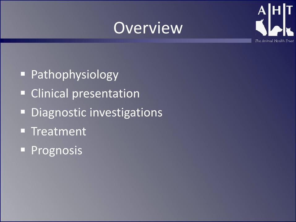
What is fibrocartilaginous embolic myelopathy (FCEM)?
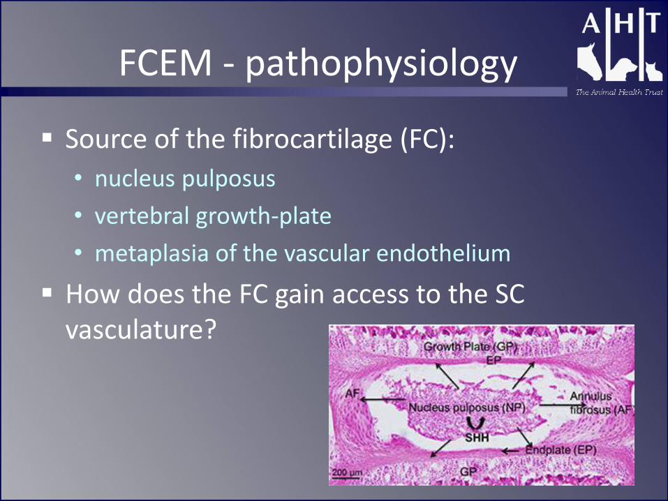
FCEM - pathophysiology
Source of the fibrocartilage (FC):
• nucleus pulposus
• vertebral growth-plate
• metaplasia of the vascular endothelium
How does the FC gain access to the SC vasculature?
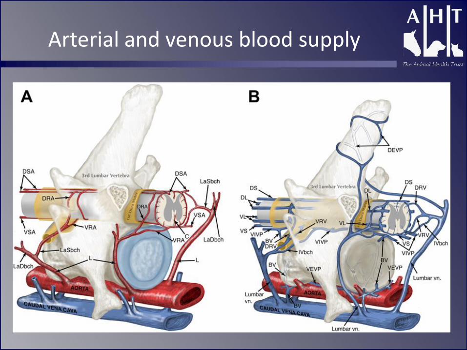
Arterial and venous blood supply
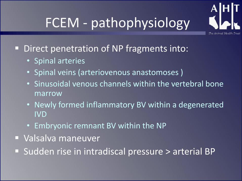
FCEM - pathophysiology
Direct penetration of NP fragments into: • Spinal arteries
• Spinal veins (arteriovenous anastomoses )
• Sinusoidal venous channels within the vertebral bone marrow
• Newly formed inflammatory BV within a degenerated IVD
• Embryonic remnant BV within the NP
Valsalva maneuver
Sudden rise in intradiscal pressure > arterial BP
FCEM - Clinical presentation
peracute (<6 hours) onset of nonprogressive and nonpainful (after the first 24 hours) and often lateralized neurological deficits (ND)
physical activity at onset of ND in up to 80% of dogs
sudden and transient hyperalgesia at the onset of ND in up to 61.5% of dogs
lateralisation of ND in 52.8% to 86.5% of dogs
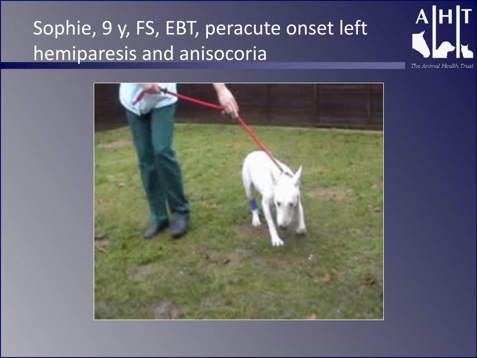
Sophie, 9 y, FS, EBT, peracute onset left hemiparesis and anisocoria
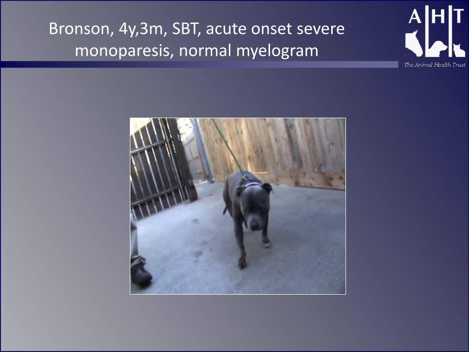
Bronson, 4y,3m, SBT, acute onset severe monoparesis, normal myelogram
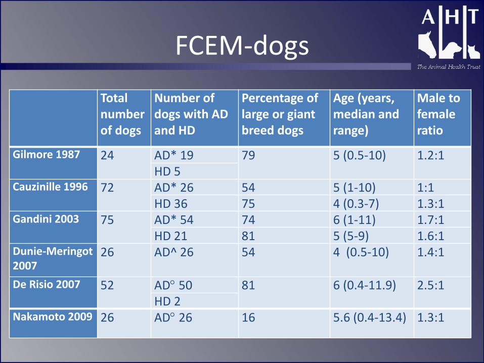
FCEM-dogs
Total number of dogs
Number of dogs with AD and HD
Percentage of large or giant breed dogs
Age (years, median and range)
Male to female ratio
Gilmore 1987 24 AD* 19 79 5 (0.5-10) 1.2:1
HD 5 Cauzinille 1996 72 AD* 26 54 5 (1-10) 1:1
HD 36 75 4 (0.3-7) 1.3:1 Gandini 2003 75 AD* 54 74 6 (1-11) 1.7:1
HD 21 81 5 (5-9) 1.6:1 Dunie-Meringot 2007
26 AD^ 26 54 4 (0.5-10) 1.4:1
De Risio 2007 52 AD 50 81 6 (0.4-11.9) 2.5:1
HD 2 Nakamoto 2009 26 AD 26 16 5.6 (0.4-13.4) 1.3:1
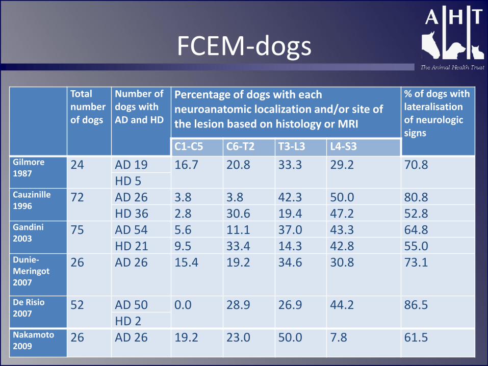
FCEM-dogs
Total number of dogs
Number of dogs with AD and HD
Percentage of dogs with each neuroanatomic localization and/or site of the lesion based on histology or MRI
% of dogs with lateralisation of neurologic signs
C1-C5 C6-T2 T3-L3 L4-S3
Gilmore 1987
24 AD 19 16.7 20.8 33.3 29.2 70.8
HD 5 Cauzinille 1996
72 AD 26 3.8 3.8 42.3 50.0 80.8
HD 36 2.8 30.6 19.4 47.2 52.8 Gandini 2003
75 AD 54 5.6 11.1 37.0 43.3 64.8
HD 21 9.5 33.4 14.3 42.8 55.0 Dunie-Meringot 2007
26 AD 26 15.4 19.2 34.6 30.8 73.1
De Risio 2007
52 AD 50 0.0 28.9 26.9 44.2 86.5
HD 2 Nakamoto 2009
26 AD 26 19.2 23.0 50.0 7.8 61.5
Differential diagnoses
IM due to other sources of emboli
Acute non compressive NP extrusion
IVD extrusion (compressive)
haemorrhage (eg, secondary to coagulopathy)
neoplasia (intra- and extramedullary)
infectious and immune-mediated focal myelitis or meningomyelitis
vertebral fracture, subluxation/luxation
FCEM- Diagnosis
Definitive diagnosis only histological
Survey radiographs • rule out vertebral fracture, sub-luxation/ luxation, neoplasia and
osteomyelitis/ discospondylitis
Myelography • rule out compressive SC disease (IVD extrusion, neoplasia)
• intramedullary pattern
CT or CT- myelography • rule out compressive SC disease (IVD extrusion, neoplasia)
• intramedullary pattern
MRI • diagnostic imaging modality of choice
CSF
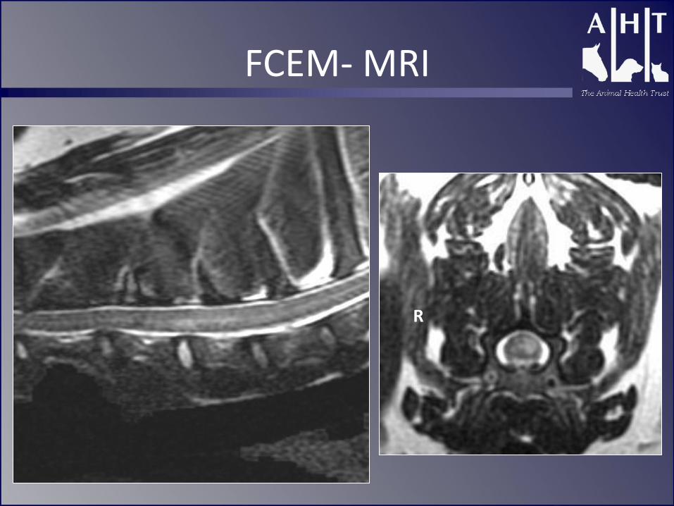
FCEM- MRI
R
FCEM- CSF
Normal
Aspecific abnormalities: • xanthochromia
• mild to moderate pleocytosis (7–84 WBC/ul)
• elevated protein concentration (reported in up to 46% of dogs with HD of FCEM and in 44-75% of dogs with AD of FCEM)
FCEM- Treatment
Nursing care
Physical rehabilitation
Neuroprotection
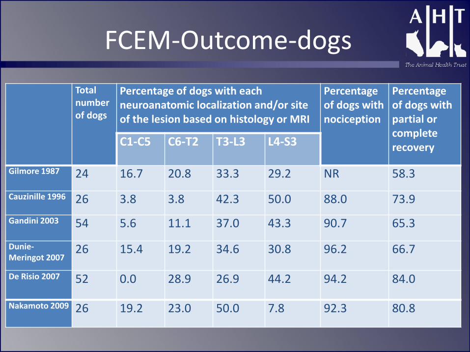
FCEM-Outcome-dogs
Total number of dogs
Percentage of dogs with each neuroanatomic localization and/or site of the lesion based on histology or MRI
Percentage of dogs with nociception
Percentage of dogs with partial or complete recovery
C1-C5 C6-T2 T3-L3 L4-S3
Gilmore 1987 24 16.7 20.8 33.3 29.2 NR 58.3
Cauzinille 1996 26 3.8 3.8 42.3 50.0 88.0 73.9
Gandini 2003 54 5.6 11.1 37.0 43.3 90.7 65.3
Dunie-Meringot 2007
26 15.4 19.2 34.6 30.8 96.2 66.7
De Risio 2007 52 0.0 28.9 26.9 44.2 94.2 84.0
Nakamoto 2009 26 19.2 23.0 50.0 7.8 92.3 80.8
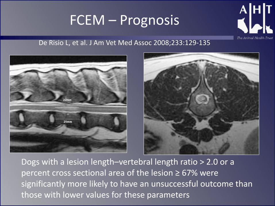
FCEM – Prognosis
De Risio L, et al. J Am Vet Med Assoc 2008;233:129-135
Dogs with a lesion length–vertebral length ratio > 2.0 or a percent cross sectional area of the lesion ≥ 67% were significantly more likely to have an unsuccessful outcome than those with lower values for these parameters
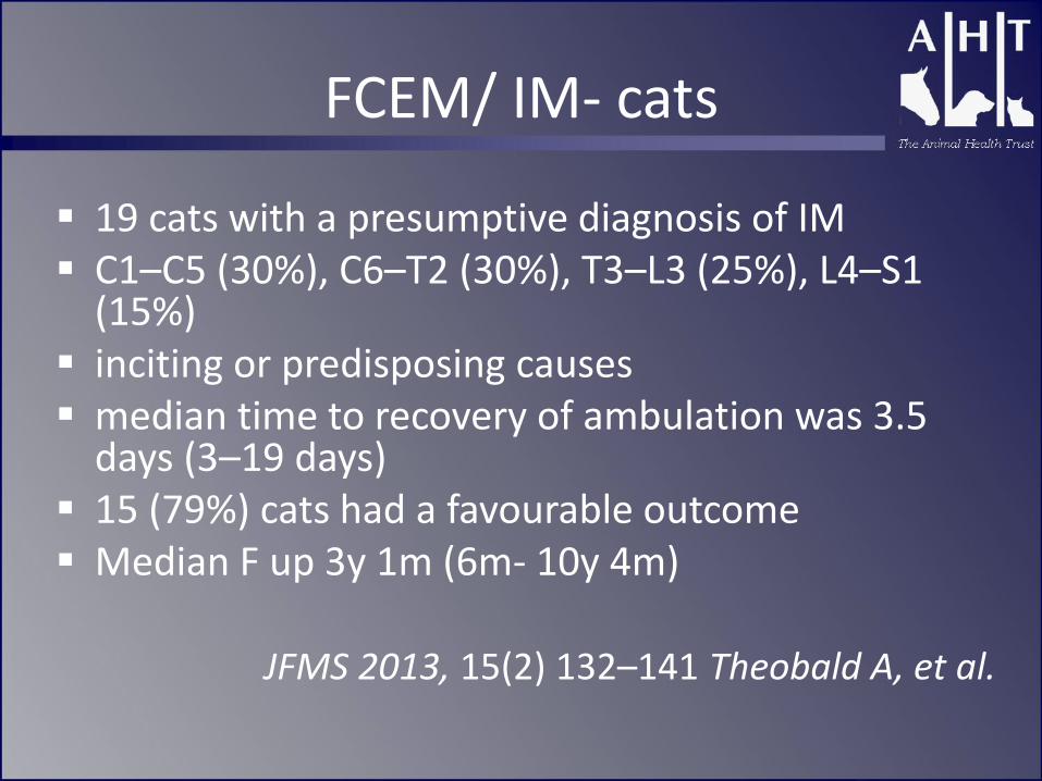
FCEM/ IM- cats
19 cats with a presumptive diagnosis of IM C1–C5 (30%), C6–T2 (30%), T3–L3 (25%), L4–S1
(15%) inciting or predisposing causes median time to recovery of ambulation was 3.5
days (3–19 days) 15 (79%) cats had a favourable outcome Median F up 3y 1m (6m- 10y 4m) JFMS 2013, 15(2) 132–141 Theobald A, et al.
Acute non compressive nucleus pulposus extrusion (ANNPE)
traumatic disc extrusion
traumatic disc prolapse
dorsolateral intervertebral disc “explosion”
high-velocity–low volume disc extrusion
Hansen type III intervertebral disc disease
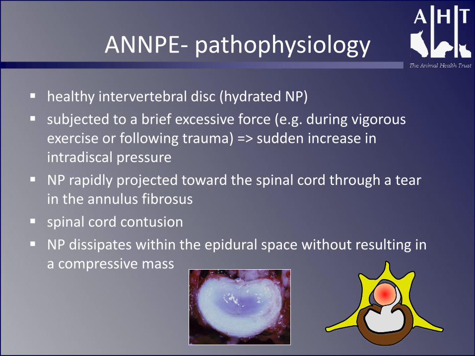
ANNPE- pathophysiology
healthy intervertebral disc (hydrated NP)
subjected to a brief excessive force (e.g. during vigorous exercise or following trauma) => sudden increase in intradiscal pressure
NP rapidly projected toward the spinal cord through a tear in the annulus fibrosus
spinal cord contusion
NP dissipates within the epidural space without resulting in a compressive mass
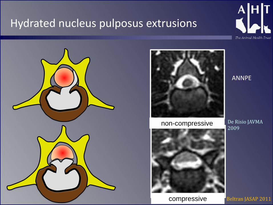
Hydrated nucleus pulposus extrusions
non-compressive
compressive Beltran JASAP 2011
De Risio JAVMA 2009
ANNPE
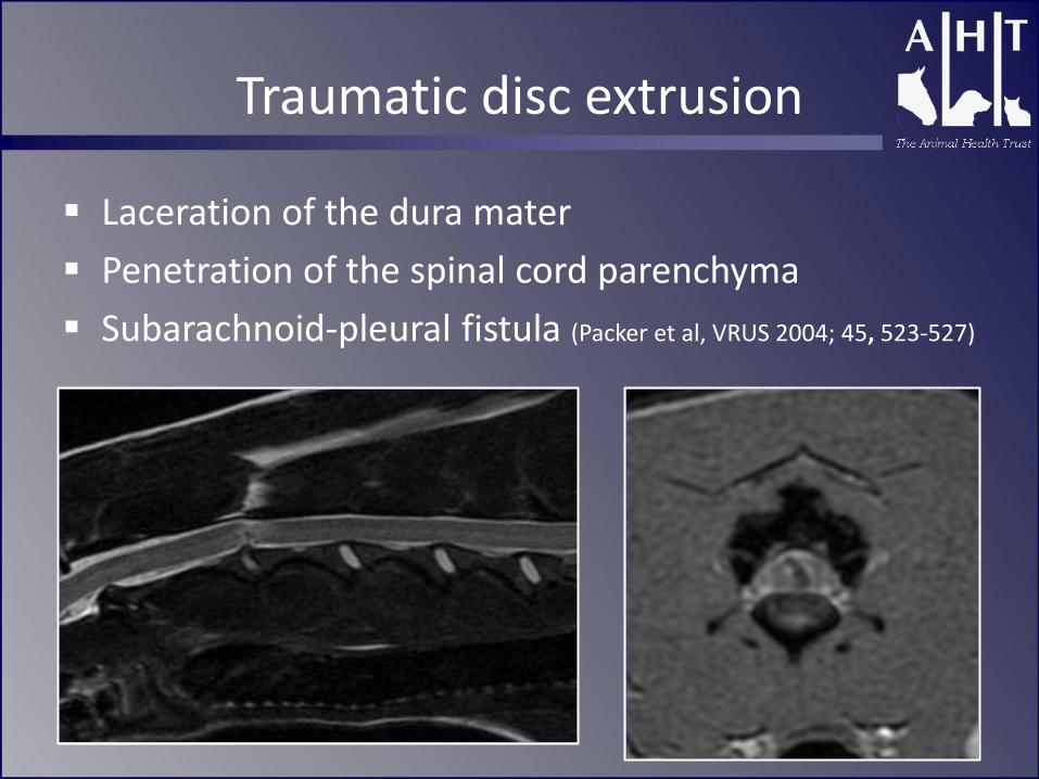
Traumatic disc extrusion
Laceration of the dura mater
Penetration of the spinal cord parenchyma
Subarachnoid-pleural fistula (Packer et al, VRUS 2004; 45, 523-527)
ANNPE- Clinical presentation
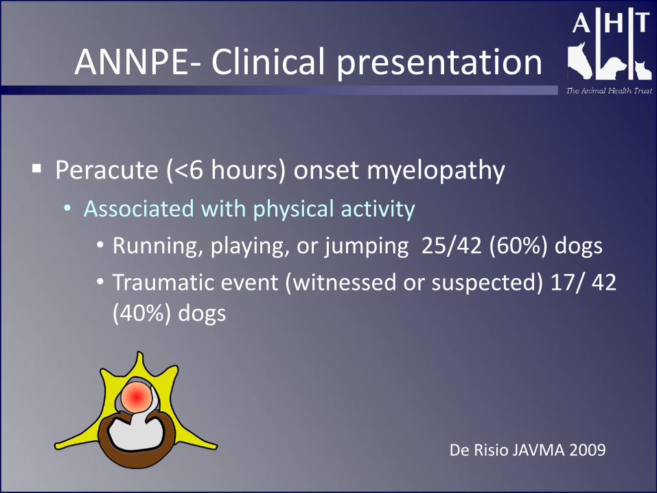
Peracute (<6 hours) onset myelopathy
• Associated with physical activity
• Running, playing, or jumping 25/42 (60%) dogs
• Traumatic event (witnessed or suspected) 17/ 42 (40%) dogs
De Risio JAVMA 2009
ANNPE- Clinical presentation
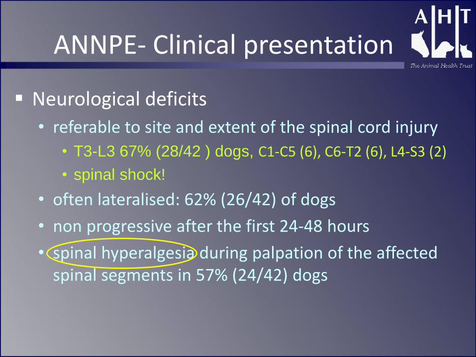
Neurological deficits
• referable to site and extent of the spinal cord injury
• T3-L3 67% (28/42 ) dogs, C1-C5 (6), C6-T2 (6), L4-S3 (2)
• spinal shock!
• often lateralised: 62% (26/42) of dogs
• non progressive after the first 24-48 hours
• spinal hyperalgesia during palpation of the affected spinal segments in 57% (24/42) dogs
3y- 5m- old, M, Boxer
peracute onset difficulty ambulating with PLs (L>R) while
playing in the garden 7 hrs before presentation
video

ANNPE- Diagnosis
Definitive diagnosis only histological
Survey radiographs • rule out vertebral fracture, sub-luxation/ luxation, neoplasia and
osteomyelitis/ discospondylitis
Myelography • rule out compressive SC disease (IVD extrusion, neoplasia)
• intramedullary pattern above a collapsed intervertebral disc space
CT or CT- myelography • rule out compressive SC disease (IVD extrusion, neoplasia)
• intramedullary pattern above a collapsed intervertebral disc space
MRI • diagnostic imaging modality of choice
CSF
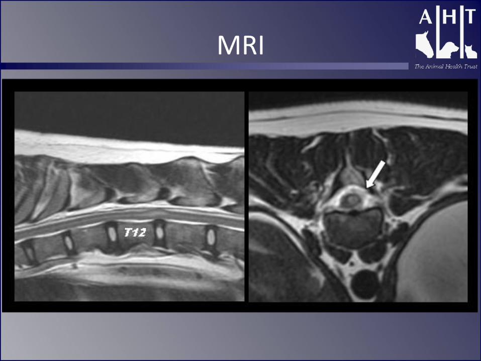
MRI
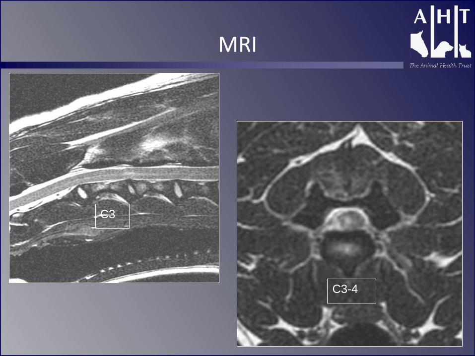
MRI
C3
C3-4
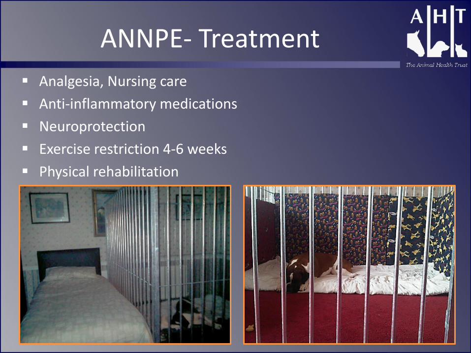
ANNPE- Treatment
Analgesia, Nursing care
Anti-inflammatory medications
Neuroprotection
Exercise restriction 4-6 weeks
Physical rehabilitation
ANNPE- Outcome
Outcome successful in 28/42 (67%) dogs and unsuccessful in 14/42 (33%) dogs
Prognostic factors: • loss of nociception
• on univariate analysis • extent of the intramedullary hyperintensity on sagittal
and transverse T2-weighted MR images
• detection of intramedullary hypointensity on T2* GE images
De Risio JAVMA 2009
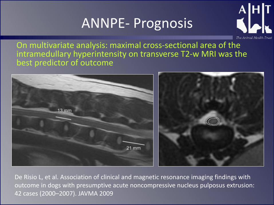
ANNPE- Prognosis
On multivariate analysis: maximal cross-sectional area of the intramedullary hyperintensity on transverse T2-w MRI was the best predictor of outcome
De Risio L, et al. Association of clinical and magnetic resonance imaging findings with outcome in dogs with presumptive acute noncompressive nucleus pulposus extrusion: 42 cases (2000–2007). JAVMA 2009
THANK YOU FOR YOUR ATTENTION