Embed Size (px)
Citation preview

Fetal Growth Fetal Growth RestrictionRestriction
FGRFGRWoman’s Hospital School of Woman’s Hospital School of Medicine Zhejing UniversityMedicine Zhejing University
He jin He jin
Definition of Definition of FGR• Growth at the 10th or less percentile
for weight of all fetuses at that gestational age or>37W<2500g
• A condition in which a fetus is unable to achieve its genetically determined potential size

FGRFGR• FGR perinatal mortality rate was 4-6
times normal fetus.
• About 22% of children with congenital malformation is accompanied by growth restriction.

small for gestational age , SGA
• Structure was normal• no malnutrition• no adverse perinatal outcomes• Relating maternal race, parity,
weight, height
Causes of FGRCauses of FGR• Maternal causes include the
following:• Chronic hypertension • Pregnancy-associated hypertension• Cyanotic heart disease• Class F or higher diabetes • Hemoglobinopathies• Autoimmune disease
Causes of FGRCauses of FGR• Maternal causes include the
following:• Protein-calorie malnutrition• Smoking• Substance abuse• Uterine malformations• Thrombophilias• Prolonged high-altitude exposure

Causes of FGRCauses of FGR• Fetal causes include the following:• Race• sex• Twin-to-twin transfusion syndrome• Multiple gestations• Trisomy 21/18/13• virus infection• Fetal alcohol syndrome

Causes of FGRCauses of FGR• Placental or umbilical cord causes
include the following:• Placental abnormalities• Chronic abruption• Placenta previa• Abnormal cord insertion• Cord anomalies
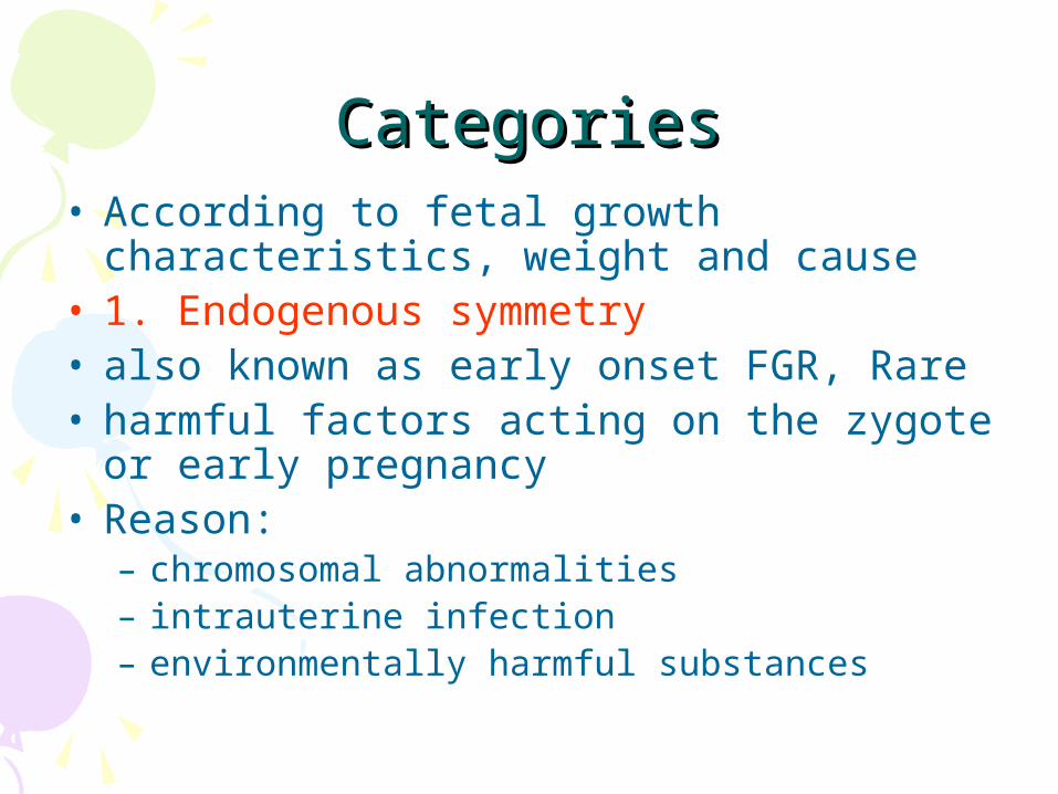
CategoriesCategories• According to fetal growth characteristics,
weight and cause• 1. Endogenous symmetry• also known as early onset FGR, Rare• harmful factors acting on the zygote or
early pregnancy• Reason:
– chromosomal abnormalities– intrauterine infection – environmentally harmful substances

CategoriesCategories• 2.Exogenous unsymmetry • harmful factors acting on second and
third trimester• most of them because the low
placental function • PIH, GDM, placenta lesions • 3. Exogenous symmetry
– One and two types mixed

Diagnosis Diagnosis
• 1. History:
• Note : there is any risk factors for FGR during this pregnancy
• Asked: appearance of FGR history

DiagnosisDiagnosis• 2. Signs and symptoms: • Continuous determination:
– fundal height, abdominal circumference and maternal weight to determine fetal growth.
• fundal height – significantly less than the corresponding
gestational age – most obvious and most easily
identifiable signs
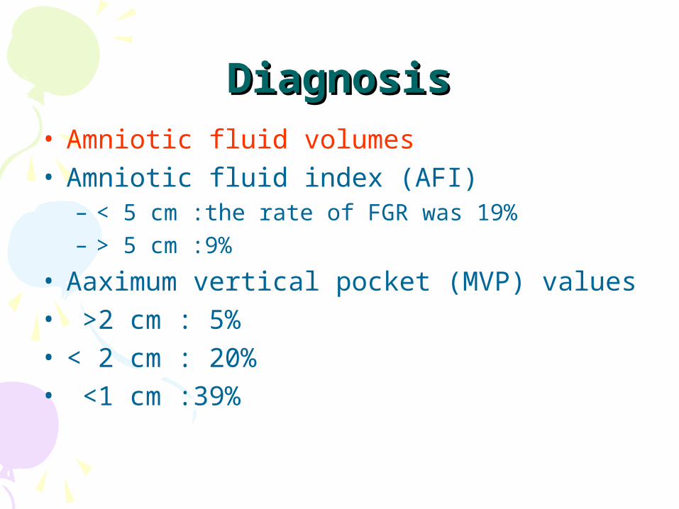
DiagnosisDiagnosis• Amniotic fluid volumes• Amniotic fluid index (AFI)
– < 5 cm :the rate of FGR was 19% – > 5 cm :9%
• Aaximum vertical pocket (MVP) values • >2 cm : 5%• < 2 cm : 20%• <1 cm :39%

DiagnosisDiagnosis• Uterine artery Doppler measurement
– contribute to the identification of fetuses at risk of FGR
• Umbilical artery Doppler measurement– absent end-diastolic velocity– reversed end-diastolic velocity– corroborates the diagnosis of FGR
• Middle cerebral artery Doppler– MCA-PSV (peak systolic velocity) is a better
predictor of FGR-associated perinatal mortality than any other single measurement
Diagnosis and Diagnosis and SurveillanceSurveillance
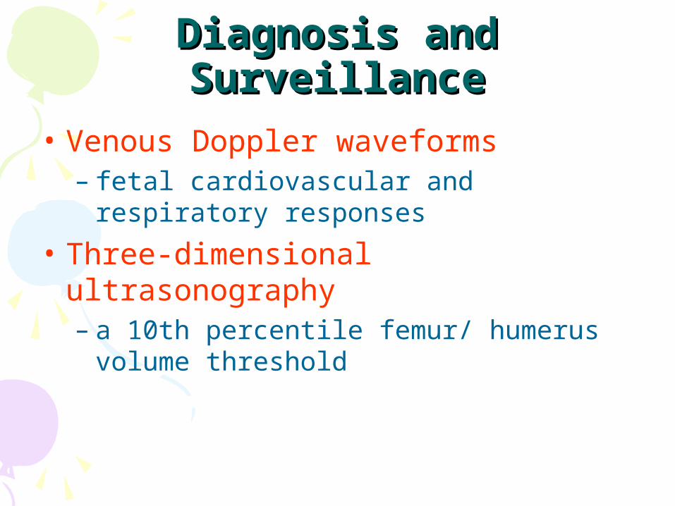
• Venous Doppler waveforms– fetal cardiovascular and respiratory
responses
• Three-dimensional ultrasonography– a 10th percentile femur/ humerus
volume threshold
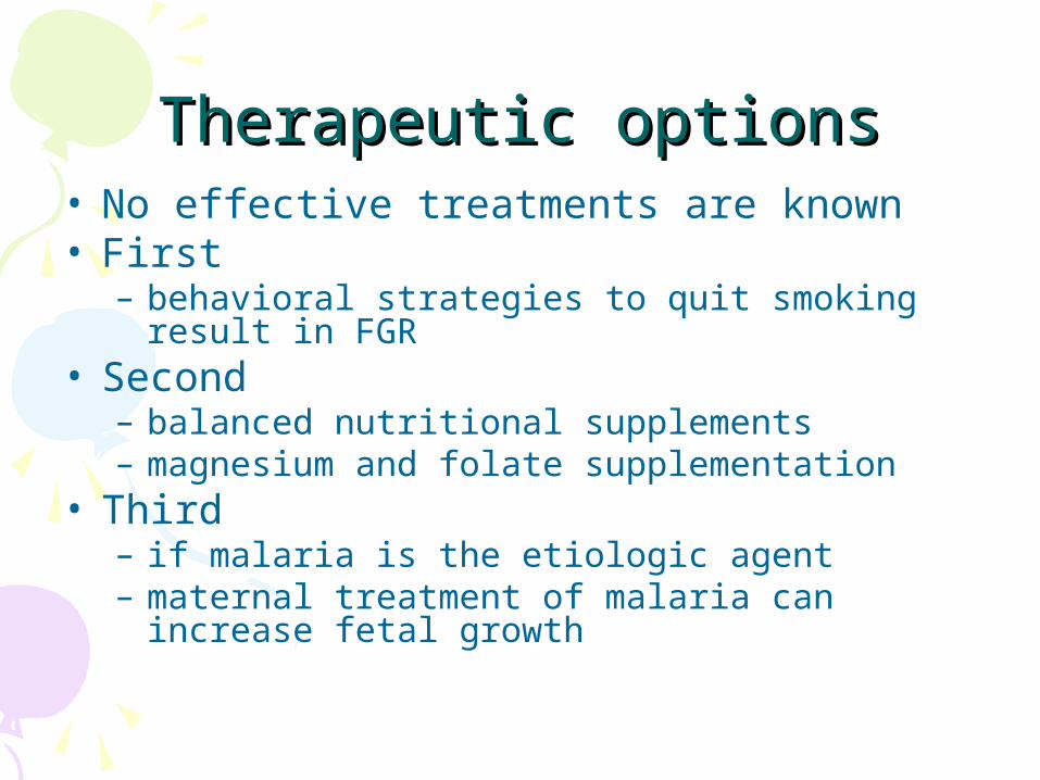
Therapeutic optionsTherapeutic options• No effective treatments are known• First
– behavioral strategies to quit smoking result in FGR
• Second– balanced nutritional supplements – magnesium and folate supplementation
• Third– if malaria is the etiologic agent– maternal treatment of malaria can increase
fetal growth
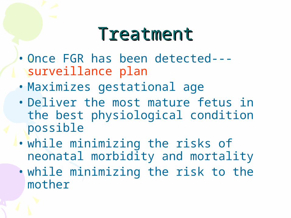
TreatmentTreatment• Once FGR has been detected---
surveillance plan• Maximizes gestational age• Deliver the most mature fetus in the
best physiological condition possible • while minimizing the risks of
neonatal morbidity and mortality• while minimizing the risk to the
mother
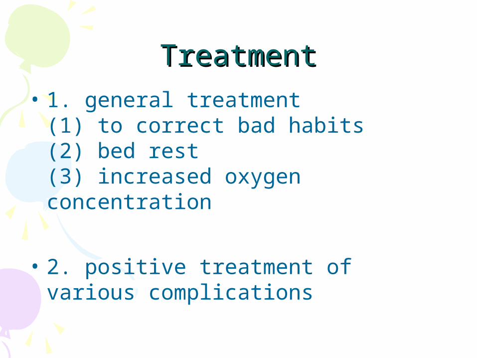
TreatmentTreatment• 1. general treatment
(1) to correct bad habits(2) bed rest(3) increased oxygen concentration
• 2. positive treatment of various complications
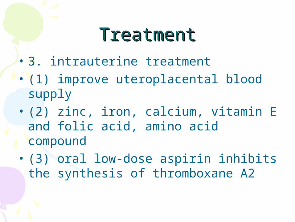
TreatmentTreatment• 3. intrauterine treatment• (1) improve uteroplacental blood
supply• (2) zinc, iron, calcium, vitamin E and
folic acid, amino acid compound • (3) oral low-dose aspirin inhibits the
synthesis of thromboxane A2

3. intrauterine treatment3. intrauterine treatment
• (4) low molecular weight heparin and low-dose aspirin may improve the outcome of FGR– but not yet widely used clinically– requires further clinical trials
• (5) the FGR fetus is expected to give birth before 34 weeks – should promote fetal lung maturity

4 obstetric management 4 obstetric management • (1) chromosomal abnormalities or severe
congenital malformations– should early termination of pregnancy.
• (2) Placental function is poor• but the treatment is effective
• continue to term– intensive care– should not exceed the expected date of
delivery
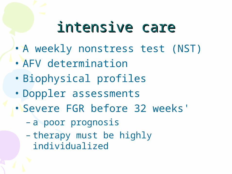
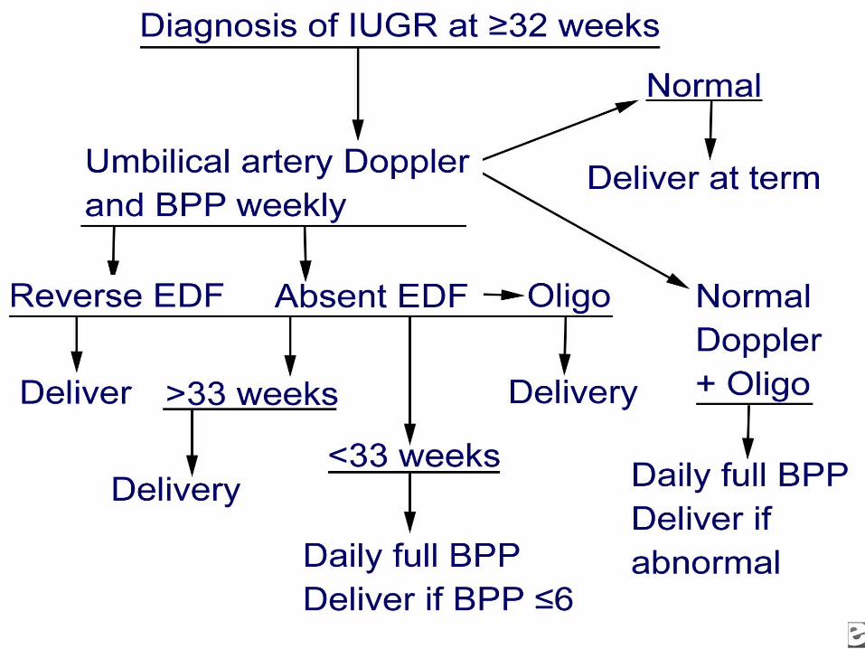
intensive careintensive care• A weekly nonstress test (NST)• AFV determination• Biophysical profiles• Doppler assessments• Severe FGR before 32 weeks'
– a poor prognosis– therapy must be highly individualized
4. obstetric management4. obstetric management• (3) termination of pregnancy:
– > 34 weeks ,a general treatment is poor– fetal distress, or stop the growth of the fetus
more than 3 weeks– pregnancy complications aggravate– < 34 weeks, has been applied to promote fetal
lung maturity
• (4) the mode of delivery : – fetal malformations– maternal complications of the severity– to evaluate fetal condition
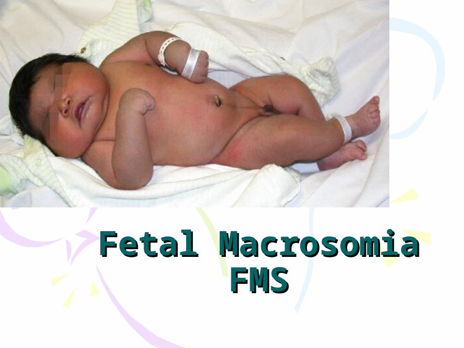
Fetal MacrosomiaFetal MacrosomiaFMSFMS
Definition of Definition of FMS• Defined in several different ways:• Birth weight of 4000-4500 g (8 lb 13
oz to 9 lb 15 oz) • Greater than 90% for gestational age• Increased dystocia, perinatal
mortality • Affects 7-15% of all pregnancies
Influencing factorsInfluencing factors• Gestational diabetes mellitus(GDM)
– class A, B, and C , 26%
• Genetics• Racial• Ethnic• Duration of gestation• Neonatal sex• Other: nutrition, parity, polyhydramnios
DiagnosisDiagnosis• Measure birth weight after delivery
– Only– retrospective
• Perinatal diagnosis difficult – often inaccurate– no risk factors can predict it accurately
enough to be used clinically– most FMS do not have identifiable risk
factors
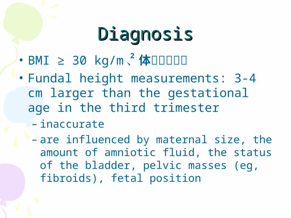
DiagnosisDiagnosis• BMI ≥ 30 kg/m 、体重增加过多• Fundal height measurements: 3-4 cm
larger than the gestational age in the third trimester – inaccurate– are influenced by maternal size, the
amount of amniotic fluid, the status of the bladder, pelvic masses (eg, fibroids), fetal position
2
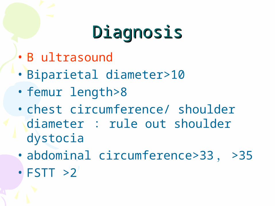
DiagnosisDiagnosis• B ultrasound• Biparietal diameter>10• femur length>8• chest circumference/ shoulder
diameter : rule out shoulder dystocia
• abdominal circumference>33 , >35• FSTT >2
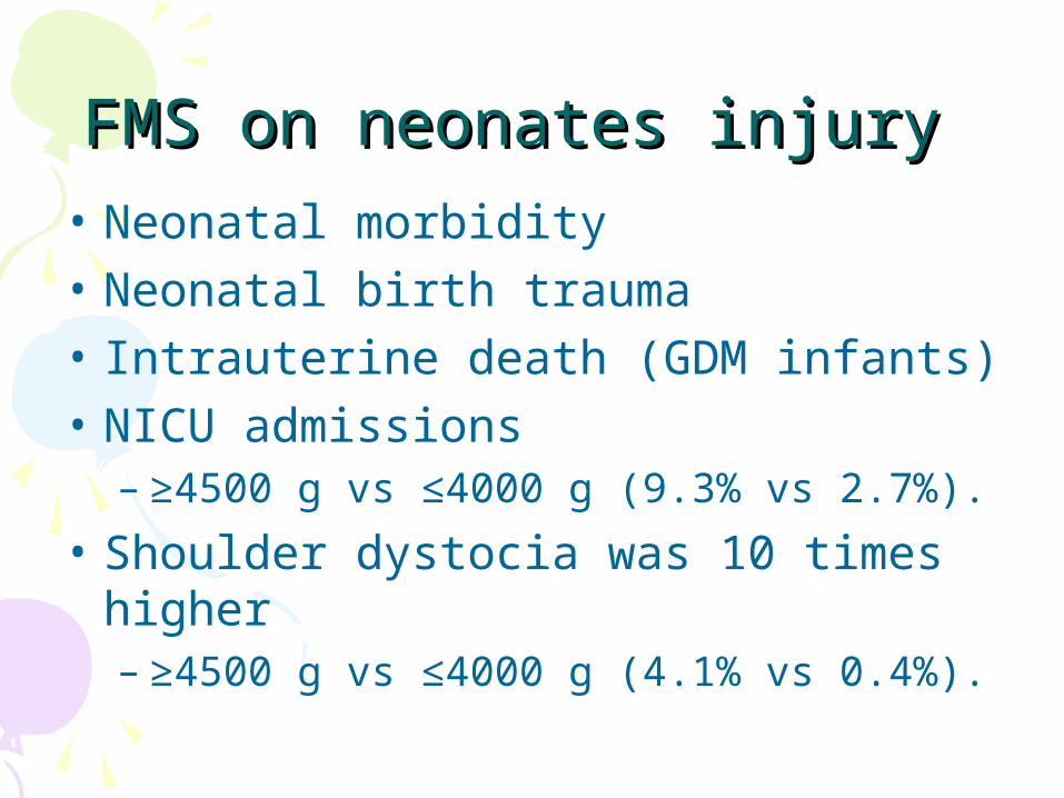
FMS on neonates injury FMS on neonates injury • Neonatal morbidity• Neonatal birth trauma• Intrauterine death (GDM infants) • NICU admissions
– ≥4500 g vs ≤4000 g (9.3% vs 2.7%).
• Shoulder dystocia was 10 times higher – ≥4500 g vs ≤4000 g (4.1% vs 0.4%).
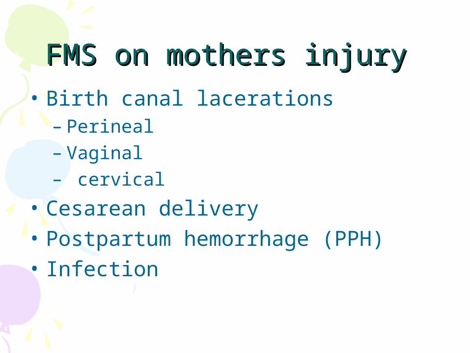
FMS on mothers injury FMS on mothers injury • Birth canal lacerations
– Perineal– Vaginal– cervical
• Cesarean delivery • Postpartum hemorrhage (PPH)• Infection
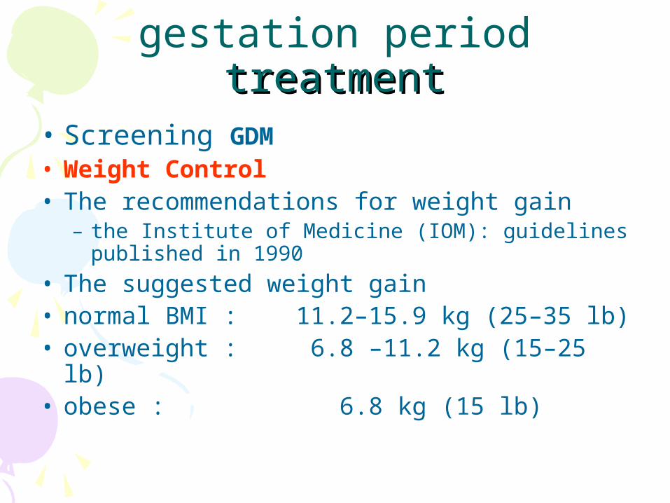
gestation period treatmenttreatment• Screening GDM• Weight Control • The recommendations for weight gain
– the Institute of Medicine (IOM): guidelines published in 1990
• The suggested weight gain • normal BMI : 11.2–15.9 kg (25–35 lb) • overweight : 6.8 –11.2 kg (15–25 lb) • obese : 6.8 kg (15 lb)
Treatment during Treatment during delivery
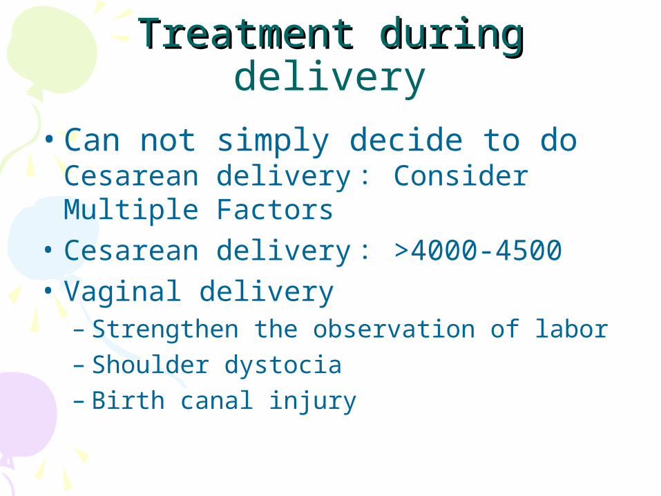
• Can not simply decide to do Cesarean delivery : Consider Multiple Factors
• Cesarean delivery : >4000-4500• Vaginal delivery
– Strengthen the observation of labor – Shoulder dystocia– Birth canal injury
NeonatalNeonatal treatmenttreatment• Fetal macrosomia• Prevention of low blood sugar
– early inleakage
• Aggressive treatment of hyperbilirubinemia– Blu-ray treatment
• Neonatal hypocalcemia – Calcium

Shoulder DystociaShoulder Dystocia
SDSD
Definition of SDDefinition of SD
• An uncommon obstetric complication of cephalic vaginal deliveries
• The fetal shoulders do not deliver after the head has emerged from the mother’s introitus
• one or both shoulders become impacted against the bones of the pelvis
• Emergency in intrapartum


Antepartum risk factors Antepartum risk factors • Listed below in order of importance:• History of SD in a prior vaginal delivery • Fetal macrosomia
– having a disproportionately large body compared to head
• Diabetes/impaired glucose tolerance • Excessive weight gain (>35 lb) • Obesity • Postterm pregnancy• 胎儿异常
Intrapartum risk factors Intrapartum risk factors • Precipitous second stage (<20 min) • Operative vaginal delivery (vacuum, forceps, or
both)• Prolonged second stage • Without regional anesthesia
– >2 h for nulliparous patients– > 1h for multiparous patients
• With regional anesthesia – >3 h for nulliparous patient– >2 h for others
• Induction of labor for impending macrosomia
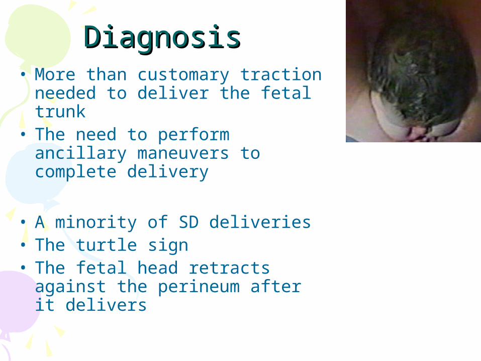
DiagnosisDiagnosis• More than customary traction
needed to deliver the fetal trunk
• The need to perform ancillary maneuvers to complete delivery
• A minority of SD deliveries • The turtle sign• The fetal head retracts against
the perineum after it delivers

TreatmentTreatment• An obstetric emergency• SD can result in significant fetal and
maternal harm if not resolved in a competent and expedient manner
• A 6-minute head-to-body interval has been demonstrated to be safe
• Beyond that time, there is increased risk – neonatal depression, acidosis, asphyxia,
central nervous system damage, or even death
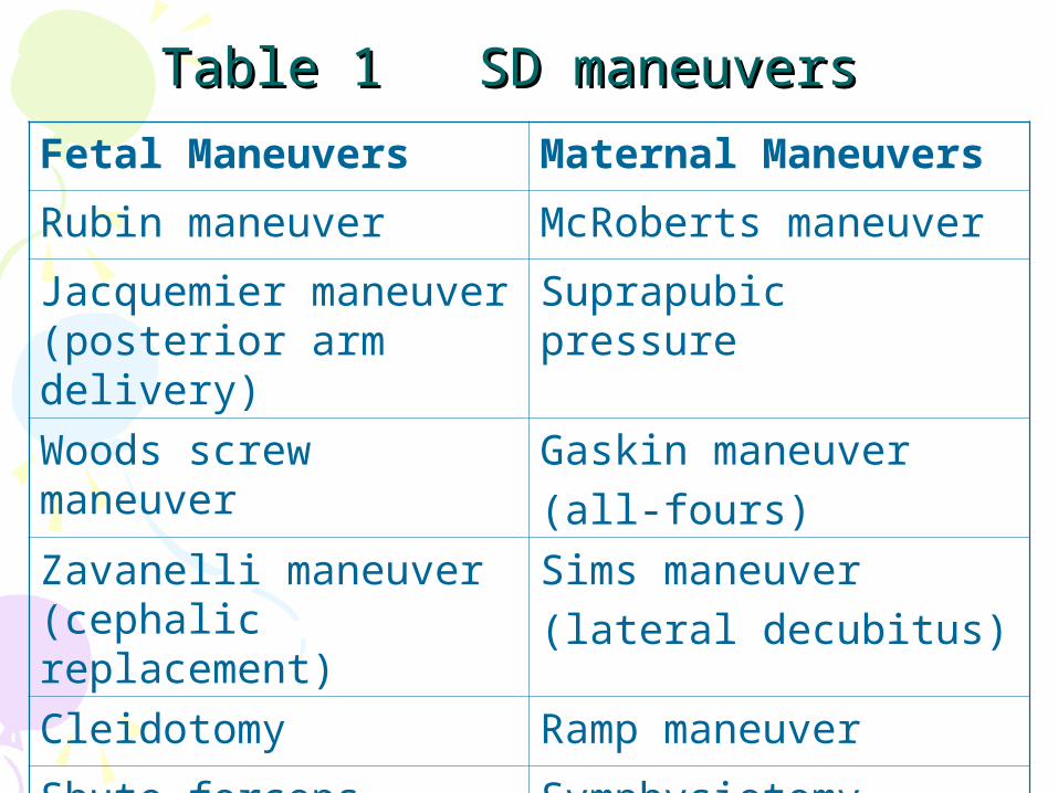
Table 1 SD maneuversTable 1 SD maneuvers Fetal Maneuvers Maternal Maneuvers
Rubin maneuver McRoberts maneuver
Jacquemier maneuver (posterior arm delivery)
Suprapubic pressure
Woods screw maneuver
Gaskin maneuver (all-fours)
Zavanelli maneuver (cephalic replacement)
Sims maneuver (lateral decubitus)
Cleidotomy Ramp maneuver
Shute forceps maneuver
Symphysiotomy

McRoberts maneuver

Suprapubic pressure

Rubin maneuver
posterior arm
delivery

Fetal DeathFetal Death
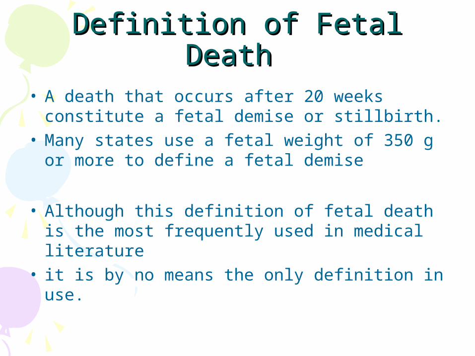
Definition of Fetal Death Definition of Fetal Death • A death that occurs after 20 weeks
constitute a fetal demise or stillbirth. • Many states use a fetal weight of 350 g or
more to define a fetal demise
• Although this definition of fetal death is the most frequently used in medical literature
• it is by no means the only definition in use.

Causes of Fetal DeathCauses of Fetal Death• The etiology of FD is unknown in 25-60%
of all cases• 1. fetal hypoxia
– The most common reason, about 50%
• maternal factors• fetal factors• Placenta• abnormal cord
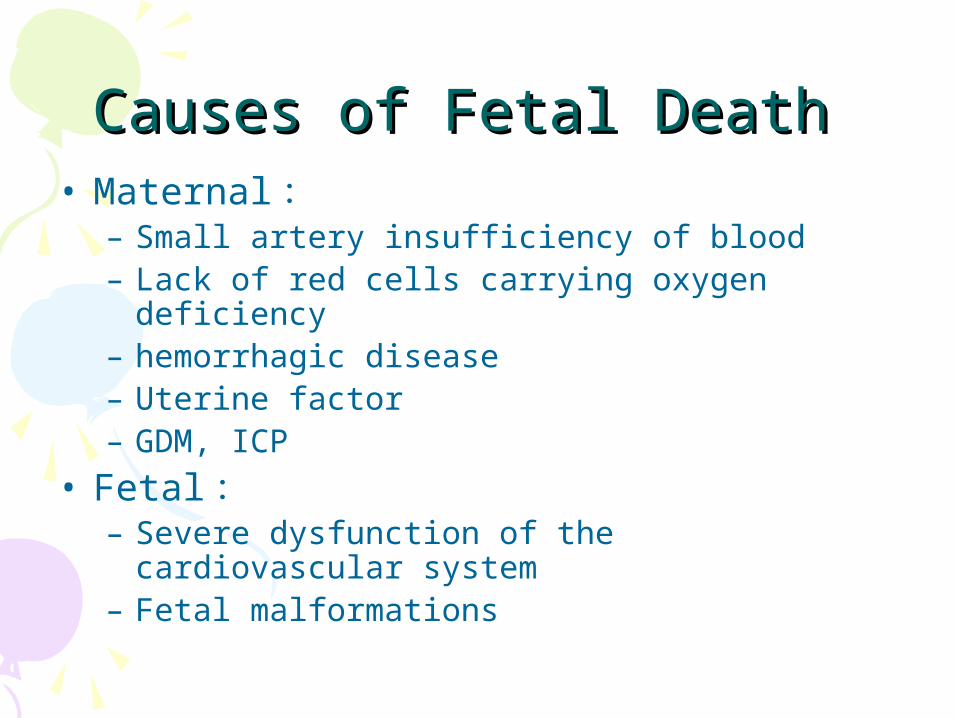
Causes of Fetal Death Causes of Fetal Death • Maternal :
– Small artery insufficiency of blood– Lack of red cells carrying oxygen deficiency– hemorrhagic disease– Uterine factor– GDM, ICP
• Fetal :– Severe dysfunction of the cardiovascular
system– Fetal malformations
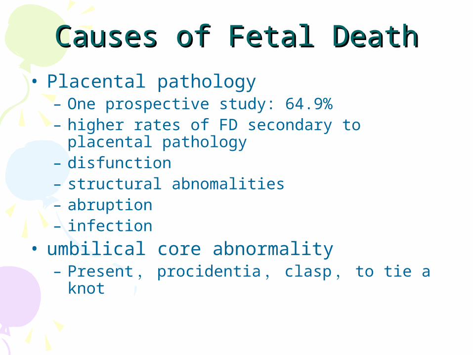
Causes of Fetal DeathCauses of Fetal Death• Placental pathology
– One prospective study: 64.9%– higher rates of FD secondary to placental
pathology– disfunction– structural abnomalities– abruption– infection
• umbilical core abnormality– Present , procidentia , clasp , to tie a knot
Causes of Fetal DeathCauses of Fetal Death• 2. Genetic mutations and chromosomal
aberrations • Parents suffering from genetic diseases• during pregnancy
– use of teratogenic drugs– exposure to radiation– chemical poisons
• Embryonic genes and chromosome aberration
• Fetal malformations, miscarriage or death

Diagnosis of Fetal Death Diagnosis of Fetal Death • History and physical examination
– limited value
• Death must be confirmed by ultrasonographic– visualization of the fetal heart – the absence of cardiac activity
• In fact, the following description is rarely– Macerated fetus– fetus compressus– fetus papyraceus
Management of Fetal Death Management of Fetal Death • Once the diagnosis has been
confirmed , the patient should be informed of her condition
• Often, allowing the mother to see the lack of cardiac activity helps her to accept the diagnosis.
• Immediate treatment – Method of least damage to the mother – Labor induction

Management of Fetal DeathManagement of Fetal Death• Medicine intra-amniotic injection • Preinduction cervical ripening followed by
intravenous oxytocin• Mifepristone and prostaglandin induction
of labor
• Patients with a history of a prior cesarean delivery should be treated cautiously – the risk of uterine rupture

Management of Fetal DeathManagement of Fetal Death• When a dead fetus has been in utero for 3-
4 weeks– Fibrinogen, blood plate levels may drop– leading to a coagulopathy– heparin therapy– Rarely: because of earlier recognition and
induction
• In some cases of twin pregnancies– induction after the death of a twin may be
delayed – to allow the viable twin to mature

Thanks four your listening