Embed Size (px)
Citation preview
REVIEW ARTICLE
Femoral vessel injuries; high mortality and low morbidity injuries
G. Ruiz • A. J. Perez-Alonso • M. Ksycki •
F. N. Mazzini • R. Gonzalo • E. Iglesias •
A. Gigena • T. Vu • Juan A. Asensio-Gonzalez
Received: 15 May 2012 / Accepted: 16 June 2012 / Published online: 1 September 2012
� Springer-Verlag 2012
Abstract Femoral vessel injuries are amongst the most
common vascular injuries admited in busy trauma centers.
The evolution of violence and the increase in penetrating
trauma from the urban battlefields of city streets has raised
the incidence of femoral vessel injuries, which account for
approximately 70% of all peripheral vascular injuries.
Despite the relatively low mortality associated with these
injuries, there is a high level of technical complexity
required for the performance of these repairs. Similarly,
they incur low mortality but are associated with signifi-
cantly high morbidity. Prompt diagnosis and treatment are
the keys to successful outcomes with the main goals of
managing ischemia time, restoring limb perfusion,
accomplishing limb salvage and instituting rehabilitation as
soon as possible.
Keywords Vascular injuries � Management of femoral
vessel injuries � Morbidity and mortality � Anatomy
Introduction
Femoral vessel injuries are amongst the most common
vascular injuries admitted in busy trauma centers. The
evolution of violence and the increase in penetrating trauma
from the urban battlefields of city streets have raised the
incidence of femoral vessel injuries, which account for
approximately 70 % of all peripheral vascular injuries.
Despite the relatively low mortality associated with these
injuries, there is a high level of technical complexity
required for the performance of their repair. Similarly, these
injuries incur low mortality but are associated with signif-
icantly high morbidity. Prompt diagnosis and treatment are
the keys to achieve successful outcomes, along with the
main goals of managing ischemia time, restoring limb
perfusion, accomplishing limb salvage and instituting
rehabilitation as soon as possible.
Anatomy
The femoral artery is a direct continuation of the external
iliac artery, located in the femoral triangle, posterior to the
inguinal ligament. Its most proximal portion, known as the
common femoral artery, courses deep to the sartorius
muscle together with the femoral vein contained within a
fibrous sheath. The lateral compartment of the femoral
sheath contains the femoral artery, the middle contains the
femoral vein, and the most medial compartment is the
femoral canal. After a short course, the common femoral
artery—which can be palpated through the skin on the
proximal aspect of the inner thigh midway between the
anterior superior iliac spine and the symphysis pubis—
divides into the deep (profunda femoris artery) and the
superficial femoral artery. The profunda femoris artery
G. Ruiz � A. J. Perez-Alonso � M. Ksycki �F. N. Mazzini � R. Gonzalo � E. Iglesias � A. Gigena � T. Vu �J. A. Asensio-Gonzalez M.D. FACS FCCM FRCS (England)
Division of Trauma Surgery and Surgical Critical Care,
Dewitt Daughtry Family Department of Surgery, Ryder Trauma
Center, University of Miami Miller School of Medicine,
1800 NW 10 Avenue Suite T-247, Miami, FL 33136-1018, USA
Present Address:J. A. Asensio-Gonzalez M.D. FACS FCCM FRCS
(England) (&)
Westchester University Medical Center, New York Medical
College, Trauma Department of Surgery, 100 Woods Road
Taylor Pavilion, Suite E137, Valhalla, NY 10595, USA
e-mail: [email protected]
123
Eur J Trauma Emerg Surg (2012) 38:359–371
DOI 10.1007/s00068-012-0206-x

traverses deep into the thigh muscles and is the main con-
tributor of blood flow to this area. The superficial femoral
artery traverses almost the entire length of the femur and
ends at the opening of the adductor magnus (also known as
Hunter’s canal) where it becomes the popliteal artery. The
main branches of the femoral artery are the pudendal artery,
the superficial epigastric artery, the circumflex iliac artery
and both the greater and lesser saphenous arteries.
The femoral vein is a continuation of the popliteal vein.
It accompanies the femoral artery and receives drainage
both from the profunda femoris vein and the greater
saphenous vein, which pierce the femoral sheath on its
anterior aspect before passing under the inguinal ligament
to become the external iliac vein.
Incidence
Traumatic injury to the femoral vessels accounts for
approximately 67 % of all vascular injuries in the military
setting. Recently, the incidence of femoral vessel injury in
the civilian arena has increased secondary to an increase in
urban violence. Penetrating injuries are reported in
approximately 88 % of these cases and are usually second-
ary to gunshot or stab wounds by knife or impalement. Less
common are blunt injuries, which account for approximately
12 % of all cases.
The incidence of these types of injuries has varied
during the major military conflicts of the past century and
has increased slightly with each conflict. During World
War I, Makin’s (1919) classical textbook [1] detailed a
31 % incidence rate reported by British surgeons, whereas
the incidence of femoral vessel injury reported by Ameri-
can surgeons was lower at 22 %. DeBakey and Simeone
[2], reported a total of 517 femoral arterial injuries for an
incidence rate of 21 % during World War II, with a very
high amputation rate of 53 %. Hughes [3] reported a
slightly higher incidence of 31 % for the Korean Conflict,
while Rich [4] reported a 35 % incidence of femoral vessel
injuries during the Vietnam Conflict.
Historical perspective and wartime experiences
The earliest documents reporting vascular injuries date
from 1600 B.C. Egyptians were known to use styptics
consisting of vegetable matter, lead sulfate and copper
sulfate to control hemorrhage, as reported in the Ebers
Papyrus [5]. In 1000 B.C., the Chinese were first to use
tight bandaging to control bleeding in the affected limb as
well as styptics for the control of hemorrhage.
However, the treatment of choice for vascular injuries
throughout the Middle Ages consisted mainly of cautery. In
1497, Hieronymus Brunschwig, also known as Jerome of
Brunswick [6], published his work on ligatures as treatment
for injuries secondary to gunshot wounds. His work was
further refined in 1552 by Ambroise Pare [7], who also
promoted the use of ligatures to control hemorrhage. Pare
[7] additionally recommended amputation above the line of
demarcation to achieve better outcomes. However, Hallo-
well [8] was first to attempt a vascular reconstruction in
1759, when he repaired a brachial artery with a suture
known as the Farrier’s stitch or veterinarian’s stitch.
Some 127 years would pass until vascular repair was
again attempted. During the latter part of the nineteenth
century, pioneers such as Jassinowsky and Postemski [9]
promoted the concept of direct vascular repair. Israel [10],
in 1883, described the first successful primary repair of a
lacerated iliac artery, while John B. Murphy [11, 12] from
Rush Medical College in Chicago completed the first suc-
cessful end-to-end anastomosis of a femoral artery in 1897.
This was later followed by the first autogenous reverse
saphenous vein graft to repair a popliteal aneurysm in 1906
by Goyanes of Spain. Despite these advances made at the
end of the nineteenth century, it would take almost three
decades before vascular repairs were again systematically
utilized.
World War I
During the first part of the World War I, vascular injuries
were typically secondary to the low velocity projectiles
used and thus limited in occurrence; however, the second
half saw the introduction of high explosive artillery with
improved, high velocity munitions. The change in the
destructive power of ordnance contributed to an increase in
mass casualties and a decrease in the number of wounded
that could be evacuated. The time between injury and
definitive surgical treatment drastically increased and
vascular repair became impractical. Although the chance
for immediate repair was lost, vascular repair was
attempted to manage false aneurysms and arteriovenous
(AV) fistulas on a delayed basis.
Limited attempts at repair of femoral vessel injuries
were made during World War I. Makins [1] reported the
British experience of femoral artery injuries consisting of
366 arterial injuries in 1,202 patients for an incidence of
31 %. American surgeons also reported 78 femoral injuries
in 344 patients for an incidence of 22 %.
World War II
The years between World War I and II showed an uneven
advance between the instruments of destruction and the
advent of surgical interventions. DeBakey and Simeone [2]
reported a total of 2,471 arterial injuries from the European
360 G. Ruiz et al.
123

theater of war; unfortunately, nearly all were treated with
ligation resulting in a very high amputation rate of 49 %. In
this study, DeBakey and Simeone reported only 81
attempts at repair, of which 78 were lateral suture repairs/
arteriographies with only three end-to-end anastomoses
performed. Of 2,471 cases of vascular injuries reported,
517 (21%) were femoral vessel injuries.
The main factors contributing to this low number of
vascular repairs included delays in transport time, lack of
training in vascular trauma for military surgeons, and rel-
ative lack of access to antibiotics; although penicillin and
sulfa had been introduced during this conflict, they were
available only in very limited supply. Other factors included
the absence of appropriate suture material, and unavail-
ability of vascular instruments. Interestingly enough, the
first vascular clamp was created by Victor Satinsky pre-
cisely during World War II.
Korean conflict
Vascular surgery advanced in leaps and bounds during the
Korean conflict thanks to several factors. Improvements in
anesthesia, the advent of antibiotics and the development of
blood transfusions greatly enhanced the success rate of vas-
cular repairs. Perhaps the single greatest factor was the
development of forward aid stations accompanied by rapid
evacuation of the wounded by helicopter, which significantly
improved transport of these patients. Similarly, surgeons such
as Hughes, Jahnke, Spencer and Howard demonstrated an
interest in developing techniques for vascular injury repair.
Several reviews from this time period showed primary
vascular repair to be gaining wide acceptance. Jahnke and
Seeley [13] reported 14 femoral artery injuries for an
incidence of 19 %. Hughes subsequently reported 95
femoral vessel injuries out 304 patients for an incidence of
31 %. These are amongst the earliest series in the literature
demonstrating a well defined approach to the management
of vascular trauma.
Vietnam conflict
Practices during the Vietnam conflict built on the previous
advances in evacuation and faster transport of the wounded.
Thus, a higher number of surgeons trained in vascular sur-
gical techniques allowed for improved outcomes and patients
that could now be followed up with on a long term basis as
the war ended. The Vietnam vascular registry created by
Dr. Norman Rich and based at the Army Medical Center of
Walter Reed allowed for long term follow-up and continued
evaluation of all vascular injuries from the Vietnam conflict.
The landmark report from Rich [4] in 1969 showed 351
femoral artery injuries out of a total 1,000 patients for an
incidence of 35 %. Unfortunately, data on femoral venous
injuries from this conflict is scarce, with ligation being the
standard of care noted in the wartime literature.
Iraq and Afghanistan
The Iraq and Afghanistan campaigns that have spanned
most of the last decade have seen significant improvements
in the overall survival of combat casualties. Increases in
survival rates can be attributed to the wide use of body armor
which has dramatically decreased torso injuries, the wide-
spread use of pre-hospital tourniquets, and the use of dam-
age control techniques along with the rapid evacuation for
definitive surgery to Landstuhl Regional Medical Center
and subsequently to the continental United States (CONUS).
The injury patterns seen in these two conflicts are
notably different than in other wars. Due to the widespread
use of body armor, the number of torso injuries has
decreased significantly, however there has been a great
increase in head injuries and distal extremity vascular
injuries. These changes in injury patterns resulted from the
departure from regular warfare to counter-insurgency
combat which involves ambush techniques and the utili-
zation of anti-personnel devices such as improvised
explosive devices (IED). The use of high explosives in
these IEDs has resulted in a very large incidence of severe
extremity injuries and in some cases, multiple amputations.
There have also been a large number of vascular injuries.
Data collected from war registries [14, 15] have shown a
general increase in the incidence of all types of vascular
injuries, which has accounted for 4 to 9 % of the total
combat casualties. When accounting for only extremity
vascular injuries it appears that most of the injured vessels
are located in the lower extremities with a reported inci-
dence of 7 % for femoral arteries, 3 % for isolated femoral
venous injuries and a combined incidence of femoral vessel
injuries accounting for 7 % of all cases. The wide use of
temporary vascular shunts as early restoration of vessel
flow, fasciotomies, and external fixation of open fractures as
methods of damage control in the field management of
vascular injuries, along with the rapid transport to Lands-
tuhl Regional Medical Center have resulted in large number
of limbs salvaged (Senior author’s personal experience)
(See Table 1).
Mechanism of injury
Penetrating trauma remains the predominant cause for the
majority of femoral vessel injuries. This was confirmed in
the series by Cargile [17], who reported an 88 % incidence
of penetrating injuries and a 12 % incidence of blunt
trauma. In Feliciano’s series [24], the incidence of pene-
trating trauma was 81 %, while Martin [16] reported that
Femoral vessel injuries 361
123

104 of 105 femoral arterial injuries were from penetrating
trauma. In Asensio’s series [21], 86 % of the patients were
admitted with penetrating injuries while 28 (14 %) sus-
tained blunt injuries.
Civilian epidemiology
The incidence of femoral vessel injury is well documented
in the civilian trauma literature. Feliciano [24] reported a
series of 220 lower extremity vascular injuries, of which
142 were of the femoral artery, for an incidence of 65 %.
Martin [16] reported 188 lower extremity injuries with 105
femoral artery injuries for an incidence of 56 %. Cargile
[17] reported data from a Parkland Hospital study, con-
sisting of 233 patients who sustained a total of 321 femoral
vessel injuries over a 17-year period. In this study, he
reported a total of 112 isolated femoral artery injuries
(48 %), 36 isolated femoral vein injuries (15 %) and 85
combined injuries for an incidence of 36 %. Femoral
venous injuries were infrequently reported in Martin’s
series [16], where out of 105 femoral vessel injuries only
three were of the femoral vein.
Asensio [21] reported a series of 204 patients treated for
298 vessel injuries during a 10 year period, for an inci-
dence of 26 % of all the vascular injuries reported for this
period at his institution (see Table 2).
Clinical presentation
The clinical presentation of patients sustaining femoral
vessel injuries ranges from severe (i.e., hemodynamic
instability and cardiopulmonary arrest requiring emergency
department thoracotomy (EDT), aortic cross-clamping and
cardiopulmonary resuscitation), to patients that are hemo-
dynamically stable (admitted with either the classical hard
signs of vascular injury secondary to distal decreased flow
and ischemia, or those that harbor soft signs of vascular
injury).
Cargile [17] reported that 87 patients (37 %) who pre-
sented with a femoral vessel injury had systolic blood
pressures \90 mmHg, while 35 patients (40 %) presented
in profound shock.
In Degiannis (37), 19 patients with femoral vessel
injuries arrived hypotensive with systolic blood pressures
\90 mmHg, 37 of 81 patients (46 %) with isolated
extremity injuries were hypotensive, and four patients
arrived in cardiopulmonary arrest requiring EDT. In
Asensio’s series [21], 11 patients (5 %) arrived in cardio-
pulmonary arrest requiring EDT; 3 (27 %) of these patients
ultimately survived.
Less commonly, the lacerated or transected femoral
artery may retract and thrombose resulting in distal limb
ischemia. Associated muscle and bone injuries are common
with penetrating injuries, therefore meticulous care should
be exercised when reducing long bone fractures. With large
tissue defects resulting from penetrating injuries, actual
hemorrhage may be present which requires prompt control
with direct pressure or tourniquet application depending on
the location of the injury. However, if the common femoral
or very proximal superficial femoral artery is involved, the
use of a tourniquet may not be feasible. In this instance,
direct pressure on or above the injury with immediate
proximal and distal surgical control is mandatory.
With blunt injuries, loss of blood flow is accompanied
with ischemic pain, and sensory and/or motor loss depend-
ing on the associated structures involved. Hemorrhage from
Table 1 Military experience
with femoral vessel injuriesConflict References Total injuries Femoral vessels Incidence (%)
WWI Makins [1] 1,191 366 30.5
WWII DeBakey and Simeone [2] 2,471 517 20.9
Korea Hughes [3] 304 95 31
Vietnam Rich [4] 1,000 351 35
Iraq and Afghanistan White [15] 1,570 268 17
Table 2 Incidence of femoral
vessel injuries in lower
extremity vascular injuries
Author(s) Years Total injuries in lower
extremities
Femoral injuries Incidence (%)
Artery Vein Total
Feliciano [24] 1988 352 142 93 235 66.7
Timberlake [26] 1990 322 – 116 (36 %) –
Bongard [20] 1990
Martin [16] 1994 188 105 21 126 67
Clouse [14] 2007 220 74 37 111 50.4
White [15] 2011 94 44 (46.9 %) – 44
362 G. Ruiz et al.
123

blunt injury is rare but may occur secondary to associated
orthopedic injuries resulting in partial or complete lacera-
tion or transection of the vessel.
In Cargile’s [17] series, 90 of 233 patients (40 %) pre-
sented with hard signs of vascular injury requiring imme-
diate surgical intervention. Despite a significant number of
these patients presenting with hard signs, preoperative
angiography was performed in 106 patients. Degiannis (37)
and associates relied more on clinical examination, noting
that 70 % of their patients presented with an ischemic
extremity.
In Asensio’s series [21], the majority of patients pre-
sented with hard signs of vascular injury; 48 % of his
patients presented with distal ischemia, 43 % presented
with absent or diminished pulses, and 29 % presented with
an expanding hematoma. In this series, the reliance on
clinical examination alone for detection of femoral vessel
injuries reduced the incidence of preoperative angiography
to 15 %. Fewer patients presented with soft signs of vas-
cular injury: peripheral nerve deficits accounted for 10 %
and proximity injuries for 7 %.
Diagnosis
The need for diagnostic workup is limited in penetrating
trauma but frequently can clarify a confusing clinical pic-
ture with blunt injuries. The bifurcation of the common and
superficial femoral artery is superficial enough to allow
clinical diagnosis by inspection and palpation. Penetrating
injury with tissue destruction and active hemorrhage needs
no diagnostic workup. Patients that have sustained blunt
trauma or penetrating injury without active bleeding benefit
from a thorough clinical exam initially. Changes in the
clinical examination of the femoral, popliteal, posterior
tibial or dorsalis pedis arteries and their respective pulses
and alterations in the sensory and motor exam may indicate
an injured vessel [18]. Tight or painful compartments and
associated orthopedic injuries warrant further investigation.
The first and easiest diagnostic test to perform is the ankle-
brachial index (ABI) exam. This exam is performed by
measuring the systolic blood pressure at the ankle, which is
then divided by the brachial artery systolic pressure. A
difference\0.9 is considered suspicious for the presence of
vascular injury and warrants further imaging either with
computed tomographic (CT) angiography or formal angi-
ography [19] (see Table 3).
Surgical management
Prior to admission to the operating room (OR) arrange-
ments for blood to be immediately available are required.
Every possible effort should be made to avoid hypother-
mia. Broad-spectrum antibiotics should be administered
prior to the commencement of the procedure. In preparing
the patient for surgery, the lower abdomen, both groins and
both lower extremities should be prepared and draped for
possible involvement.
Initial control of an exsanguinating vessel is obtained by
direct compression, followed by obtaining proximal and
distal control. This may be obtained by direct dissection
down onto the common femoral artery though a longitudi-
nal incision, overlying its course from the inguinal ligament
distally to the area of injury [16]. More proximal control
may be obtained by gaining control of the external iliac
artery, either by performing an exploratory laparotomy with
cross-clamping of the vessel in the pelvis or by transecting
the inguinal ligament through a muscle-splitting, lower
quadrant incision carried down to the retroperitoneum. With
this approach, the vessels can be controlled without entry
into the peritoneum [21]. The profunda femoris artery is
exposed through the same incision employed to expose the
common femoral artery. The superficial femoral artery is
dissected proximally and posterolaterally to identify the
origin of the profunda femoris artery.
With inflow controlled, a careful dissection along the
anatomic path limits secondary injury to the artery and
avoids secondary hemorrhage from neighboring vessels.
Vascular clamps, silastic vessel loops and ocular magnifi-
cation in the form of Loupes are essential tools necessary
for obtaining definitive control of the injured vessel [21].
Meticulous dissection of the femoral vessels is of the utmost
importance, especially for the femoral veins, as they tend to
be delicate, have a propensity to bleed significantly and are
easily injured iatrogenically. To obtain both proximal and
distal control we recommend use of 30�, 45� or 60� angled
DeBakey clamps for both the femoral arteries and veins. If
venorrhaphy of the superficial femoral vein is feasible, the
use of small, partial occlusion Cooley or Satinsky clamps is
preferred (see Fig. 1). The profunda femoris artery should
Table 3 Hard and soft signs of vascular injury at admission
Hard
Signs of distal ischemia
Absent or diminished pulses
Expanding hematoma
Palpable thrill
Pulsatile bleeding
Bruit
Soft
Capillary refill [3 s
Peripheral nerve deficit
Proximity injury
Moderate bleeding (limited)
Femoral vessel injuries 363
123

be controlled with special profunda vascular clamps. Argyle
shunts should always be available to temporarily restore
perfusion to the common or superficial femoral arteries and
as an adjunct to damage control (see Figs. 1, 2).
Primary repair
Arteriorrhaphy should be performed with 4–0 or 5–0
polypropylene monofilament sutures in either an inter-
rupted or running fashion. Whichever method chosen, great
care must be taken to avoid narrowing the artery and
causing stenosis [19]. Fogarty catheters should be sized
appropriately and used to clear the distal and proximal
femoral artery of potential clots; this should be performed
prior to completion of the repair or anastomosis. As with
any injury, damaged or devitalized segments of the vessel
must be debrided, even if this renders primary repair no
longer a feasible option. End-to-end anastomosis is an
option following proper debridement, as long as there is
adequate length to perform the repair without any undue
tension whatsoever (see Figs. 3, 4, 5, 6, 7, 8, 9, 10, 11, 12,
13, 14, 15, 16, 17, 18, 19, 20, 21, and 22).
Fig. 1 One of Professor Asensio’s vascular trays. Notice 60� angled
DeBakey clamps (1, 2), and profunda femoris clamps (3, 4). Also
notice Satinsky clamp (5)
Fig. 2 Argyle shunts
Fig. 3 Anterior–posterior (AP) view of an open right femur fracture
associated with a distal femoral arterial injury just above Hunter’s
canal. Note missile fragments within the wound (arrows)
Fig. 4 Lateral view of an open left femur fracture with associated
femoral arterial and venous injury. Note comminuted bone fragments
which often act as secondary missiles (arrow)
364 G. Ruiz et al.
123

Grafts
Interposition grafts must be utilized when there has been
significant vessel destruction and primary end-to-end
anastomosis is thus not possible. PTFE is an excellent
choice for a replacement conduit, as it is available in var-
ious sizes and has moderate resistance to infection in
contaminated fields.
Autogenous reverse saphenous vein grafts have been
traditionally advocated as the preferred conduit due to their
long term patency and resistance to infection. Feliciano
[24] reported on a prospective study of the use of PTFE and
concluded that it had good long term patency. Asensio [21]
reported for his series that the use of PTFE was associated
with increased mortality only because it was employed on
patients who had sustained greater injury severity, multiple
Fig. 5 Emergency department resuscitative thoracotomy with aortic
cross clamping and open cardiopulmonary resuscitation required in a
patient that sustained an exsanguinating combined gunshot wound to
the right superficial femoral artery (SFA) and vein (SFV)
Fig. 6 After a successful emergency department thoracotomy the
patient was transported to the operating room while digital control of
the femoral vessels was applied along with simultaneous intravascular
volume replacement with crystalloids, blood and blood products.
Figure shows proximal and distal control of a right superficial femoral
artery (SFA) and superficial femoral vein (SFV)
Fig. 7 Same patient showing placement of an 8-mm PTFE graft in
the right superficial femoral artery (SFA) and a primary venorrhaphy
of the superficial femoral vein (SFV). The patient required thigh and
foreleg four-compartment fasciotomies which were initially covered
with cadaveric xenografts and subsequently closed on the fifth
postoperative day
Fig. 8 Patient after emergency department resuscitative thoracotomy
Fig. 9 Young man that sustained an exsanguinating gunshot wound
secondary to a high velocity handgun missile which lacerated his right
superficial femoral artery (SFA) and right superficial femoral vein
(SFV). The patient was rapidly transported to the operating room, and
a 10 Fr Argyle shunt placed to rapidly restore blood flow. The patient
also sustained a comminuted right femur fracture
Femoral vessel injuries 365
123

associated injuries and presented in shock. He recom-
mended its use in patients who presented with hypother-
mia, coagulopathy, acidosis and increased requirements of
blood and blood products, given its availability and
because it significantly decreases operative time.
The senior author of this manuscript has subsequently
evolved towards the utilization of ringed PTFE grafts (see
Figs. 3, 4, 5, 6, 7, 8, 9, 10, 11, 12, 13, 14, 15, 16, 17, 18, 19,
20, 21, and 22).
Fasciotomies and Shunting
Patients sustaining extremity vascular injuries are at risk for
the development of compartment syndromes depending on
the length of ischemia and associated tissue destruction.
The consequences of missing the diagnosis are dire, with
the patient often losing entire muscle compartments, if not
the entire extremity. Even after successful revasculariza-
tion, the systemic complications of the reperfusion syn-
drome, its effects on the kidney and its resulting electrolyte
abnormalities and acidosis can result in significant mor-
bidity [24]. The performance of fasciotomies on the re-
vascularized limb allows for swelling and edema to occur
without vascular compromise, thus ensuring limb salvage.
Compartment fasciotomies should be performed via sepa-
rate incisions (see Table 4). They should be complete fas-
ciotomies. Thigh fasciotomies including lateral and medial
incisions may also be necessary (see Figs. 3, 4, 5, 6, 7, 8, 9,
10, 11, 12, 13, 14, 15, 16, 17, 18, 19, 20, 21, and 22).
Venous injuries
Injuries to the femoral vein are life threatening, though not
to the extent of their arterial counterparts, and are usually
Fig. 10 The same patient after the shunt has been removed and the
orthopedic surgeons have performed an open reduction and internal
fixation (ORIF) with a reamed femoral rod. The Fogarty Bulldog
clamps are controlling the SFV while the 45� and 60� angle DeBakey
clamps are controlling the superficial femoral artery (SFA)
Fig. 11 The same patient after ligation of the superficial femoral
vein. The patient has undergone an end-to-end superficial femoral
artery interposition graft with an autogenous reverse saphenous vein
graft. Note femoral rod bridging the open femoral fracture
Fig. 12 The same patient. Intraoperative arteriogram demonstrates
excellent flow in the interposition graft. Notice the absence of
interlocking screw in the distal femoral rod
Fig. 13 The same patient. Intraoperative completion arteriogram
showing excellent blood flow in the interposition graft as well as in
the tibioperoneal trunk and shank vessels. Note absence of interlock-
ing screw in the distal femoral rod
366 G. Ruiz et al.
123
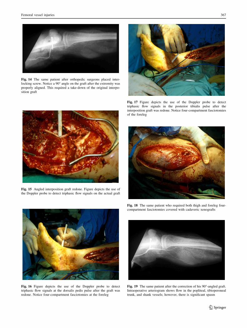
Fig. 14 The same patient after orthopedic surgeons placed inter-
locking screw. Notice a 90� angle on the graft after the extremity was
properly aligned. This required a take-down of the original interpo-
sition graft
Fig. 15 Angled interposition graft redone. Figure depicts the use of
the Doppler probe to detect triphasic flow signals on the actual graft
Fig. 16 Figure depicts the use of the Doppler probe to detect
triphasic flow signals at the dorsalis pedis pulse after the graft was
redone. Notice four-compartment fasciotomies at the foreleg
Fig. 17 Figure depicts the use of the Doppler probe to detect
triphasic flow signals in the posterior tibialis pulse after the
interposition graft was redone. Notice four-compartment fasciotomies
of the foreleg
Fig. 18 The same patient who required both thigh and foreleg four-
compartment fasciotomies covered with cadaveric xenografts
Fig. 19 The same patient after the correction of his 90�-angled graft.
Intraoperative arteriogram shows flow in the popliteal, tibioperoneal
trunk, and shank vessels; however, there is significant spasm
Femoral vessel injuries 367
123

ligated in the face of associated massive arterial hemor-
rhage. Ligation, although a reasonable approach, is fraught
withthe risk of developing significant complications that
are prevalent with ligation of the major paths of venous
return. Venous thrombosis places the patient at risk for clot
propagation and potentially the development of pulmonary
emboli requiring long term anticoagulation, if the patient
survives. The development of venous insufficiency can be
debilitating, and leads to disability and increased infectious
complications throughout the postoperative course. The
resulting lower extremity edema can be devastating for
previously physically active patients [21]. Repair of the
femoral vein is encouraged in appropriate situations when
the patient’s survival is not in question.
Several studies including Nypaver [25], Timberlake
[26], Kerstein [27], and Zamir et al. [33] agree on major
vein repair when feasible. This is also recommended by
Asensio [21]. The surgical repair of venous injuries is
dependent on several factors: limb salvage, patient safety
and associated complications. It is noted that the presence
of an associated venous injury in addition to an arterial
injury greatly increases limb morbidity [28, 32]. If war-
ranted, the vein should be repaired first, with simple lac-
erations repaired with a running technique. If debridement
is necessary, a tension-free end-to-end anastomosis is also
an option, although this is very rarely performed. If there is
extensive damage and a more involved repair is required,
especially when there has been extensive destruction of the
venous system or for important veins such as the common
femoral vein, a shunt can also be placed to temporarily re-
establish outflow as a life-saving measure, and/or in
anticipation of performing a vein–vein bypass [29–31, 34]
(see Figs. 23, 24, 25, and 26).
Outcomes and mortality
The advances in diagnosis and techniques for injuries to the
femoral artery have shown significant improvements in
patient survivals that are close to 95 % as reported by
Asensio [21], with a concomitant decrease in the amount of
amputations and a general improvement on the functional
outcomes. The morbidity has remained high, despite
improvement in detection, operative techniques and fol-
low-up. Asensio [21], in his study of 298 femoral injuries
in the span of more than a decade, identified and
Fig. 20 The same patient with closed fasciotomies
Fig. 21 Figure depicts the same patient with primarily closed right
foreleg fasciotomies and healed left anterolateral thoracotomy
incision
Fig. 22 Depicts the same patient ambulating. Physical therapy is of
the utmost importance in these patients
368 G. Ruiz et al.
123

Table 4 Surgical management of femoral vessel injuries
Author Total
femoral
injuries
Procedure
Lateral
suture
Ligation End-to-end Autogenous PTFE Vein
patch
Other Amputation
Phifer
[33]
Artery 25 7 (28 %) – 8 (32 %) 9 (36 %) 1 (4 %) – –
Vein 25 14 (56 %) 6 (24 %) 2 (8 %) 2 (8 %) 1 (4 %) – –
Feliciano
[24]
Artery 142 10 (7 %) 21
(14.8 %)
40
(28.2 %)
– 59 (41.5 %) 6
(4.2 %)
6
Vein 93 49 (52.7) 19
(20.4 %)
7 (7.5 %) – 18 (19.3 %) –
Timberlake
[26]
Artery – – – – – – – –
Vein 116 45
(38.8 %)
71
(61.2 %)
– – – – –
Cargile [17] Artery 190 34
(17.9 %)
2 (1 %) 81
(42.6 %)
66 (34.7 %) 1 (0.5 %) 6 (3.2 %) – 11
Vein 131 69
(52.7 %)
12 (9.2 %) 15
(11.4 %)
22 (16.8 %) 3 (2.3 %) 70
(7.6 %)
–
Martin [16] Artery 105 25
(23.8 %)
– – 25 (23.8 %) 55
(52.4 %)
– – 1
Vein 21 10 (47 %) 6 (28.6 %) – 1 (4.8 %) 4 (19 %) – –
Asensio [21] Artery 204 53 (26 %) 13 (6.4 %) – 108
(52.9 %)
21
(10.3 %)
9 (4.4 %) – 6
Vein 94 41
(43.6 %)
49
(52.1 %)
– 4 (4.3 %) – – –
White [15] Artery 44 8 (18.2 %) 7 (15.9 %) – 23 (52.3 %) – 6
(13.6 %)
– –
Vein – – – – – – – –
Clouse [14] Artery 74 14
(18.9 %)
9 (12.2 %) – 46 (62.2 %) 4 (5.4 %) – 1
(1.3 %)
–
Vein 37 12
(32.4 %)
11
(29.7 %)
– 14 (37.9 %) – – –
Fig. 23 Figure depicts a complex venorrhaphy of the right common
femoral vein (CFV). The common femoral vein should always be
repaired if at all possible. Ligation frequently results in significant
complications and possibly limb loss
Fig. 24 Figure depicts a superficial femoral arterial injury interpo-
sition graft with an autogenous reverse saphenous vein graft being
shown with Gerald forceps. Arrows also depict a superficial femoral
vein interposition graft with non-reversed autogenous saphenous
vein
Femoral vessel injuries 369
123

statistically validated predictors of outcome. These include
a GCS \8, the need for emergency intubation, as well as
the need for Emergency Department Thoracotomy (EDT)
and Injury Severity Score (ISS) [25 which are significant
predictors of mortality. Hypotension, hypothermia and
coagulopathy, as well as the use of PTFE or the presence of
an associated abdominal injury or bony fracture are also
associated with high mortality and wound sepsis [21]. No
other maneuvers other than prompt detection, quick vas-
cular control and sound surgical technique are likely to
decrease the morbidity and mortality of femoral vessel
injuries [22, 23].
Conclusions
Femoral vessel injuries are the most common peripheral
vascular injuries encountered in both the civilian and
military arenas. Throughout history they have evolved
from rare and highly lethal to frequent with low mortality,
but highly morbid injuries.
Clinical evaluation is often sufficient with penetrating
injuries, however, blunt injures require a combined clinical
and invasive approach to defining the area and type of
injury. Advances in vascular grafts and surgical instru-
ments has made the repair of these injures easier and faster,
but the associated morbidities caused by prolonged ische-
mia and the complications resulting from ligation of the
venous system are still significant. When suspected, rapid
transport together with rapid surgical control of hemor-
rhage and restoration of flow remain the mainstay of
femoral vessel injury management. When damage control
is initiated, shunted vessels should be accompanied by
fasciotomies of the associated lower extremity and ade-
quate DVT prophylaxis. With prompt identification and
rapid surgical intervention along with aggressive postop-
erative care, patients should be able to survive if every
possible precaution is undertaken to minimize the high
complication rates resulting from these injuries.
Conflict of interest None.
References
1. Makins GH. The Bradshaw lecture on gunshot injuries to the
arteries: delivered before the Royal College of Surgeons of
England. Br Med J. 1913;2(2764):1569–77.
2. DeBakey ME, Simeone F. Battle injuries of the arteries in WWII:
an analysis of 2,471 cases. Ann Surg. 1946;123:534–79.
3. Hughes CW. Acute vascular trauma in Korean War casualties; an
analysis of 180 cases. Surg Gynecol Obstet. 1954;99:91–100.
4. Rich NM. Vascular trauma in Vietnam. J Cardiovasc Surg
(Torino). 1970;11(5):368–77.
Fig. 25 Figure depicts a high velocity gunshot wound which caused
combined arterial and venous injuries to the left common superficial
and profunda femoris arteries and vein. Arrows show both the
profunda femoris artery and vein controlled with profunda femoris
clamps
Fig. 26 Same patient showing the common femoral (CFA), super-
ficial femoral (SFA) and profunda femoris (PFA) arteries controlled
after massive exsanguinating hemorrhage. Similarly, the left common
femoral vein (CFV) and superficial femoral vein (SFV) and profunda
femoris vein (PFV) are also controlled. This patient arrived in
profound shock with a systolic pressure of 60 and was rapidly
transported to the operating room with digital control. He was
acidotic, hypothermic and coagulopathic. Given his severely com-
promised hemodynamic status, we initially performed damage
control, but were unable to shunt his common femoral vein (CFV),
he thus required ligation of his common femoral (CFV), superficial
femoral (SFV) and profunda femoris vein (PFV) as a life saving
procedure. He also required an 8-mm ringed PTFE common femoral
(CFA) to distal femoral (SFA) interposition graft just above the
Hunter’s canal. Unfortunately the profunda femoris artery (PFA) was
severely damaged and could not be incorporated as a profundaplasty
in the proximal common femoral artery (CFA) anastomosis. The
patient required thigh and foreleg fasciotomies which were later
closed. The patient survived and has resumed work as a senior officer
in a counterterrorist team
370 G. Ruiz et al.
123

5. Von Klein CH. The Medical Features of the Papyrus Ebers. J Am
Med Assoc. 1905 Dec.
6. Tubbs RS, Bosmia AN. Hieronymus Brunshwig (c. 1450–1513):
his life and contributions to surgery. Childs Nerv Syst. 2011;
28(4):629–32.
7. Kocher MS. Early limb salvage: open tibia fractures of Ambroise
Pare (1510–1590) and Percivall Pott (1714–1789). World J Surg.
1997;21(1):116–22.
8. Rich NM, Mattox KL, Hirshberg A. Vascular trauma. 2nd ed.
Philadelphia: Elsevier Saunders; 2004.
9. Rich NM. Vascular trauma historical notes. Perspect Vasc Surg
Endovasc Ther. 2011;23(1):7–12.
10. Cohen SM. The surgical management of vascular trauma.
Overseas Postgrad Med J. 1946;1(1):15–33.
11. Hufnagel CA. Acute vascular trauma. Fundamentals of man-
agement. Postgrad Med. 1966;39(1):81–92.
12. Bergentz SE, Bergqvist D, Ericsson BF. Vascular trauma: a
review. Acta Chir Scand. 1983;149(1):1–10.
13. Jahnke EJ Jr. The surgery of acute vascular injuries; a report of 77
cases. Mil Surg. 1953;112(4):249–51.
14. Fox CJ, Patel B, Clouse WD. Update on wartime vascular injury.
Perspect Vasc Surg Endovasc Ther. 2011;23(1):13–25. doi:
10.1177/1531003511400625.
15. White JM, Stannard A, Burkhardt GE, Eastridge BJ, Blackbourne
LH, Rasmussen TE. The epidemiology of vascular injury in the
wars in Iraq and Afghanistan. Ann Surg. 2011;253:1184–9.
16. Martin LC, McKenney MG, Sosa JL, Ginzburg E, Puente I,
Sleeman D, et al. Management of lower extremity arterial trauma.
J Trauma. 1994;37(4):591–8. (discussion 8–9).
17. Cargile JS, Hunt JL, Purdue GF. Acute trauma of the femoral
artery and vein. J Trauma. 1992;32(3):364–70. (discussion 70–1).
18. Frykberg ER. Advances in the diagnosis and treatment of extremity
vascular trauma. Surg Clin North Am. 1995;75(2):207–23.
19. Modrall JG, Weaver FA, Yellin AE. Diagnosis and management
of penetrating vascular trauma and the injured extremity. Emerg
Med Clin North Am. 1998;16(1):129–44.
20. Lee JT, Bongard FS. Iliac vessel injuries. Surg Clin North Am.
2002;82(1):21–48.
21. Asensio JA, Kuncir EJ, Garcia-Nunez LM, Petrone P. Femoral
vessel injuries: analysis of factors predictive of outcomes. J Am
Coll Surg. 2006;203(4):512–20.
22. Baker WE, Wassermann J. Unsuspected vascular trauma: blunt
arterial injuries. Emerg Med Clin North Am. 2004;22(4):1081–98.
23. Carter SL, McKenzie JG, Hess DR Jr. Blunt trauma to the
common femoral artery. J Trauma. 1981;21(2):178–9.
24. Feliciano DV, Herrskowite K, O’Gorman RB, et al. Management
of vascular injuries in the lower extremities. J Trauma. 1988;25:
319–28.
25. Nypaver TJ, Schuler JJ, McDonnell P, Ellenby MI, Montalvo J,
Baraniewski H, Piano G. Long-term results of venous recon-
struction after vascular trauma in civilian practice. J Vasc Surg.
1992;16(5):762–8.
26. Timberlake GA, O’Connell RC, Kerstein MD. Venous injury: to
repair or lıgate, the dilemma. J Vasc Surg. 1986;4(6):553–8.
27. Timberlake GA, Kerstein MD. Venous injury: to repair or lıgate,
the dilemma revisited. Am Surg. 1995;61(2):139–45.
28. Adebo O. Limb salvage in peripheral vascular trauma. West Afr J
Med. 1996;15(3):139–42.
29. Choudry R, Schmieder F, Blebea J, Goldberg A. Temporary
femoral artery bifurcation shunting following penetrating trauma.
J Vasc Surg. 2009;49(3):779–81.
30. Bhargava JS, Kumar R, Singh RB, Makkar A. Civilian vascular
trauma: an experience of 54 cases. J Indian Med Assoc. 1996;
94(2):47–9.
31. Fisher MM. Trauma and peripheral vascular disease. Ind Med
Surg. 1952;21(11):538–42.
32. Degiannis F, Levy RD, Velmahos GC, et al. Penetrating injuries
to the femoral artery. Br J Surg. 1995;82:492–3.
33. Zamir A, Weaver FA, Rosenthal RE, et al. Combined skeletal and
vascular injuries of the lower extremities. Am Surg. 1984;50:
189–97.
34. Field CK, Senkowsky J, Hollier LH, Kvamme P, Saroyan RM,
Rice JC, et al. Fasciotomy in vascular trauma: is it too much, too
often? Am Surg. 1994;60(6):409–11.
Femoral vessel injuries 371
123