Embed Size (px)
Citation preview
May 6 -9, 2015Michigan State UniversityEast Lansing, Michiganwww.feis-2.org
Femtosecond Electron Imaging and SpectroscopyFEIS-2
Scientific Committee
Organizing Committee
Thomas Devereaux (Stanford/SLAC) Valery Dolgashev (SLAC) Ludek Frank (Brno) Jim Freericks (Georgetown) Peter Hawkes (Toulouse) Jom Luiten (Eindhoven) Margaret Murnane (Colorado/JILA) Jerry Nolen (ANL) Hrvoje Petek (Pittsburgh) Harald Rose (Ulm) John Spence (Arizona State) Weishi Wan (LBNL) Dao Xiang (Shanghai Jiao Tong University) Linda Young (Argonne) Ahmed Zewail (Caltech) Jie Zhang (Shanghai Jiao Tong University) Yimei Zhu (BNL)
Ultrafast Science : Chong-Yu Ruan (Michigan State) Marcos Dantus (Michigan State)Condensed Matter Physics : Phillip Duxbury (Michigan State)Aberrations and Martin Berz (Michigan State)Non-Linear Dynamics : Kyoko Makino (Michigan State)Electron Diffraction and Marty Crimp (Michigan State)Microscopy :
Sponsors

Wednesday 6‐May Session: UED/UEM and ultrafast X‐ray experiments
12:30‐1:25 Registration
1:25‐1:30 Welcome
1:30‐2:00 Geoffrey Campbell LLNLExperiments and simulations of phase transformations at similar length and time
scales
2:00‐2:30 Xijie Wang SLAC Development of ultrafast diffraction and imaging at SLAC
2:30‐3:00 Jason Tenboer Wisconsin‐MilwaukeeMaking movies of biological macromolecules using time‐resolved serial
femtosecond crystallography
3:00‐3:30 German Sciaini Waterloo Ultrabright femtosecond electron sources for the study of ultrafast structural
dynamics
3:30‐4:00 Coffee break
4:00‐4:30 Patrik Grychtol Colorado/JILABright circularly polarized soft X‐Ray high harmonics for X‐Ray magnetic circular
dichroism.
4:30‐5:00 Gilles Doumy Argonne Applications of laser streaking at X‐ray free electron lasers
5:00‐5:15 Zhaohan He MichiganA laser‐plasma based particle accelerator for ultrafast electron diffraction
applications
5:15‐5:45 Chong‐Yu Ruan Michigan State High‐brightness beamlines for ultrafast microdiffraction and imaging
5:55 Transition to bus
6:00‐7:30 Reception at Broad Museum
Thursday 7‐May Session: Quantum dynamics and phase transitions I
8:00‐8:30 Keiichiro Nasu IMSS,KEK Concepts and perspectives on photoinduced structural phase transitions
8:30‐9:00 Shinichiro Iwai Tohoku Strong field effects on organic conductors induced by 1.5‐cycle infrared light pulse
9:00‐9:30 Thomas Devereaux Stanford/SLAC Floquet‐Bloch states and photo‐Induced chiral edge modes in monolayer TMDCs
9:30:10:00 Hermann Durr Stanford/SLACImaging the ultrafast spin‐lattice motion during ultrafast demagnetization of
ferromagnets
10:00‐10:30 Coffee break
10:30‐11:00 Jim Freericks Georgetown Pump/probe ARPES in electron‐phonon coupled metals
11:00:11:15 Bin Hwang Michigan State Optimal ultrafast laser pulse‐shaping to direct photo‐induced phase transitions
11:15‐11:30 Faran Zhou Michigan State Nonequilibrium quantum dynamics in TMD materials
11:30‐11:45 Junjie Li BNL Measuring charge and orbital ordering dynamics in layered manganites
11:45‐12:00 Tatiana Konstantinova Stonybrook/BNL Ultrafast structural dynamics in Bi2Sr2CaCu2O8+d under polarized
photoexcitation
12:00‐2:00 Lunch break
Session: Surfaces and nanosystems
2:00‐2:30 Hrvoje Petek PittsburghUltrafast photoemission microscopy: Imaging of electromagnetic fields on the
femto‐nano scale
2:30‐3:00 Christoph Lienau OldenburgAbove‐threshold ionization and wavepacket dynamics of Rydberg electrons bound
to their image potential in a single metallic nanostructure
FEIS‐2 Schedule

3:00‐3:30 Xuan Wang IOP, CASLattice dynamics in Au thin film and nanoparticles, and PbSe quantum dot studied
by ultrafast electron diffraction
3:30‐3:45 Giulia Mancini EPFLCharacterization and light‐induced dynamics of alkanethiol‐capped gold
nanoparticles supracrystals by small‐angle ultrafast electron diffraction
3:45‐4:00 Sebastian Schramm Göttingen Developing ultrafast low‐energy electron diffraction
4:00‐4:30 Coffee break
Session: Quantum dynamics and phase transitions II
4:30‐5:00 Emanuel Gull Michigan Diagrammatic Monte Carlo for real‐time propagation
5:00‐5:15 Andrey Antipov Michigan Exact transient dynamics of the Anderson impurity
5:15‐5:30 Luca Piazza EPFL Simultaneous observation of the quantization and the interference pattern of a
plasmonic near‐field
6:00 Bus to Banquet, board at front door of Radisson
6:30‐10:00 Banquet at Michigan Princess
Friday 8‐May Session: Electron optics and beam dynamics
8:00‐8:30 Harald Rose Ulm Holographic imaging and optical sectioning in the aberration‐corrected STEM
8:30‐9:00 Martin Berz Michigan State End‐to‐End simulation of nonlinear UEM beam dynamics
9:00‐9:30 Ludek Frank ISI ASCR Ultra‐low‐energy SEM and STEM
9:30:10:00 Phil Duxbury Michigan State Photoemission and beam physics of space charge dominated electron bunches
10:00‐10:30 Coffee break
10:30‐11:00 Jie Chen SJTU Mapping transient electric fields with picosecond electron bunches
11:00:11:15 He Zhang JLab Traceless totally symmetric tensor based fast multipole method for space charge
field calculation
11:15‐11:30 Gregory Hirsch Hirsch Scientific Bright and durable field emission source derived from refractory‐metal Taylor
cones
11:30‐11:45 Andreas Schroeder UI‐Chicago Oriented single‐crystal photocathodes: A route to high‐quality electron pulses
‐
12:00‐2:00 Lunch break
Session: UED/UEM development
2:00‐2:30 Pietro Musumeci UCLA RF photoinjector based MeV electron microscopy
2:30‐3:00 Dao Xiang SJTU MeV UED/UEM development at Shanghai Jiao Tong University
3:00‐3:30 Valery Dolgashev SLAC Attosecond Diagnostics of Muti‐GeV Electron Beams Using W‐Band Deflectors
3:30‐4:00 Sascha Schäfer Göttingen Ultrafast transmission electron microscopy based on a laser‐driven Schottky field
emitter
4:00‐4:30 Coffee break
Session: Femtosecond pulse shaping and control
4:30‐5:00 Josef Frisch SLAC RF Locking of femtosecond lasers
5:00‐5:30 Marcos Dantus Michigan State Coherent control of femtosecond pulses and their applications
5:30‐6:00 Thomas Weinacht Stony Brook Electronic and nuclear dynamics in strong field ionization
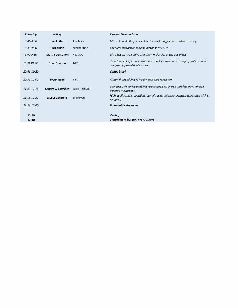
Saturday 9‐May Session: New horizons
8:00‐8:30 Jom Luiten Eindhoven Ultracold and ultrafast electron beams for diffraction and microscopy
8:30‐9:00 Rick Kirian Arizona State Coherent diffractive imaging methods at XFELs
9:00‐9:30 Martin Centurion Nebraska Ultrafast electron diffraction from molecules in the gas phase
9:30‐10:00 Renu Sharma NIST Development of in‐situ environment cell for dynamical imaging and chemical
analysis of gas‐solid interactions
10:00‐10:30 Coffee break
10:30‐11:00 Bryan Reed IDES (Tutorial) Modifying TEMs for high time resolution
11:00‐11:15 Sergey V. Baryshev Euclid TechLabsCompact GHz device enabling stroboscopic laser‐free ultrafast transmission
electron microscopy
11:15‐11:30 Jasper van Rens EindhovenHigh quality, high repetition rate, ultrashort electron bunches generated with an
RF cavity
11:30‐12:00 Roundtable discussion
12:00 Closing
12:30 Transition to bus for Ford Museum
FEIS-2
EXACT TRANSIENT DYNAMICS OF THEANDERSON IMPURITY
Andrey Antipov, Qiaoyuan Dong, Emanuel Gull
University of Michigan
We study dynamics of a single Anderson impurity model sub-ject to voltage and thermal quenches. We develop a hybridizationexpansion diagrammatic Monte Carlo algorithm to describe theexact dynamics of the problem. By including the initial correla-tions into the problem we describe the destruction of the Kondostate and infer the characteristic time scales of the problem. Aninterplay between different time scales of spin and charge exci-tations of the strongly correlated setup as visible in the ultra-fastresponse is in the focus of our study. We compare our results withfrequently employed non-crossing and one-crossing approxima-tions.
——————————————–
COMPACT GHZ DEVICE ENABLINGSTROBOSCOPIC LASER-FREE ULTRAFASTTRANSMISSION ELECTRON MICROSCOPY
Gwanghui Ha1, Jiaqi Qiu1, Chunguang Jing1, SergeyAntipov1, June W. Lau2, Yimei Zhu3, and Sergey V.
Baryshev1
1. Euclid TechLabs ; 2. Materials Science and EngineeringDivision, National Institute of Standards and Technology ;3. Department of Condensed Matter Physics and Materials
Science, Brookhaven National Laboratory
Time-resolved experiments looking at fundamental processes ofatomic/molecular motion and interaction set challenging require-ments: lateral resolution ∼ 1 A and time resolution ∼ 100 fs. Acombined resolution metrics called space-time resolution (STR)must reach ∼ 10−23 m·s or smaller. X-ray and electron based me-thods are best candidates to push the boundaries of STR due tothe smallest wavelength of the primary probe. Currently, x-ray in-struments such as free electron lasers and high harmonic genera-tion laser microscopes have STR between 10−22 and 10−20 m·s.However, these have limited accessibility, either because of ex-orbitant equipment cost or meager beam time allocations. Trans-mission electron microscope (TEM), being the gold standardtechnique in spatially-resolved imaging, cannot resolve dynamicevents in its default form. Until recently, TEM has been ruledout as a viable time-resolved technique. Successful integrationof lasers with TEM created truly new fields called ultrafast anddynamic electron microscopy (UTEM and DTEM). Neverthe-less, laser (repetition rate) and photocathode (quantum efficien-cy) performance limitations and cost seriously limit applicationsand ubiquity of UTEM and DTEM. Alternatively, a pre-existingelectron direct current (dc) beam can be blanked/unblanked withsome periodicity and temporal structure of an electron pulse inthe sequence. A device and a method for producing ultrashortelectron pulses with GHz repetition rates via pulsing a dc be-am will be presented. These are based on an electromagnetic-mechanical pulser (EMMP) that consists of a series of transverse
deflecting cavities and magnetic quadrupoles. The EMMP modu-lates and chops the incoming dc beam and converts it into femto-to pico-second electron pulse sequences at > 1 GHz repetitionrates, as well as controllably manipulates the resulting pulses inthe phase space. The EMMP is largely tunable, i.e. temporal pul-se length and repetition rate are both tunable. A generalized ma-trix description of the EMMP in thin lens approximation will bepresented together with particle tracking results. When appliedto TEM, a GHz stroboscopic TEM with STR metrics between10−23-10−20 m·s can be realized. Such stroboscopic microscopecan be simply based on a traditional TEM platform with a ther-mionic or field emission electron source with no additional laserand photocathode needed.
——————————————–
EXPERIMENTS AND SIMULATIONS OF PHASETRANSFORMATIONS AT SIMILAR LENGTH AND
TIME SCALES
Geoffrey H. Campbell, Tae-Wook Heo, Joseph T.McKeown, Linh Nguyen, Robert E. Rudd, and Melissa K.
Santala
Lawrence Livermore National Laboratory
We have developed time resolved in situ observation methods ba-sed on the Dynamic Transmission Electron Microscope (DTEM)to characterize solid - solid and solid - liquid phase transformati-ons in a range of systems, including phase change materials, se-miconductors, and metallic alloys and intermetallics. The DTEMprovides data from which phase change kinetics can be preciselymeasured in the nanosecond and nanometer regimes. We use ato-mistic simulations to explore nucleation and growth rates of newphases for comparison with the experimental measurements. Wealso use atomistic simulations and experimental measurementsto parameterize phase field simulations to capture microstructuredevelopment during the phase transformation and its interactionwith pre-existing structure. We will discuss insights brought bythe synergy of the experiments with the simulations. This workperformed under the auspices of the US Department of Energy,Office of Basic Energy Sciences by Lawrence Livermore Natio-nal Laboratory under contract DE-AC52- 07NA27344.
——————————————–

ULTRAFAST ELECTRON DIFFRACTION FROMMOLECULES IN THE GAS PHASE
Martin Centurion
University of Nebraska - Lincoln
Ultrafast electron diffraction can in principle be used to obser-ve the structure of a molecule as it undergoes a photoreaction,providing images of the initial, intermediate and final states withatomic resolution. Achieving this goal requires overcoming twomajor challenges: The first challenge is to retrieve the structuredirectly from the diffraction pattern for molecules that are in thegas phase, and thus have random orientation, and the second isto achieve sufficient temporal resolution to observe the relevantdynamics. The random orientation of molecules can be overco-me by diffracting from laser-aligned molecules. We have shownthat by using femtosecond laser pulses to impulsively align themolecules, it is possible to capture a field-free diffraction pat-tern while the molecules are transiently aligned. First experimentwere done on a static molecular structure, and we have recentlydemonstrated imaging of a short lived excited state, with a re-solution of about 1 ps. The second challenge is to improve thetemporal resolution to the regime of 100 fs in order to map thestructural changes as the molecule transitions from the groundto the excited states. This means not only delivering sufficient-ly short pulses on the target, but also compensating the effect ofthe velocity mismatch of electrons and laser as they traverse thesample. We are pursuing two approaches for this, one is to useRF compression of 100 keV electron pulses in combination witha tilted laser pulse excitation to overcome the velocity mismatch,the second is to use relativistic (MeV) electron pulses where thevelocity mismatch is negligible.
——————————————–
MAPPING TRANSIENT ELECTRIC FIELDS WITHPICOSECOND ELECTRON BUNCHES
Jie Chen
Shanghai Jiao Tong University
Transient electric field (TEF), which is an important but hardlyexplored parameter of laser plasmas, can now be diagnosed expe-rimentally with combined ultrafast temporal resolution and fieldsensitivity using femtosecond to picosecond electron or protonpulses as probes. However, it is challenging to simultaneouslyrecord both the global and local TEF features owing to poor highspatial resolution. We present a combined diagnostic of electronschlieren-type radiography and the Abel inversion method to di-rectly measure the 3D TEFs using simultaneous 80-micron spa-tial and 3.7-picosecod temporal resolutions. We show that theTEFs, which are induced by femtosecond laser pulses illumina-tion on an aluminum foil at 1.9E12 W/cm2 and are on the orderof 1E5 V/m, display a unique “peak-valley” structure along theoutgoing direction of the emitted electron cloud, a feature thathas never been reported. The field map also naturally leads to aquantitative characterization of the transient density distributionof the electron cloud, which has a charge density of -0.04 C/m3
and expands at a speed of 4E6 m/s. By combining the micronspatial and picosecond temporal resolutions, this type of radio-graphy using charged particle pulses should enable the mappingof various fast-evolving field structures, such as those found inplasma-based particle accelerators.
——————————————–
COHERENT CONTROL OF FEMTOSECONDPULSES AND THEIR APPLICATIONS
Marcos Dantus
Michigan State University
Femtosecond pulses allow the unprecedented opportunity to pro-be matter on a timescale that is faster than atomic motion, as re-cognized by the 1999 Nobel Prize for Femtochemistry. The nextfrontier is to elucidate and control laser-matter interactions. Thistalk will present efforts in the Dantus group towards achievingthis ambitious goal. The first step in this quest is to control thelaser source itself. I will describe how coherent control has ledto automated pulse compression down to near single-cycle 3.8 fspulses. The talk will cover applications related to (a) improvingnonlinear optical microscopy by using shaped laser pulses; (b)nanoplasmonic dephasing in single, paired, and dendritic chainsof nanoparticles; and (c) standoff detection of explosives.
——————————————–
FLOQUET-BLOCH STATES ANDPHOTO-INDUCED CHIRAL EDGE MODES IN
MONOLAYER TMDCS
Tom Devereaux
Stanford and SLAC National Accelerator Laboratory
In this talk I will discuss manipulation of electron dynamics viaoff-resonant application of light, via dynamical breaking of sym-metries & dynamical modulation of the electronic band struc-ture. I will present detailed predictions of dynamic band struc-ture manipulation in pump-probe angle-resolved photoemissionresponse in transition metal dichalcogenides. The results showa dynamical breaking of valley degeneracy/TRS via circularly-polarized light, a scheme to achieve a condensed-matter realizati-on of light-induced chiral edge modes for weak pump fields, andthe development of topological Wannier Stark physics at strongpump fields. This provides a rich phase diagram of light-inducedFloquet topological phases.
——————————————–
ATTOSECOND DIAGNOSTICS OF MUTI-GEVELECTRON BEAMS USING W-BAND
DEFLECTORS
Valery Dolgashev, Paul Emma, Massimo Dal Forno,Jeffrey Neilson
SLAC National Accelerator Laboratory
Performance of the LCLS free electron laser is determined by theproperties of extremely short electron bunches. The multi-GeVelectron bunches in LCLS are less than 100 fs long. Optimiza-tion of beam properties and understanding of free-electron laseroperation require electron beam diagnostics with time resolutionof less than 10 fs. These were achieved by using the 2-m-longX-band RF deflector. As for now, the performance and utility ofthis deflector remains unchallenged. We propose the next gene-ration of this time-resolved beam diagnostic with improvementsin time resolution by an order of magnitude, possibly down to afew hundred attoseconds at 15 GeV. We expect that, as with thecurrent X-band deflector, it will allow smooth commissioning,operation and further improvement of future free electron lasers.This 10-fold increase of the timing resolution could, in principal,be achieved by scaling the existing X-band system. In that case,the new deflector system would be ∼20 meter long and poweredby ten 50 MW X-band klystrons. We see this as an impracticalsolution and instead propose to increase the operating frequencyof the deflector from 11 GHz to ∼90 GHz. A 1-meter-long de-flector could provide 10-times better temporal resolution down to∼0.4 fs, or less. We envision two practical options: one poweredby ∼10-MW 90-GHz rf source, and another one, passive: powe-red by the bunch’s self-wake fields. In this talk we will reviewSLAC’s experience with X-band deflectors and 100 GHz high-gradient accelerating structures, then will discuss the potential of90 GHz rf deflectors.
——————————————–
APPLICATIONS OF LASER STREAKING ATX-RAY FREE ELECTRON LASERS
Gilles Doumy
Argonne National Laboratory
X-ray radiation has been long used to address selectively atomsand to yield structural information with atomic precision. Theadvent of X-ray Free Electron Lasers (XFEL) is revolutionizingthe field of time resolved x-ray techniques. The availability oftunable pulses ranging from the soft to the hard x-ray region,and lasting only few tens of femtoseconds, or perhaps less, isenabling access to unprecedented temporal resolution. However,knowledge of the temporal properties of the x-ray pulses is poor,and synchronization to external sources introduces a timing jit-ter that dominates the fast dynamics and needs to be correctedfor every shot. Manipulating electrons generated by x-ray indu-ced ionization and subsequent relaxation processes represents apowerful concept to gain access to those properties. Specifical-ly, using laser streaking techniques developed by the attosecondcommunity, one can measure the pulse duration, and possibly im-prove the temporal resolution of pump probe experiments whereelectrons are collected to follow the processes by use of a self-referencing measurement. Illustration is presented following Au-ger decay in the time domain.
——————————————–
IMAGING THE ULTRAFAST SPIN-LATTICEMOTION DURING ULTRAFAST ALL-OPTICAL
SWITCHING OF FERROMAGNETS
Hermann Durr
SLAC National Accelerator Laboratory
Understanding the ultrafast interplay between charge, magneticand lattice degrees of freedom is central to gaining control ofcondensed matter phenomena as diverse as insulator-metal tran-sitions and magnetic switching. Although magnetism in metallicsystems is expected to couple only weakly to phonons, the ob-served ultrafast demagnetization and all-optical magnetic swit-ching demonstrate just the opposite. Femtosecond soft x-ray pul-ses from the Linac Coherent Light Source, offer the unique op-portunity to image in realtime the ultrafast spin dynamics thatleads to magnetization reversal. Hard x-rays and fs electron pul-ses enable first glimpses at the laser-induced lattice motion reve-aling unexpected spin-lattice relaxation channels. Understandingthe evolving spin-lattice motion on the fs time and nm lengthscales associated with the exchange interaction opens new possi-bilities of engineering angular momentum relaxation channels inmagnetic systems.
[1] E. Beaurepaire, et al., Phys. Rev. Lett, 76, 4250 (1996).[2] C.D. Stanciu et al., Phys. Rev. Lett, 99, 047601 (2007).[3] S. Mangin, et al. Nature Materials 13, 286 (2014).[4] C. E. Graves, A. H. Reid et al., Nature Materials, 12, 293(2013).
——————————————–
PHOTOEMISSION AND BEAM PHYSICS OFSPACE-CHARGE DOMINATED ELECTRON
BUNCHES
Phillip M. Duxbury, Jenni Portman, Chong-Yu Ruan,Martin Berz, Kyoko Makino, He Zhang, Zhensheng Tao
Michigan State University
Efficient computational methods to treat space charge effects areessential to accurate simulation of electron bunches with highelectron density. High electron density is typical at the photoca-thode, in RF cavities and at the focal plane, where N-particle si-mulations are critical. Stochastic space charge effects occurringat photoemission place a fundamental limitation on UEM andUED resolution. By combining N-particle simulations and ana-lytic Gaussian calculations we provide estimates of the practicallimits of UEM and UED resolution as a function of beam energy.
[1] Z. Tao, et al., Journal of Applied Physics 111(4), 044316, pa-ges 1-10 (2012).[2] J. Portman, et al., Applied Physics Letters 103, 253115 (2013)[3] J. Portman, et al., J. Appl. Phys. 116, 174302 (2014).[4] He Zhang, Jenni Portman, Zhensheng Tao, Phillip Duxbury,Chong-Yu Ruan, Kyoko Makino and Martin Berz. “The Diffe-rential Algebra Based Multiple Level Fast Multipole Algorithmfor 3D Space Charge Field Calculation and Photoemission Si-mulation”. Proceedings of the 9th International Conference onCharged Particle Optics, Brno September, 2014.
——————————————–

ULTRA-LOW-ENERGY SEM AND STEM
Ludek Frank
ISI ASCR, Brno, Czech Republic
The general trend for reducing the energies of primary electronsin electron microscopy, which is promising from the point ofview of desirable enhancement of the scattering rate of electronsin targets, had faced a gradual deterioration of the image resolu-tion. Biasing the sample to a high negative voltage and makingthe electrons arbitrarily slow solely on and inside the sample hasshown itself to be far more feasible than originally expected. Themost important thing is that the fundamental aberration coeffi-cients (spherical and chromatic) of a combination of an objectivelens and an immersion electrostatic lens formed by the biasedsample decrease with the decreasing landing energy of the elec-trons. As a result, the spot size in scanning systems may beco-me nearly independent of the landing energy of the electrons.The requirements placed on samples are strict but feasible: bulksamples observed in the reflection mode have to be flat (but notpolished), while thin films for the transmission mode should notexceed 10 nm in thickness. Detection of signal electrons is grea-tly facilitated by acceleration of both reflected and transmittedelectrons in the field of the biased sample and their collimationtoward the optical axis. Naturally, the reflected axial ray usual-ly escapes detection through the detector bore, so special arran-gements are needed (such as separated primary and signal co-lumns with a beam splitter in LEEM). The interaction of slowelectrons is not only more intensive than that at standard ener-gies, but even scattering phenomena appear which are not other-wise available. The wavelength of the electrons approaches theinteratomic distances, so diffraction and interference phenome-na become sources of image contrasts even in signals acquiredwith single-channel detectors in the SEM. Moreover, nontrivialdistribution of the local density of states close above the vacu-um level makes the electron reflectivity dependent on energy ina way specific to a crystallographic system and its orientation, soreflectivity may serve as a fingerprint of the local crystallinity.STEM observation of 2D crystals at units of eV and measure-ment of their reflectivity and transmissivity provide informationsupporting calculations of their electronic structure. Biologicalspecimens in the form of ultrathin sections exhibit very high con-trasts showing complete structure details without the addition ofthe heavy metal salts that highlight only some of the details. Thebenefits of very-low-energy EM are still being uncovered afterhaving been in routine use for several years.
——————————————–
PUMP/PROBE ARPES IN ELECTRON-PHONONCOUPLED METALS
Jim Freericks1, Lex Kemper2, Brian Moritz3, MichaelSentef4 and Tom Devereaux3
1. Georgetown University ; 2. Lawrence BerkeleyLaboratory ; 3. Stanford University and SLAC ; 4.
Hamburg University
In this talk, I will summarize recent work we have done in stu-dying the properties of electron-phonon coupled metals that areexamined with pump/probe photoemission studies. In the lowfluence regime, one can map time-dependent data within the so-called “phonon window”(where the relaxation has a bottleneckand the system relaxes slowly) onto the equilibrium dynamics.As the fluence is increased, the nonequilibrium driving starts tochange the density of states and the scattering, but it does so in away that preserves the integrated strength of the electron-phononcoupling due to a nonequilibrium sum rule. When one goes intothe superconducting state, a moderate fluence can be employedto directly excite the Higgs (or amplitude) mode of the super-conductivity, whose dynamic oscillations are correlated with thetransiently evolving superconducting gap. We will also discussimplications of this work for experiments.
——————————————–
RF LOCKING OF FEMTOSECOND LASERS
Josef Frisch, Karl Gumerlock, Justin May, Steve Smith
SLAC National Accelerator Laboratory
In RF gun based femtosecond electron imaging systems manyexperiments require low noise timing control between a pumplaser and the electron beam. As the beam time is influenced byboth the RF gun photocathode laser and the gun RF fields, pre-cision timing control between the laser and the gun RF fields isneeded. We discuss RF based laser timing control, and the systembeing used in the SLAC ASTA femtosecond electron diffractionsystem. This system has been used for 100 femtosecond scaletiming experiments.
——————————————–
BRIGHT CIRCULARLY POLARIZED SOFT X-RAYHIGH HARMONICS FOR X-RAY MAGNETIC
CIRCULAR DICHROISM
Patrick Grychtol
JILA - University of Colorado
Ultrafast light sources based on the high-harmonic up-conversionof femtosecond laser pulses have been successfully employed toaccess resonantly enhanced magnetic contrast at the M absorp-tion edges of the 3d ferromagnets Fe, Co and Ni in a table-topexperimental setup. Thus, it has been possible to study element-specific dynamics in magnetic materials at femtosecond time sca-les in a laboratory environment, providing a wealth of opportu-nities for a greater fundamental understanding of correlated phe-nomena in solid-state matter. However, these investigations haveso far been limited to linear polarized higher harmonics, sincemost techniques by which circular soft x-rays can be generatedare highly inefficient reducing the photon flux to a level unfitfor scientific applications. Besides presenting key findings of ourultrafast studies on charge and spin dynamics, we introduce asimple setup which allows for the efficient generation of circularharmonics bright enough for x-ray magnetic circular dichroic.Our work thus represents a critical advance that makes possibleelement-specific imaging and spectroscopy of multiple elementssimultaneously in magnetic and other chiral media with very highspatial and temporal resolution, using tabletop-scale setups.
——————————————–
DIAGRAMMATIC MONTE CARLO FORREAL-TIME PROPAGATION
Emanuel Gull, Guy Cohen, Andrey Antipov
University of Michigan, Columbia University
We present diagrammatic Monte Carlo methods for the real-timedynamics of quantum impurities, introduce the concept of bold-line Monte Carlo methods, and present a formalism to obtainspectral functions from an auxiliary lead formalism. Using thesemethods we show results for nonequilitrium systems, includingthe voltage splitting of the Kondo peak.
——————————————–
A LASER-PLASMA BASED PARTICLEACCELERATOR FOR ULTRAFAST ELECTRON
DIFFRACTION APPLICATIONS
Zhaohan He1, John Nees1, Roye Clarke2, KarlKrushelnick1, and Alec Thomas1
1. Center for Ultrafast Optical Science, University ofMichigan ; 2. Department of Physics, School of LSA,
University of Michigan
Femtosecond bunches of electrons with relativistic to ultra-relativistic energies can routinely be produced by laser plasmawakefield accelerators (LWFA) driven by 10-100 TW, high peakpower laser systems. Scaling the electron energy down to sub-relativistic and MeV level is possible using a sub-TW laser sy-stem. Such an electron source can be a potential candidate for
ultrafast electron diffraction (UED) applications, due to the int-rinsic short bunch duration and perfect synchronization with theoptical pump. We will present results of transmission electrondiffraction from a crystalline sample, using LWFA electrons dri-ven by 8-mJ, 35-fs laser pulses at 0.5 kHz. The accelerated elec-trons are collimated with a solenoid magnetic lens. Single-shotdiffraction patterns are obtained from a single-crystal gold foil ina proof-of-principle experiment to demonstrate the beam quali-ty and charge. Temporal characterization of the electron bunchesusing a non-equilibrium plasma reveals the linear energy chirp,which can be utilized for further electron bunch compression ortime-resolved measurement in streak mode. Preliminary pumpprobe studies on gold foil will also be presented.
——————————————–
BRIGHT AND DURABLE FIELD EMISSIONSOURCE DERIVED FROM REFRACTORY-METAL
TAYLOR CONES
Gregory Hirsch
Hirsch Scientific
A novel method for in-situ creation of field emission tips havingdesirable operational characteristics is described. In this process,the end of a refractory metal wire is melted in vacuum using afocused laser beam. A high positive potential simultaneously ap-plied to the wire is used to electrostatically form a Taylor conefrom the liquid meniscus on the wire end. This experimental con-figuration can be regarded as an unusual type of Liquid Metal IonSource (LMIS). Upon cessation of the laser power, the Taylor co-ne freezes almost instantaneously. Extremely high radiative andconductive cooling assures that solidification of the cone is ex-ceptionally rapid, resulting in a frozen structure having a shapeand surface smoothness very closely matching that of the initi-al liquid Taylor cone. Very uniform and sharp metal cones havebeen generated from a variety of refractory metals having mel-ting points as high as tungsten. The frozen Taylor cones can besubsequently employed as field emitters by simply reversing thepolarity of the applied voltage. This type of field emission sourcemay find widespread application for both continuously operatingelectron guns, as well as with pulsed sources. A feature of thistechnology is the ability to rapidly regenerate the frozen Taylorcones after they undergo some degradation during use. When em-ployed for ultrafast applications using laser-assisted field emissi-on, the capability to readily restore the emitter to pristine conditi-on in-situ may permit the attainment of emission currents near orabove damage threshold, without undue concern regarding redu-ced field-emitter lifetimes. The large characteristic opening angleof Taylor cone structures (near 98.6 degrees full angle) results inelectron emission being electrostatically directed into a relative-ly low-divergence forward directed beam. This may be advanta-geous for achieving low emittance and bright electron sources.The current state of this technology will be described, as well asplans for further development.
——————————————–
OPTIMAL ULTRAFAST LASER PULSE-SHAPINGTO DIRECT PHOTO-INDUCED PHASE
TRANSITIONS
Bin Hwang, Jenni Portman, Phillip Duxbury
Michigan State University
Photo-induced phase transitions (PIPT) in quantum and/or com-plex materials are the epitome of challenging non-equilibriummany-body phenomena, that also have a wide range of potenti-al applications. We present a computational approach to findingoptimal ultrafast laser pulse shapes to control the outcome ofpump-probe PIPT experiments. The Krotov approach for Quan-tum optimal control theory (QOCT) is combined with a KeldyshGreen’s function calculation to describe experimental outcomessuch as photoemission, transient single particle density of statesand optical responses. Results for a simple model charge densitywave system will be presented.
——————————————–
STRONG FIELD EFFECTS ON ORGANICCONDUCTORS INDUCED BY 1.5-CYCLE
INFRARED LIGHT PULSE
Shinichiro Iwai
Department of Physics, Tohoku University
Ultrafast control of conduction and magnetic properties in stron-gly correlated systems has been extensively studied from the per-spective of photoinduced insulator-to-metal transitions, or, equi-valently, the “melting” of electronic orders in Mott insulators,charge ordered systems, and charge/spin density wave materials.On the other hand, the development of strong electric fields (>MV/cm) of few-cycle optical pulses and recent theoretical stu-dies using dynamical mean-field theory (DMFT) [1] suggest thatextreme non-equilibrium electronic states such as charge locali-zation, negative temperatures, and repulsive-attractive conversi-on can be achieved. Among those highly non-equilibrium pheno-mena, reducing the intersite transfer integral t by modulating thesite energy under high-frequency [ω >t/(h)] continuous-wave(CW) and pulsed AC electric fields E(ω) have attracted attentionfor > 30 years[2, 3]. Such an intense modulation of the elec-tronic structure driven by a strong electric field, referred to as“dynamical localization”, provides a new strategy for control-ling charge motion in strongly correlated materials, which have acompeting energy balance between the on-site or intersite Cou-lomb repulsion and t. Here we describe a charge localization in-duced by the 9.3 MV/cm instantaneous electric field of a 1.5 cy-cle (7 fs) infrared pulse in an organic conductors alpha-(ET)2I3(ET; bis[ethylenedithio]-tetra-thiafulvalene)[4], (TMTTF)2AsF6
(TMTTF; tetramethyltetrathiafulvalene). In alpha–(ET)2I3, a lar-ge reflectivity change of > 25% and a coherent charge oscillationalong the time axis reflect the opening of the charge ordering gapin the metallic phase. This optical freezing of charges, which isthe reverse of the photoinduced melting of electronic orders, isattributed to the 10% reduction of t driven by the strong, high-frequency electric field. In (TMTTF)2 AsF6, such reduction of thas been demonstrated by the analysis of the transient reflectivi-ty spectra using Drude-model. Dependence on the field intensityand the direction of electric field can be also understood in termsof dynamical localization.
[1] H. Aoki, N. Tsuji, M. Eckstein, M. Kollar, T. Oka, P. Werner,Rev. Mod., Phys. 86, 779, (2014).[2] D. H. Dunlap, and V. M. Kenkre, Phys. Rev. B 34, 3625-3633(1986).[3] K. Nishioka and K. Yonemitsu, J. Phys. Soc. Jpn., 83,024706(2014).[4] T. Ishikawa, S. Iwai et al., Nature commun. 5, 5528(2014).
——————————————–
COHERENT DIFFRACTIVE IMAGING METHODSAT XFELS
Richard A Kirian
Arizona State University
In this talk I will discuss some unique ways in which coherentdiffraction data from bioparticles in nanocrystal, solution, andgas phases can be obtained at x-ray free- electron lasers (XFELs).For example, the continuum of diffraction intensities that are ob-served when protein nanocrystals are illuminated with coherentfemtosecond XFEL pulses gives rise to information in regionsbetween the usual Bragg peaks. Such information can be usedto determine diffraction phases without prior information, andwithout resolution restrictions. Similarly, intensity correlationscan be observed in what might appear to be isotropic solutionscattering, because XFELs effectively freeze atomic motion andcapture intensity fluctuations that are weak and persist only fora brief period of time. Intensity correlations contain a wealth ofstructural information from hydrated biomolecules at room tem-perature, well beyond the level of information that conventionalsolution scattering contains. The key to gathering the needed da-ta at XFELs for these methods and others often lies in the sam-ple delivery method. Numerous innovations have been requiredin order to replenish precious sample rapidly, since in most bio-imaging schemes each target is vaporized and must be rapidlyreplaced with a new one before the next XFEL pulse arrives. Li-quid jets, for example, were key to the development of methodssuch as serial femtosecond crystallography and sub-picosecondtime- resolved wide-angle solution scattering. As time permits,I will discuss some of our recent efforts to improve on sampledelivery methods, with a look toward the upcoming generationof XFELs that will produce pulses at MHz repetition rates.
——————————————–
ULTRAFAST STRUCTURAL DYNAMICS INBI2SR2CACU2O8+δ UNDER POLARIZED
PHOTOEXCITATION
T.Konstantinova1,2, L.Wu1, J.Li1, A.H.Reid3, X.Wang3
and Y.Zhu3
1. Brookhaven National Laboratory ; 2. Stony BrookUniversity ; 3. SLAC National Accelerator Laboratory
We report the structural dynamics of high-Tc superconductorBi2Sr2CaCu2O8+δ (Bi 2212) induced by femtosecond 1.5eVphotoexcitation using ultrafast electron diffraction (UED). A pro-nounced feature of Bi 2212, similar to other high-Tc cuprates, isits ab-plane anisotropy of electronic properties (a superconduc-ting gap below Tc and a pseudogap in the normal state) due tothe symmetry of d-wave with nodes near the bisectors of Cu-O bonds. The electron coupling to half-breathing in-plane Cu-Ostretching and out-of-plane O-buckling phonon modes is one ofthe possible explanations of electron glueing in a superconduc-ting state. Thus, the strength of the coupling is predicted to havethe d-wave symmetry as well. In current research the use of thepump beam polarized along a- and b- axes and at 45 degree tothem allows to investigate the anisotropy of lattice response tothe photoexcitation. The lattice dynamics along different crystalaxes has been examined while separating signals from the basiccrystal structure and from the incommensurate modulation alongb-axis. This separation helps us to distinguish between electro-nic and lattice origin of the structure modulation, which is stillan open question. The dynamics was monitored up to 25 ps afterexcitation with 0.4 ps resolution. We will present the intensitychange for the diffraction peaks, corresponding to dynamics ofdifferent directionalities of the crystal structure under the pho-toexcitation with variable polarization, as well as the analysisof the intensity trend. Detailed data analysis and challenges inquantification of our RF-MeV UED experiments will be discus-sed. Work at Brookhaven National Laboratory was supported bythe US DOE, Office of Science, Basic Energy Sciences, underContract No. DE-SC0012704.
——————————————–
DEVELOPMENTS OF 4D-TRANSMISSIONELECTRON MICROSCOPE AND ANISOTROPIC
LATTICE DYNAMICS OF MULTI-WALLEDCARBON NANOTUBES
Jianqi Li, Shuaishuai Sun, Gaolong Cao, Huaixin Yang,Zhongwen Li, Huanfang Tian
Institute of Physics, Chinese Academy of Science
Recent advances in the four-dimensional ultrafast transmissi-on electron microscope (4D-UTEM) with combined spatial andtemporal resolutions have made it possible to directly visualizestructural dynamics of materials at the atomic level. In this pa-per, we report on our development on a 4D-UTEM which can beoperated properly on either the photo-emission or the thermionicmode. We demonstrate its ability to obtain sequences of snaps-hots with high spatial and temporal resolutions in the study of lat-tice dynamics of the multi-walled carbon nanotubes (MWCNTs).This investigation provides an atomic level description of remar-kable anisotropic lattice dynamics at the picosecond timescales.
Moreover, our UTEM measurements clearly reveal that distin-guishable lattice relaxations appear in intra-tubular sheets on anultrafast timescale of a few picoseconds and after then an evidentlattice expansion along the radical direction. These anisotropicbehaviors in the MWCNTs are considered arising from the varie-ty of chemical bonding, i.e. the weak van der Waals bonding bet-ween the tubular planes and the strong covalent sp2-hybridizedbonds in the tubular sheets.
——————————————–
MEASURING CHARGE AND ORBITALORDERING DYNAMICS IN LAYERED
MANGANITES
J. Li1, P. Zhu1,2, L. Wu1, T. Konstantinova1, J. Tao1, J.P.Hill1, X.J. Wang1,3 and Y. Zhu1
1. Brookhaven National Laboratory ; 2. ShanghaiJiaoTong University ; 3. SLAC National Accelerator
Laboratory
We report a study of the dynamics of electronic order in a pho-toinduced phase transition in bi-layered LaSr2Mn2O7 using timeresolved MeV electron diffraction at 77K. We found that meltingof the orbital order (OO) takes place with a similar time constantof the charge order (CO). The time constant and order parameterduring the phase transition is investigated at various sample tem-perature and pump fluence. No substantial difference in dynamicbehavior is observed. Saturation and threshold fluence are obtai-ned by fitting the order parameter dependence on pump fluence.By quantitative comparison between experimental electron dif-fraction patterns and simulations based on dynamic diffractiontheory, we found that the charge disproportionation on the twoMn ions is very small and the intensity of the CO peaks can bemostly attributed to the secondary harmonics of orbital orderingin our MeV-UED measurements. We further analyzed the influ-ence of different phonon models on diffraction intensities in thesystem. The results suggest that Jahn-Teller distortion domina-tes the photo-induced structure phase transition. Our study shedslight on the various and often intertwined degrees of freedomand their responses to external perturbations in strongly corre-lated electron systems. The authors would like to thank S.-W.Cheong for providing the samples. This research was supportedby the US Department of Energy, Basic Energy Sciences, underContract No: DE-SC0012704.
——————————————–

ABOVE-THRESHOLD IONIZATION ANDWAVEPACKET DYNAMICS OF RYDBERGELECTRONS BOUND TO THEIR IMAGE
POTENTIAL IN A SINGLE METALLICNANOSTRUCTURE
Jrg Robin1,2, Jan Vogelsang1,2, Benedek Nagy1,2, PetraGross1,2, Christoph Lienau1,2
1. Institute of Physics, Carl von Ossietzky University ;2. Center of Inferface Science, Carl von Ossietzky
University
In atomic and molecular physics, the study of strong-field pheno-mena has been a major research focus since many years, inclu-ding a wealth of fundamental processes such as high-harmonicgeneration, attosecond streaking and the generation of attosecondelectron wavepackets for tomographic imaging of molecular or-bitals. During the last few years, single metallic nanostructureshave developed into a veritable testbed for translating conceptsfrom strong-field physics into the realms of nano-optics and so-lid state science. In this talk, I will discuss recent experimentalof our own group probing the dynamics of photoemission fromsharp metallic nanotapers driven by intense few-cycle laser pul-ses. I will demonstrate how to use to optical near-field at the ta-per apex to accelerate and steer ultrashort electron pulses [1] andhow to manipulate such electron pulses by means of the carrier-envelope phase of the driving laser [2]. I will then discuss recentexperimental studies of two-color photoemission from a gold ta-per in which we observed, for the first time to our knowledge,the coherent dynamics of Rydberg electron wavepackets, boundto their image potentials and nanolocalized to a single nanostruc-ture. When illuminating a sharp gold tip with a an apex radiusof 10 nm with a sequence of two time-delayed pulses centered at600 nm and 1.6 m, we observe pronounced temporal modulationsin the photoemission yield. The periodicity of this modulation isin the range of a few tens of fs and depends on the time-orderingof the pulses. The origin of these oscillations is revealed by recor-ding intensity-dependent kinetic energy spectra of the releasedphotoelectrons for optical excitation of the tip with two temporal-ly overlapping visible (600 nm) and near-infrared (1600 nm) pul-ses. The photoelectron spectra are governed by above-thresholdionization and reveal a rich and surprising fine-structure. Our re-sults provide strong evidence that the field enhancement at the tipapex confines the Rydberg orbits locally in all three dimensionsto a volume of a few nm3 only and results in high ATI orders andlarge kinetic energy photoelectrons for laser intensities well be-low the destruction level of the metallic nanostructure. Thus theydemonstrate a new level of quantum control over the motion ofelectrons in strong nanolocalized laser fields and open the doorto time-resolved photoelectron spectroscopy of single nanostruc-tures with ultrahigh time resolution.
[1] D. J. Park, et al. Strong Field Acceleration and Steering ofUltrafast Electron Pulses from a Sharp Metallic Nanotip. Phys.Rev. Lett. 109, 244803 (2012).[2] B. Piglosiewicz et al. Carrier-envelope phase effects on thestrong-field photoemission of electrons from metallic nanostruc-tures. Nature Photon., 8, 37-42 (2014).
——————————————–
ULTRACOLD AND ULTRAFAST ELECTRONBEAMS FOR DIFFRACTION AND MICROSCOPY
Jom Luiten
Eindhoven University of Technology
At Eindhoven University of Technology we are developing newmethods for generating beams for ultrafast electron diffracti-on and microscopy. By femtosecond photoionization of a laser-cooled gas we produce highly charged, highly coherent electronbunches, which ultimately should enable single-shot electron dif-fraction of macromolecules. Using a simple graphite sample werecently generated the first diffraction patterns with the ultracoldelectron source, which provided direct evidence that the sourcehas the required coherence. We are currently characterizing thelongitudinal phase space of the ultracold electron bunches andwe are preparing crystalline samples of the 2D membrane prote-in hydrophobin. Progress will be reported. By implementing anultrafast, RF-cavity-based beam blanker in a FEI Tecnai TEMwe will realize ‘stroboscopic’ ultrafast electron microscopy withsubstantially improved performance in terms of spatial resolu-tion, temporal resolution, and data acquisition time. A special-ly designed compact and energy efficient RF cavity is currentlybeing prepared for installation in the TEM. Progress will be re-ported.
——————————————–
CHARACTERIZATION AND LIGHT-INDUCEDDYNAMICS OF ALKANETHIOL-CAPPED GOLD
NANOPARTICLES SUPRACRYSTALS BYSMALL-ANGLE ULTRAFAST ELECTRON
DIFFRACTION
Giulia Mancini
EPFL, Lausanne, Switzerland
Monodisperse nanoparticles coated with organic ligands formtwo- and three-dimensional supracrystals, which can be regardedas new complex materials, with applications in optoelectronicsand catalysis [1]. The knowledge of the arrangement of the na-noparticles core and their ligands, and its dynamical control uponphoto-irradiation, represents the next frontier in the full charac-terization of these supracrystals. Here we present a femtosecond(fs)-resolved electron diffraction study of a two-dimensional su-pracrystal of alkanethiol-coated gold nanoparticles with femtose-cond time, sub-nanometer spatial resolution and sensitivity downto the carbon and hydrogen atoms in the ligands [2, 3]. Com-bining dynamical diffraction experiments with static real-spaceimaging and simulations, we performed ultrafast-electron dif-fractive imaging obtaining a fs/A-resolved movie of the evolu-tion of the gold NPs and their organic ligands. We show thatboth disordering and ordering dynamics of the supracrystal canbe achieved upon ultrafast laser excitation, demonstrating thatself-organization can be triggered by light. These results revealthe role of thermal disorder in the supracrystal and the ability ofultrafast photo-excitation to induce an ordering transition of theorganic ligands in the picosecond (ps) time scale.
[1] G. Schmid, Nanoparticles: From Theory to Application(Wiley-VCH, 2006).[2] G. F. Mancini et al., Nucl. Instrum. Methods Phys. Res. A691, 113-122 (2012).[3] G. F. Mancini et al., submitted to Nature (2015).
——————————————–
RF PHOTOINJECTOR BASED MEV ELECTRONMICROSCOPY
P. Musumeci
UCLA Department of Physics and Astronomy
Using relativistic electrons from RF photoinjectors offers thepossibility to improve temporal resolution of single-shot trans-mission electron microscopes by three orders of magnitude overcurrent state of the art. Here we discuss our progress in develo-ping a new kind of tool based on this high brightness electronsource. In particular we’ll present our recent results in the con-denser lens stage and our plans to further expand the capabilitiesof the instrument.
——————————————–
CONCEPTS AND PERSPECTIVES ONPHOTOINDUCED STRUCTURAL PHASE
TRANSITIONS
Keiichiro Nasu
IMSS, KEK
We, at first, briefly see the early experimental history of photo-induced structural phase transition study, in connection with theTTF-CA, being the mother material, connotes this new and exci-ting non-equilibrium critical phenomenon triggered only by thevisible lights irradiation. [1-3] We will also see recent develop-ments in the way of observation, not only by the visible lightspectroscopy, but also by the electron beam spectroscopy andSTM methods in connection with the recent discovery of photo-induced “ Diaphite (sp3) ” domain in the graphite crystal, whichoriginally has only the sp2 bonds. [4] Finally, we see the elec-tronic excited domain formation, not by the direct visible lightexcitations, but by the large amplitude lattice displacement pul-ses ( THz pulses ) and their nonlinear propagations.
[1] K.Nasu, Photo-induced phase transitions, ( World Scientific,Singapore, 2004).[2] K. Nasu, Rep. on Prog. Phys. ( IOP, London ) 67(2004)1607-1662.[3] K. Yonemitsu and K. Nasu, Physics Report ( Elsevier ) 465(2008) 1-60.[4] K. Nasu, Eur. Phys. J. B, Colloquium, 75(2010) 415-430.
——————————————–

ULTRAFAST PHOTOEMISSION MICROSCOPY:IMAGING OF ELECTROMAGNETIC FIELDS ON
THE FEMTO-NANO SCALE
Hrvoje Petek
Department of Physics and Astronomy, University ofPittsburgh
By combining ultrafast laser photo excitation with photoelectronemission microscopy we achieve simultaneous femtosecond tem-poral and nanometer spatial resolution of electromagnetic fieldsat nanostructured metal-semiconductor surfaces. A variety of na-no structured surfaces are formed by deposition of Ag films onSi(001) substrates under UHV conditions. The nanostructuredsamples can be imaged with <10 nm spatial resolution eitherby photoemission electron microscopy (PEEM) using UV laseror Hg lamp excitation sources, or by low energy electron mi-croscopy (LEEM). Femtosecond laser excitation in the UV-VISenergy range excites coherent electromagnetic modes of the sam-ple, which are dominated by the localized and propagating sur-face plasmons. The regions of the sample with high field enhan-cement emit photoelectrons by nonlinear two- or three-photonphotoemission process. The photoelectrons are imaged by thePEEM optics to generate spatial maps of the plasmonic field en-hancement in the sample. Tuning of the delay between identi-cal femtosecond pump-and probe pulses generates movies of thespatiotemporal evolution of the plasmon fields. I will cover theprinciples of time-resolved PEEM imaging of surface electroma-gnetic fields and report on recent measurements with a broadlytunable femtosecond laser source.
[1] Kubo, A., et al., Femtosecond Imaging of Surface PlasmonDynamics in a Nanostructured Silver Film. Nano Lett. 5, 1123(2005).[2] Kubo, A., et al., H. Femtosecond microscopy of localized andpropagating surface plasmons in silver gratings. J. Phys. B: At.Mol. Opt. Phys. 40, S259 (2007).[3] Zhang, et al., T. Imaging of surface plasmon polariton fieldsexcited at a nanometer-scale slit. Phys. Rev. B 84, 245442 (2011).
——————————————–
SIMULTANEOUS OBSERVATION OF THEQUANTIZATION AND THE INTERFERENCEPATTERN OF A PLASMONIC NEAR-FIELD
L. Piazza1, T.T.A. Lummen1, E. Quinonez2, Y.Murooka1, B.W. Reed3, B. Barwick2, F. Carbone1
1. EPFL, Lausanne, Switzerland ; 2. Trinity College ;3. LLNL
Surface plasmon polaritons can confine electromagnetic fieldsin subwavelength spaces and are of interest for photonics, op-tical data storage devices and biosensing applications. In analo-gy to photons, they exhibit wave-particle duality, whose differentaspects have recently been observed in separate tailored experi-ments. Here we demonstrate the ability of ultrafast transmissionelectron microscopy to simultaneously image both the spatial in-terference and the quantization of such confined plasmonic fields.Our experiments are accomplished by spatiotemporally overlap-ping electron and light pulses on a single nanowire suspended ona graphene film. The resulting energy exchange between singleelectrons and the quanta of the photoinduced near-field is imagedsynchronously with its spatial interference pattern. This metho-dology enables the control and visualization of plasmonic fieldsat the nanoscale, providing a promising tool for understandingthe fundamental properties of confined electromagnetic fieldsand the development of advanced photonic circuits.
——————————————–
(TUTORIAL) MODIFYING TEMS FOR HIGHTIME RESOLUTION
Bryan W. Reed and Daniel J. Masiel
Integrated Dynamic Electron Solutions, Inc.
This tutorial will describe modifications that convert a conventio-nal TEM to a stroboscopic ultrafast TEM (UTEM), single-shot ormovie-mode Dynamic TEM (DTEM), or hybrid instrument ca-pable of both modes of operation (UDTEM). Piping pulsed lasersinto a microscope is just one step in this process. Experimen-tal demands on reliability, stability, performance, and usabilitymean that every aspect of the modification-laser ports, cathodeselection, column modifications, vibration isolation, laser beamprofile and alignment control, data acquisition, electronics, andsoftware-must be optimized not just individually but also in thecontext of the full integrated system. Without such attention todetail, instrument development time can easily stretch into years,and the resulting instrument can be frustratingly saddled withmundane yet crucial usability issues. Such issues can be avoidedwith forethought and good system-level design, yielding a com-plex, flexible, high-performance, yet relatively easy-to-use in-strument. This is especially true for movie-mode DTEM, whichrequires both an advanced arbitrary-waveform laser system anda perfectly synchronized high-speed deflector system. In closing,we will discuss nanosecond/microsecond-scale pulse shaping,complementing the discussion of femtosecond/picosecond-scalepulse shaping also taking place at this workshop.
——————————————–

HIGH-QUALITY, HIGH-REPETITION RATE,ULTRASHORT ELECTRON BUNCHES
GENERATED WITH AN RF-CAVITY
Jasper van Rens1, Wouter Verhoeven1, Erik Kieft2, JimFranssen1, Peter Mutsaers1 and Jom Luiten1
1. Coherence & Quantum Technology Group, EindhovenUniversity of Technology ; 2. FEI Company
In collaboration with FEI Company, we are studying the possi-bility of using microwave TM110 streak cavities in combinationwith a slit, to chop a continuous electron beam into 100 fs elec-tron bunches. We have shown that this can be done with minimalincrease in transverse emittance and longitudinal energy spread.Furthermore, these bunches are created at a repetition rate of 3GHz. Accurately synchronized to a mode-locked laser system,this allows for high-frequency pump-probe experiments with thebeam quality of high-end electron microscopes.
At Eindhoven University of Technology, we will soon implementsuch a cavity in a 200 keV Tecnai, which should result in high-frequency ultrafast (S)TEM with sub-ps time-resolution whilemaintaining the atomic spatial resolution of the TEM.
——————————————–
HOLOGRAPHIC IMAGING AND OPTICALSECTIONING IN THE
ABERRATION-CORRECTED STEM
Harald H Rose
Ulm University
The correction of spherical aberration enables efficient hologra-phic imaging in the scanning transmission electron microscope(STEM) if the energy width of the incident electron beam is suf-ficiently reduced. Holographic imaging implies that the Fouriertransform of the image is linearly related with the elastic scatte-ring amplitude of the object. Effective optical sectioning can berealized in the aberration-corrected STEM by employing “holo-graphic” phase-contrast imaging. This imaging mode requires asegmented bright-field detector and a Fresnel phase plate whichcan be formed with a sufficient degree of accuracy by adjustingappropriately the third-order spherical aberration and the defo-cus of the corrected objective lens. By subtracting the signals ofthe annular detector segments covering the region of destructiveinterference of the scattered wave with the non-scattered wavefrom that recorded by the annular segments covering the regi-ons of constructive interference, we obtain a pure phase contrastimage which may be conceived as a holographic image becausethe terms of the intensity which depend quadratic on the scat-tering amplitude cancel out. Theoretical results will be presen-ted which demonstrate the feasibility of the proposed method. Inparticular, the method enables the transfer of spatial frequenciesover a large range which exceeds significantly that of conventio-nal phase contrast imaging.
——————————————–
HIGH-BRIGHTNESS BEAMS FOR ULTRAFASTMICRODIFFRACTION AND IMAGING
Chong-Yu Ruan
Department of Physics and Astronomy, Michigan StateUniversity
Currently the ultrafast electron diffraction (UED) with 103-105
electrons per pulse has achieved sub-picosecond temporal reso-lution and atomic resolution. However, direct ultrafast imagingof a nanometer scale specimen through coherent single-particlediffraction has not been achieved largely due to insufficient in-tensity when tuned to a coherence length that matches the si-ze of the specimen under the projected phase space density. Asource-limited performance can be delivered by proper and fle-xible electron optical designs to rotate the phase space so as tooptimize the performance limited only to the Liouvilles theoremconstraint, which is ultimately subject to the brightness of theelectron sources preserved during the production of the electronbeam. Utilizing a recently implemented high-brightness electronsource under a DC-gun linear acceleration field, we test the per-formance of such a beamline for ultrafast electron microdiffrac-tion and coherence imaging. We demonstrate the feasibilities ofsingle-shot mirodiffraction on a single micrometer-sized domainin Highly Ordered Pyrolytic Graphite (HOPG) and coherent dif-fractive imaging of 10 nm scale charge-ordered domain structu-res in single-crystal complex materials, as qualified by the mea-sured brightness at the sample plane. These initial results showthat source-limited performance even from a sub-relativistic elec-tron beamline can drastically improve the current performance ofultrafast electron imaging and diffraction.
——————————————–

ULTRAFAST TRANSMISSION ELECTRONMICROSCOPY BASED ON A LASER-DRIVEN
SCHOTTKY FIELD EMITTER
Armin Feist, Katharina E. Echternkamp, Reiner Bormann,Nara Rubiano da Silva, Marcel Moller, Jakob Schauss,Sergey V. Yalunin, Sascha Schafer, and Claus Ropers
4th Physical Institute, University of Gottingen
Ultrafast transmission electron microscopy (UTEM) is a pro-mising technique, which utilizes sub-picosecond electron pulseswithin an electron microscope to resolve ultrafast laser-inducedprocesses on a nanometer length scale [1]. The potential of thisapproach crucially depends on the implementation of advancedlaser-driven electron sources which make full use of the high-quality electron optics within a TEM. Here, we present recentdevelopments and first applications of the Gottingen UTEM in-strument, which is based on the custom modification of a JEOL2100F Schottky field emission TEM. The laser-triggered nanos-copic electron source employs localized single-photon photoe-mission from a tip-shaped ZrO/W(100) emitter. Highly cohe-rent ultrashort electron pulses with a normalized emittance of5 nm·mrad are generated, enabling ultrafast electron imagingwith phase contrast and time-resolved local probing. Specifical-ly, at the sample position, we obtain electron focal spot diame-ters down to 3 nm with a temporal pulse width of 300 fs (full-width-at-half-maximum) and a spectral bandwidth of 0.6 eV. Ina first application, we utilize the nanoscopic probing capabilitiesof our microscope to investigate the inelastic scattering of freeelectrons with confined light [2,3]. Specifically, the energy spec-trum of electrons traversing an optical near-field develops intoa comb of spectral side-bands, corresponding to the absorptionand emission of multiple photons with the interaction strengthgoverned by the local near-field amplitude. We experimentallydemonstrate the quantum coherence of this process by observingRabi oscillations in the population of spectral side-bands as afunction of the amplitude of the optical driving field [4]. Suchan interaction constitutes a coherent optical phase-modulation ofthe incident electron wave function, and we discuss its applica-tion for the longitudinal manipulation of electron densities onattosecond time scales.
[1] A. H. Zewail, Science 328, 187 (2010).[2] B. Barwick et al., Nature 462, 902 (2009).[3] F. J. Garcıa de Abajo et al., Nano Lett. 10, 1859 (2010).[4] A. Feist et al., Nature, accepted for publication (2015).
——————————————–
DEVELOPING ULTRAFAST LOW-ENERGYELECTRON DIFFRACTION
Sebastian Schramm, Simon Schweda, Gero Storeck,Max Gulde, Sascha Schafer, Claus Ropers
IV. Physical Institute - Solids and Nanostructures,University of Gottingen
Time-resolved high-energy electron diffraction experiments ha-ve unraveled ultrafast structural and electronic processes in bulkmedia [1]. Ultrafast diffraction with low-energy electrons wouldbe ideal to investigate the structural dynamics of quasi-two-dimensional systems such as ultrathin films and surfaces. Ho-wever, the realization of such experiments remains demanding,due to the challenges in creating suitable electron pulses [2].Here, we discuss the development of ultrafast low-energy elec-tron diffraction (ULEED) with first applications in transmissionand reflection geometries [3]. Low-energy electron pulses of few-picoseconds duration are generated with a laser-driven tungstenneedle emitter and are employed in a laser-pump/electron pro-be scheme. In a first application, we study the structural dyna-mics of an ordered polymer adsorbate on free-standing grapheneby ULEED in transmission [3]. We give a detailed account ofthe characteristic time scales of the superstructure dynamics, in-cluding the energy transfer from the graphene to the adsorbatelayer, the loss crystalline order in the polymer, and the formationof amorphous components at extended spatial frequencies. Fur-thermore, we extend ULEED to a backscattering geometry bydeveloping a miniaturized laser-driven photoelectron source. Atlow electron energies around 100 eV, we can resolve the near-ly commensurate charge density wave (CDW) [4] at a 1T-TaS2
surface, which demonstrates the capability of ULEED to accesscomplex electronic and structural dynamics at surfaces.
[1] G. Sciaini & R.J.D. Miller, Rep. Prog. Phys. 74, 096101(2011).[2] A. Paarmann, M. Gulde, M. Muller, S. Schafer, S. Schweda,M. Maiti, C. Xu, T. Hohage, F. Schenk, C. Ropers, and R. Ern-storfer, Journal of Applied Physics 112, 113109 (2012).[3] M. Gulde et al., Science 345, 200 (2014).[4] M. Eichberger et al., Nature 468, 799 (2010).
——————————————–
ORIENTED SINGLE-CRYSTALPHOTOCATHODES: A ROUTE TO
HIGH-QUALITY ELECTRON PULSES
W. Andreas Schroeder, T. Li, and B.L. Rickman
University of Illinois at Chicago
The generation of ultrashort electron pulses with a high spatialquality (i.e., high transverse brightness) from laser-driven photo-cathode electron guns is primarily dependent upon two factors:(i) areal pulse charge density limitations due to screening of theacceleration field (the short-pulse Child’s law), and (ii) the rmstransverse momentum of the emitted electrons. While the formeris mainly influenced by the photo-gun’s operational parameters,a density functional theory (DFT) based analysis of photoemis-sion has revealed the fundamental role that the band structure ofa planar photocathode material has on momentum distribution ofits emitted electrons. This DFT-based analysis is in good agree-ment with our solenoid-scan measurements of the rms transversemomentum of electrons emitted from ten elemental metal photo-cathodes (Ag, Be, Cr, Cu, Mo, Nb, Sn, Ta, V, and W) - measure-ments which deviate significantly from prior theoretical predic-tions for many of the metals. Importantly, our analysis indicatesthat photoemission from hole-like (negative dispersion) electro-nic states with a low transverse effective mass is preferred for thegeneration of electron pulses with intrinsically low divergence.Moreover, the rms transverse momentum of electrons emittedfrom such hole-like states is not strongly dependent upon tem-perature. As a result, Group Vb (Nb, Ta, and V) metal photo-cathodes are preferred over Ag, Be, Cu and Sn, and those fromGroup VIb (Cr, Mo, and W). Further and in agreement with priorexperimental work, the DFT-based analysis also shows that manyelemental metals have strong work function anisotropy with re-spect to crystal orientation, which should lead to the photoemis-sion of spatially inhomogeneous electron pulses from planar po-lycrystalline metal photocathodes as each micro-crystalline facewill generally have different emission characteristics (i.e., effi-ciency and rms transverse momentum). Consequently, significantimprovements in the homogeneity and transverse brightness ofultrashort electron pulses for diffraction and imaging applicati-ons appear to be possible through the use of single crystal photo-cathodes with a selected orientation
——————————————–
ULTRABRIGHT FEMTOSECOND ELECTRONSOURCES FOR THE STUDY OF ULTRAFAST
STRUCTURAL DYNAMICS
German Sciaini1,2,3, Meng Gao2,3, Cheng Lu2,3, HubertJean-Ruel2,3, Lai Chung Liu2,3, Alexander Marx3, Ken
Onda4, Shin-ya Koshihara4, Yoshiaki Nakano5,Xiangfeng Shao5, Takaaki Hiramatsu6, Gunzi Saito6,
Hideki Yamochi5, Ryan R. Cooney2,3, GustavoMoriena2,3, R. J. Dwayne Miller2,3
1. University of Waterloo, Canada ; 2. University ofToronto, Canada ; 3. Max Planck Institute for the Structureand Dynamics of Matter, Germany ; 4. Tokyo Institute ofTechnology, Japan ; 5. Kyoto University, Japan ; 6. Meijo
University, Japan
Current ultrafast structural techniques provide the temporal andspatial resolutions required for the stroboscopic observation ofatoms in motion. In regards to femtosecond electron sources, dif-ferent compression approaches have made it possible the genera-tion of ultrashort and ultrabright electron pulses. With an effec-tive brightness only one hundredfold below that of fs-hard X-rayFree Electron Lasers, ultrabright femtosecond electron sourceshave reveal unprecedented results in the study of photoinducedultrafast structural dynamics [1, 2]. I will present a brief over-view of field along with a recent femtosecond electron diffraction(FED) study of the photoinduced insulator-to-metal phase tran-sition of organic charge-transfer salt (EDO-TTF)2PF6 [3]. Here,we implemented a low repetition rate (10 Hz) and ultra-brightfemtosecond electron source in order to avoid cumulative heatingand photo degradation effects and obtain a movie of the relevantmolecular motions driving this photo-induced insulator-to-metalphase transition. We were able to record time-delayed diffractionpatterns that allow us to identify time-dependent changes overhundreds of Bragg peaks. Model refinement calculations indica-te the formation of a transient intermediate structure (TIS) in theearly stage of charge delocalization (during the initial 2 ps). Themolecular motions driving the formation of TIS were found tobe distinct from those that, assisted by thermal relaxation, con-vert the system into a metallic-like state on the 100-ps timescale.These findings illustrate the potential of ultrabright femtosecondelectron sources for capturing the primary processes governingstructural dynamics with atomic resolution in labile systems re-levant to chemistry and biology.
[1] G. Sciaini and R. J. Dwayne Miller. Rep. Prog. Phys. 74,096101 (2011).[2] R. J. D. Miller. Science 343, 1108 (2014).[3] M. Gao, C. Lu, H. Jean-Ruel, L. C. Liu, A. Marx, K. On-da, S-y. Koshihara, Y. Nakano, X. Shao, T. Hiramatsu, G. Saito,H. Yamochi, R. R. Cooney, G. Moriena, G. Sciaini and R.J.D.Miller. Nature 496, 343 (2013).
——————————————–
DEVELOPMENT OF IN-SITU ENVIRONMENTCELL FOR DYNAMICAL IMAGING ANDCHEMICAL ANALYSIS OF GAS-SOLID
INTERACTIONS
Renu Sharma
Center for Nanoscale Science and Technology-NIST
In recent years the environmental transmission scanning electronmicroscope (ESTEM) has been successfully employed to elu-cidate the structural and chemical changes occurring in the ca-talyst nanoparticles under reactive environments. While atomic-resolution images and the combination of high spatial and ener-gy resolution is ideally suited to distinguish between active andinactive catalyst particles and identify active surfaces for gas ad-sorption, unambiguous data can only obtained from the area un-der observation. This lack of global information available fromTEM measurements is generally compensated for by using other,ensemble measurement techniques such as x-ray or neutron dif-fraction, x-ray photoelectron spectroscopy, infrared spectrosco-py, Raman spectroscopy etc. However, it is almost impossibleto create identical experimental conditions in two separate in-struments to make measurements that can be directly compa-red. Moreover, ambiguities in ESTEM studies may arise fromthe unknown effects of the incident electron beam and uncer-tainty of the sample temperature. We have designed and built aunique platform that allows us to concurrently measure atomic-scale and micro-scale changes occurring in samples subjectedto identical reactive environmental conditions by incorporatinga Raman Spectrometer on the ESTEM. We have used this cor-relative microscopy platform i) to measure the temperature from60 µm2 area using Raman shifts, ii) to investigate light/matter in-teractions iii) as a heating source, iii) for concurrent optical andelectron spectroscopy such as cathodoluminescence, EELS andRaman. Details of the design, function, and capabilities will beillustrated with results obtained from in situ combinatorial mea-surements.
——————————————–
MAKING MOVIES OF BIOLOGICALMACROMOLECULES USING TIME-RESOLVEDSERIAL FEMTOSECOND CRYSTALLOGRAPHY
Jason Tenboer1, Shibom Basu2, Nadia Zatsepin3,Kanupriya Pande1, Despina Milathianaki4, MatthiasFrank5, Mark Hunter5, Sebastien Boutet4, Garth J.Williams4, Jason E. Koglin4, Dominik Oberthuer6,Michael Heymann7, Christopher Kupitz2, Chelsie
Conrad2, Jesse Coe2, Shatabdi Roy-Chowdhury2, UweWeierstall3, Daniel James3, Dingjie Wang3, ThomasGrant8, Anton Barty7, Oleksandr Yefanov7, JenniferScales1, Cornelius Gati6,7, Carolin Seuring6, Vukica
Srajer9, Robert Henning9, Peter Schwander1, RaimundFromme2, Abbas Ourmazd1, Keith Moffat9,10, Jasper J.Van Thor11, John C. H. Spence3, Petra Fromme2, Henry
N. Chapman6,7, Marius Schmidt1
1. Physics Department, University of Wisconsin ; 2.Department of Chemistry and Biochemistry, Arizona State
University ; 3. Department of Physics, Arizona StateUniversity ; 4. Linac Coherent Light Source, SLAC
National Accelerator Laboratory ; 5. Lawrence LivermoreNational Laboratory ; 6. Centre for Ultrafast Imaging,
University of Hamburg ; 7. Center for Free Electron LaserScience, Deutsches Elektronen Synchrotron DESY ; 8.
Hauptman-Woodward Institute, State University of NewYork at Buffalo ; 9. Center for Advanced RadiationSources, University of Chicago ; 10. Department of
Biochemistry and Molecular Biology and Institute forBiophysical Dynamics, University of Chicago ; 11.Faculty of Natural Sciences, Life Sciences, Imperial
College
The method of time-resolved serial femtosecond crystallography(TR-SFX) provides a technique to record reaction intermedia-tes at atomic spatial resolution and femtosecond time resolution,enabling the construction of an ultrafast movie of biological ma-cromolecules in action. We present results from the model systemphotoactive yellow protein (a bacterial blue light photoreceptor),demonstrating that TR-SFX can produce difference electron den-sity maps of exquisite quality to 1.6 angstroms resolution with alevel of reaction initiation not possible using conventional 3rd ge-neration synchrotron X-ray sources (Tenboer et al., 2014). Thishigh level of reaction initiation stems from the ability of an X-rayfree electron laser to obtain diffraction images from single mi-crocrystals. These results pave the way toward probing excitingfemtosecond intermediates, such as the cis to trans isomerizationthat is characteristic of many photo-reactive proteins.
——————————————–
DEVELOPMENT OF ULTRAFAST DIFFRACTIONAND IMAGING AT SLAC
X.J. Wang
SLAC National Accelerator Laboratory
SLAC launched the Ultrafast Electron Diffraction and Imaging(UED&UEM) initiative with the objective of developing theworld leading ultrafast electron scattering instrumentation, com-plementary to the X-ray Free Electron Laser - Linac CoherentLight Source (LCLS). The objective of the SLAC initiative isto develop a UED&UEM facility will possess unique capabili-ties that enable Grand Challenge science in chemistry, materialscience, physics and biology. In addition, the ability to couplethe UED&UEM measurements with linac-based intense THz andX-ray FEL pump pulses will open new scientific opportunities.The SLAC UED&UEM facility will take advantage of the recentdevelopments in high-brightness ultrafast electron sources, high-field magnets and electron detection. It will provide direct ac-cess to atomic coordinates with temporal resolution down to 100fs and even below in the diffraction (UED) mode. The ultrafastimaging capabilities of the SLAC UEM will represent a para-digm shift compared to present day facilities, and it can achieve10-nanometer and 10-picosecond resolution in single-shot mo-de. To realize high temporal resolution required for the SLACUED&UEM facility, a MeV high-brightness electron beam ge-nerated by a photocathode RF gun will be employed. This allowsmore electrons to be packed into each bunch, offering single-shotcapabilities similar to those of x-rays from LCLS. A further im-portant advantage of relativistic beams is that they eliminate thevelocity mismatch between the electromagnetic pump pulses andthe electron probe beam. This mismatch limits the time resoluti-on of ultrafast dynamics for dilute samples, such as gas and liquidsamples. In addition to the higher temporal resolution, MeV elec-trons can penetrate thicker samples. Finally, the higher electronbeam energy leads to a larger elastic scattering cross section anda decrease in the inelastic scattering cross sections, increasing thediffraction signal and reducing inelastic scattering.
——————————————–
LATTICE DYNAMICS IN AU THIN FILM ANDNANOPARTICLES, AND PBSE QUANTUM DOT
STUDIED BY ULTRAFAST ELECTRONDIFFRACTION
Xuan Wang1], Jun Zhou2, Matthew Gorfien2, Dong Li2,Jim Cao2
1. Institute of Physics, Chinese Academy of Science ; 2.Florida State University & National High Magnetic Field
Lab
Ultrafast electron diffraction (UED) can simultaneously monitorboth coherent and random motion of lattice, and therefore provi-des a power tool to study lattice dynamics with sub-picosecondtemporal resolution. In this talk, I will present several such ex-amples in nanometer scale devices. In Au thin film and nano-particles, we found that the thermal expansion was always aheadof the thermalization process of the lattice, indicating the contri-bution from the electron thermal stress. By carefully fitting thevibration in Au thin film in picosecond time scale, the electro-nic Gruneisen constant of Au was deduced. In PbSe quantum dot(QD), the thermalization process of lattice followed by heat dif-fusion to the substrate was observed. The thermalization processof lattice could be well fitted by a mono-exponential curve witha time-constant about 2ps showing no sign of phonon bottleneckeffect. The heat diffusion process was also very fast with a time-constant about 30ps, which was due to the big surface to volumeratio and good contact between QD and the substrate.
——————————————–
ELECTRONIC AND NUCLEAR DYNAMICS INSTRONG FIELD IONIZATION
Peter Sandor, Arthur Zhao, Vincent Tagliamonti, TamasRozgonyi and Thomas Weinacht
Stony Brook University
Strong field ionization is an important tool for studying molecu-lar structure and dynamics. This talk will focus on experimentsthat study strong field ionization with few cycle laser pulses,using velocity map imaging of electrons and ions in coincidenceto understand the coupled electron and nuclear dynamics duringthe ionization process.
——————————————–
MEV UED/UEM DEVELOPMENT AT SHANGHAIJIAO TONG UNIVERSITY
Dao Xiang
Shanghai Jiao Tong University
Historically particle accelerators are instrumental for high energyphysics (accelerator based colliders) and photon science (accele-rator based synchrotron light sources and free electron lasers).Now there is growing interest in applying accelerator technologyto solve the grand challenge in probing matter at ultrafast tem-poral and ultrasmall spatial scales. In this talk I will present thestatus and future plans for the MeV ultrafast electron diffractionand microscopy facility being constructed at Shanghai Jiao TongUniversity.
——————————————–
TRACELESS TOTALLY SYMMETRIC TENSORBASED FAST MULTIPOLE METHOD FOR SPACE
CHARGE FIELD CALCULATION
He Zhang1, He Huang2, Rui Li1, Jie Chen1, Li-Shi Luo2
1. Jefferson Lab ; 2. Old Dominion University
The fast multipole method (FMM) is widely used to calculate theCoulomb interaction between a huge amount of charged partic-les. The efficiency of FMM scales with O(N) for N particleswith any arbitrary distribution. Hence it is apposite for problemswith complicated charge distribution or geometry. We develo-ped a new FMM code for Coulomb interaction. In the new codethe Coulomb potential is expressed as traceless totally symme-tric tensors in Cartesian coordinates, in which the number of in-dependent element is only 2n + 1 for the nth rank, while it is3n for a general Cartesian tensor. The significant reduce of in-dependent element number in Cartesian tensor helps to achievehigh efficiency in the new algorithm. Result of test runs for spacecharge field calculation and multi-particle tracking using the newalgorithm will be presented.
——————————————–
NONEQUILIBRIUM QUANTUM DYNAMICS INTMD MATERIALS
Faran Zhou1, Tzong-Ru T. Han1, Christos D.Malliakas2,3, Phillip M. Duxbury1, Subhendra D.
Mahanti1, Mercouri G. Kanatzidis2,3, Anna Turnbull1,Tianyin Sun1, Chong-Yu Ruan1
1. Department of Physics and Astronomy, Michigan StateUniversity ; 2. Department of Chemistry, Northwestern
University ; 3. Materials Science Division, ArgonneNational Laboratory
Characterizing and understanding the emergence of multiple ma-croscopically ordered electronic phases over subtle tuning oftemperature, pressure and chemical doping has been a long-standing central issue for complex materials research. Unlikeclassical systems, phase transition of many- body quantum sy-stems can be driven nonthermally by tuning the electron-phononand electron- electron interactions without involving excessivelattice entropy, which may be enhanced by low-dimensionality.Identifying the origins for the competitive or cooperative emer-gence of various interaction-driven functional states and their tu-ning by doping, temperature, strain, or electrostatic and magneticfields is of vital importance for elucidating the basic physics andin order to leverage their enormous technological potential. Westudy transition-metal dichalcogenide (TMD) materials, whichhost an assortment of intriguing electronic phases, including va-rious textured charge-density orders, as well as recently repor-ted superconducting phase under chemical doping, and pressu-re. Using femtosecond infrared pulses a succession of electro-nic phase transitions are successfully triggered in 1T-TaS2, anda temperature-optical-density phase diagram is constructed, andsubstantiated with the dynamics of metastable states highlightingthe cooperation and competition through which the macrosco-pic quantum orders emerge. The optical-doping-induced chan-ges highlight the previously undisclosed interaction-driven dy-namics and hidden states, in contrast to thermally driven phasetransformations. The methodology introduced here can be gene-rally applied to survey the complex energy landscape in stronglycorrelated electron systems, avoiding the difficulty of electrosta-tic gating or confounding effects due to defects and/or disorder.In particular, the observation of robust non-thermal switching atmeso-scales and at ultrafast timescales, provides a platform fordesigning high-speed low-energy consumption nano-photonicsand electronics devices.
——————————————–

List of Participants
Last Name First Name E-mail AffiliationAntipov Andrey [email protected] University of MichiganBaryshev Sergey V. [email protected] Euclid Techlabs LLCBerz Martin [email protected] Michigan State UniversityCampbell Geoffrey [email protected] Lawrence Livermore National LaboratoryCao Jim [email protected] Florida State UniversityCenturion Martin [email protected] University of NebraskaChen Jie [email protected] Shanghai Jiao Tong UniversityCrimp Martin [email protected] Michigan State UniversityDantus Marcos [email protected] Michigan State UniversityDevereaux Thomas [email protected] Stanford/SLACDolgashev Valery [email protected] SLAC National Accelerator LaboratoryDoumy Gilles [email protected] Argonne National LaboratoryDu Liang [email protected] The University of Texas at AustinDunlap Bret [email protected] Michigan State UniversityDurr Hermann [email protected] Stanford/SLACDuxbury Phillip [email protected] Michigan State UniversityFedurin Mikhail [email protected] Brookhaven National LaboratoryFreericks James [email protected] Georgetown UniversityFrisch Josef [email protected] SLAC National Accelerator LaboratoryGorfien Matthew [email protected] Florida State University/NHMFLGrychtol Patrik [email protected] University of Colorado/JILAGull Emanuel [email protected] University of MichiganGunaratne Tissa [email protected] Clark-MXR, Inc.Han Tzong-Ru T. [email protected] Michigan State UniversityHe Zhaohan [email protected] University of MichiganHipple Robert [email protected] Michigan State UniversityHirsch Gregory [email protected] Hirsch ScientificHuang He [email protected] Jefferson Lab/Old Dominion UniversityHwang Bin [email protected] Michigan State UniversityIwai Shinichiro [email protected] Tohoku UniversityJagasia Ravi [email protected] Michigan State UniversityKieft Erik [email protected] FEI CompanyKirian Richard [email protected] Arizona State UniversityKonstantinova Tatiana [email protected] Brookhaven National Laboratory/Stony Brook Univ.Li Dong [email protected] Florida State UniversityLi Jianqi [email protected] IOP, Chinese Academy of SciencesLi Junjie [email protected] Brookhaven National LaboratoryLienau Christoph [email protected] Universität OldenburgLoseth Ben [email protected] Michigan State UniversityLudek Frank [email protected] Institute of Scientific InstrumentsLuiten Jom [email protected] Eindhoven University of TechnologyLuo Didi [email protected] Michigan State UniversityMahanti Bhanu [email protected] Michigan State UniversityMakino Kyoko [email protected] Michigan State UniversityMancini Giulia [email protected] EPFLMankos Marian [email protected] Electron Optica, Inc.Mendez Joshua [email protected] Florida State University/NHMFLMunro Eric [email protected] Munro's Electron Beam Software LtdMusumeci Pietro [email protected] University of California - Los AngelesNasu Keiichiro [email protected] IMSS, KEKPetek Hrvoje [email protected] University of PittsburghPetruk Ariel Alcides [email protected] University of WaterlooPiazza Luca [email protected] EPFLPichugin Kostyantyn [email protected] University of Waterloo
Last Name First Name E-mail AffiliationQiu Jiaqi [email protected] Euclid Techlabs LLCRahmani Hamidreza [email protected] Florida State University/NHMFLReed Bryan [email protected] Integrated Dynamic Electron Solutions, Inc.Rose Harald [email protected] University of UlmRuan Chong-Yu [email protected] Michigan State UniversitySchäfer Sascha [email protected] University of Göttingen Schramm Sebastian [email protected] University of Göttingen Schroeder Andreas [email protected] University of Illinois - ChicagoSciaini German [email protected] University of WaterlooSharma Renu [email protected] National Institute of Standards and TechnologySu Yang [email protected] Michigan State UniversitySun Kai [email protected] University of MichiganSun Tianyin [email protected] Michigan State UniversityTenboer Jason [email protected] University of Wisconsin - MilwaukeeThomas Alec [email protected] University of Michiganvan Rens Jasper [email protected] Eindhoven University of TechnologyVerhoeven Wouter [email protected] Eindhoven University of TechnologyWang Xijie [email protected] SLAC National Accelerator LaboratoryWang Xuan [email protected] IOP, Chinese Academy of SciencesWeinacht Thomas [email protected] Stony Brook UniversityWilliams Joe [email protected] Michigan State UniversityXiang Dao [email protected] Shanghai Jiao Tong UniversityXiang Xukun [email protected] Michigan State UniversityYang Huaixin [email protected] IOP, Chinese Academy of SciencesZhang He [email protected] Jefferson LabZhou Faran [email protected] Michigan State UniversityZhu Yimei [email protected] Brookhaven National Laboratory

Participating partners