Embed Size (px)
Citation preview
Feasibility study of the iterative x-rayphase retrieval algorithm
Fanbo Meng,1 Hong Liu,1,* and Xizeng Wu2
1Center for Bioengineering and School of Electrical and Computer Engineering,University of Oklahoma, Norman, Oklahoma 73019, USA
2Department of Radiology, University of Alabama at Birmingham, Birmingham, Alabama 35233, USA
*Corresponding author: [email protected]
Received 25 September 2008; accepted 6 November 2008;posted 14 November 2008 (Doc. ID 101906); published 19 December 2008
An iterative phase retrieval algorithm was previously investigated for in-line x-ray phase imaging.Through detailed theoretical analysis and computer simulations, we now discuss the limitations, robust-ness, and efficiency of the algorithm. The iterative algorithm was proved robust against imaging noisebut sensitive to the variations of several system parameters. It is also efficient in terms of calculationtime. It was shown that the algorithm can be applied to phase retrieval based on one phase-contrastimage and one attenuation image, or two phase-contrast images; in both cases, the two images canbe obtained either by one detector in two exposures, or by two detectors in only one exposure as inthe dual-detector scheme. © 2009 Optical Society of America
OCIS codes: 340.7440, 100.5070.
1. Introduction
A. Background
An x ray, or an electromagnetic wave in general, ischaracterized by three parameters: amplitude, fre-quency, and phase, all of which are subject to altera-tion when passing through an object. However,diagnostic x-ray imaging has been employing almostonly the change in amplitude since the discovery of xrays by Röentgen more than a century ago. This ismainly due to the fact that all x-ray detectors can re-cord only the amplitude information as a result of theextremely high frequency. X-ray frequency change orphase shift can be detected only after they are en-coded into amplitude information through wave in-terference (it is well known that the superpositionof two coherent waves with intensities I1 and I2, re-spectively, and a phase differenceΔφwill result in anintensity I1 þ I2 þ 2 cosðΔφÞ ffiffiffiffiffiffiffiffiffi
I1I2p
). This is relativelydifficult to implement for x rays due to the extremely
small wavelength and, hence, weak interferenceeffect.
Despite the difficulties, recent years have wit-nessed surging research interests in phase-sensitivex-ray imaging techniques. The reason lies partly intheir potential high sensitivity to ailments in soft tis-sues like human breasts, which can induce only verysmall image contrast in amplitude-based imaging.One of the phase-sensitive imaging techniques,in-line phase imaging (or propagation based phaseimaging) [1–33], is particularly suitable for clinicalx-ray imaging applications because it can be imple-mented with polychromatic sources such as micro-focus x-ray tubes that are compact and relativelyreadily available.
Several groups have presented [16,25,34,35] theirformulations to quantify the formation of phase con-trast. These formulas were derived based on certainassumptions and simplifications so that the phaseterm can be explicitly solved. These theories havebeen used to retrieve the phase maps of severalsimple models such as (a) “weak” objects (where bothattenuation and phase shift are small), (b) “pure-phase” objects (where attenuation is constant), (c)
0003-6935/09/010091-08$15.00/0© 2009 Optical Society of America
1 January 2009 / Vol. 48, No. 1 / APPLIED OPTICS 91
“homogeneous” objects (where the object consists ofonly one material), and (d) soft tissues and highkV where the phase-attenuation duality [27] holds.However, studies on general objects with nontrivialand uncorrelated distributions of phase and attenua-tion are less successful so far with these methods.A more general and concise formulation [16,25]
was presented for in-line x-ray phase imaging forma-tion and retrieval. This formula was simplified by ne-glecting the last two terms for direct retrieval. Then,in a recent publication [36], it was pointed out that,by keeping one of the two terms and applying itera-tive retrieval, one can deal with more complicatedsamples, achieve higher precision, and avoid “phaseambiguity.”We continue to study the feasibility of theiterative retrieval algorithm by investigating its lim-itations, robustness, and efficiency and presentingtwo variant phase imaging techniques employingthis retrieval algorithm.
B. General Theory
As defined in the x-ray phase-sensitive imaging lit-erature, the purpose of phase retrieval is to obtainthe two-dimensional distribution of the phase shiftφð~ρÞ (~ρ ¼ ðx; yÞ is the two-dimensional coordinate per-pendicular to the propagating direction z of the x-raywave) induced by the object under investigation,which is described by its optical transmission func-tion Tð~ρÞ ¼ Að~ρÞ expðiφð~ρÞÞ.Utilizing a microfocus x-ray source, the typical ex-
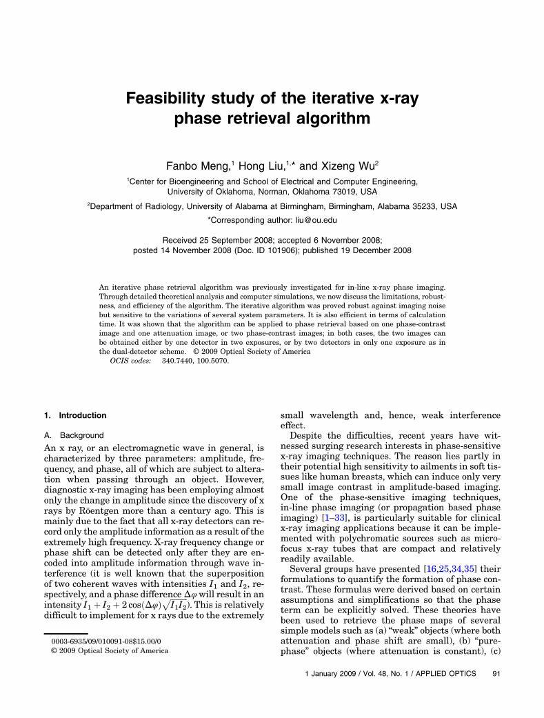
perimental setup of in-line x-ray phase imaging takesa magnification configuration, as is shown in Fig. 1.With a source–object distance (SOD) R1 and an ob-ject–image distance (OID) R2, the geometric magnifi-cation can be calculated as M ¼ ðR1 þ R2Þ=R1.Using the notion “backprojected image”
Ibpð~ρÞ ¼ M2IðM~ρÞ, the formulation of phase imagingby Wu and Liu [16,25] can be written as
F̂ ½Ibpð~ρÞ� ¼ I0 · OTF�~uM
��cosðαu2ÞF̂ ½A2ð~ρÞ�
þ 2 sinðαu2ÞF̂ ½A2ð~ρÞϕð~ρÞ�
þ iλR2
2Msinðαu2Þ~u · F̂ ½∇A2ð~ρÞ�
þ iλR2
Mcosðαu2Þ~u · F̂ ½ϕð~ρÞ∇A2ð~ρÞ�
�; ð1Þ
where I0 is the incident x-ray intensity on the objectplane, λ is the wavelength of the x ray,~u is the spatialfrequency, α ¼ πλR2=M, F̂ ½·� means Fourier trans-form (FT), and the total optical transfer function(OTF),
OTF�~uM
�¼ OTFG:U:
�~uM
�· OTFdet
�~uM
�; ð2Þ
is the product of the OTFs of the x-ray source and thedetector. Equation (1) shows that the phase contrastchanges as the propagation distance R2 increases,
making possible phase retrieval from two imagestaken with different R2, say, an attenuation-basedimage I1 with R2 ¼ 0 and a phase-contrast imageI2 with R2 > 0 taken in two exposures. This is themost basic scheme for implementing an in-line phaseretrieval experiment, therefore, we focus our discus-sion on this simple configuration until Section 5,where we extend the iterative algorithm to two morecomplicated schemes.
It is also worth noting that, in a recent study byGuigay et al. [33], a mixed theoretical model was pre-sented as a generation of the transport of intensityequation (TIE) and the contrast transfer function(CTF) approaches and was validated by imaging athree dimensional biological sample with strong at-tenuation and phase shift. It could be proved by sim-ple derivation that this mixed model is equivalent toEq. (1), so the results of Guigay et al. also proved thevalidity and applicability of Eq. (1).
Reducing Eq. (1) to the TIE approach takes theassumption cosðαu2Þ ¼ 1 and sinðαu2Þ ¼ αu2. There-fore, one of the advantages of our theory over the TIEapproach is its applicability in cases when αu2 ≪ 1does not hold, i.e., when there is low photon energy,large propagation distance, or fine structures in theobject under investigation.
C. Iterative Retrieval Algorithm
Equation (1) was at first simplified by neglecting thethird and the fourth terms within brackets [16], how-ever, it was shown in a recent study [36] that, whilethe third term is negligible, the fourth term (T4) iscomparable to the second one and should be keptfor higher precision. Based on this conclusion, weproposed an iterative algorithm to solve this equa-tion [36]. The idea is to treat T4 as a correction,which is calculated using the retrieved phase mapafter each iteration and then fed back to the nextiteration of phase retrieval. The algorithm can berepresented as
ALGORITHM IterAlgo1
F̂ ½A2� ¼ ~I1;
F̂ ½A2ϕnþ1� ¼~I2 − cosðαu2ÞF̂ ½A2� − T4ðA2;ϕn;R2Þ
2 sinðαu2Þ ; ð3Þ
Fig. 1. Schematic experimental configuration of the in-line x-rayphase imaging. The distance between the x-ray source and the ob-ject plane is R1, and that between the object and the image planesis R2. The geometric magnification can then be calculated asM ¼ ðR1 þ R2Þ=R1.
92 APPLIED OPTICS / Vol. 48, No. 1 / 1 January 2009
where ~I ¼ F̂ ½Ibp�=ðI0 · OTFÞ is the normalized and de-convoluted image, and subscripts n and nþ 1 areiteration counts.In addition to higher precision, another advantage
of this new iterative algorithm is to solve the “phaseambiguity problem” resulted from the neglect of T4by properly choosing a “region of zero phase” (RZP)[32]. From the view of phase contrast, “phase ambi-guity” means the dependence of the phase-contrastimage on the choice of the zero-point of the phasemap; from the view of phase retrieval, it meansthe indeterminable division by sinðαu2Þ at ~u ¼ 0.The detailed algorithm is as follows [32]:First, the attenuation map A2 is calculated from
the attenuation-based image I1 by deconvolution ofthe OTF of the detector. (Note that OTFG:U: ¼ 1 forthe attenuation-based image).Then, an initial distribution of A2ϕ is chosen. We
used A2ϕ ¼ 0 in our tests. It was proved that the al-gorithm does not require a precise estimation.Here begins the iteration. Step one, calculate T4
using the A2ϕ distribution.Step two, retrieve A2ϕ according to Eq. (3) using
the phase-contrast image I2 and the T4 calculatedin the previous step. For ~u ¼ 0, let the division bysinðαu2Þ equal an arbitrary value, say, 0.Step three, calculate the average value of the re-
trieved A2ϕ in the RZP and subtract this averagefrom A2ϕ.Then repeat the iteration from step one to three
until a steady distribution of A2ϕ is obtained.Dividing A2ϕ by A2 gives the phase map ϕ.
2. Assumptions and Limitations
The phase imaging formulation [Eq. (1)] is based onseveral assumptions on the experimental conditions.These assumptions, in turn, determine to which ex-perimental systems this formulation can apply.Therefore, to clarify the applicability of the theory,it is necessary to discuss in depth these assumptions.
A. Fresnel Approximation
Most existing phase retrieval algorithms and simu-lation programs for in-line phase x-ray imaging, in-cluding Eq. (1), are based on the Fresnel diffractionintegral formula, which is, in turn, based on theFresnel approximation, i.e.,
1rexpð−ikrÞ ≈ 1
zexp
�−ikz − ik
ρ22z
�; ð4Þ
where ~r ¼ ðx; y; zÞ, r ¼ j~rj ¼ffiffiffiffiffiffiffiffiffiffiffiffiffiffiffiffiffiffiffiffiffiffiffiffiffix2 þ y2 þ z2
p, and k ¼
2π=λ is the wavenumber. The validity of Eq. (4)requires more than ρ ≪ z. It also requests that theerror in phase due to the approximation is far lessthan π, or,
ρ4 ≪ 4λz3: ð5ÞUnder the condition of x-ray photon energy of
15keV, an OID R2 ¼ 2m, and an error tolerance in
phase term of 0:1π, Eq. (5) requires that ρ < 4mm.But from this argument one cannot draw a conclu-sion that Fresnel approximation fails for any objectof its size > 4mm in the cases of 15keV x-ray andR2 ¼ 2m. In fact, in clinical imaging the body-partsizes are often much larger and the condition ofEq. (4) does not hold in these cases. Fortunately,Eq. (4) is just a sufficient condition, not the necessarycondition for the validity of the Fresnel approxima-tion. This is because the diffracted wavefield is reallygiven by a convolution integral of the object’s trans-mission function with the Fresnel propagator:
expðiπð~ρ −~ρ0Þ2=λzÞ; ð6Þ
where ~ρ is a 2D coordinate on the diffracted wave-front and~ρ0 is on the object plane. As the separationbetween~ρ and~ρ0 gets large, the phase of the Fresnelpropagator varies very rapidly as ~ρ varies over theobject due to the extremely small x-ray wavelengths.Therefore, if the object’s transmission function variesslower than the Fresnel phase does, the contributionof these object points to the diffracted wavefield at~ρwill be averaged out and diminish. More specifically,if ~ρ0 changes to ~ρ0 þ d~ρ0, the Fresnel propagator’sphase changes by
jδϕj ≤ ð2πj~ρ −~ρ0j · jd~ρ0j=λzÞ: ð7Þ
For an object point to make contributions to the dif-fracted wavefield at ~ρ, the Fresnel phase variationshould satisfy jδϕj ≤ π, hence these contributing ob-ject points must be in the range of
j~ρ−~ρ0j ≤ λz=2jd~ρ0j: ð8Þ
Therefore, if the finest structure size of the object is σ,those contributing object points must be in the rangeof
j~ρ−~ρ0j ≤ λz2σ : ð9Þ
For example, for an x ray of 15keV, R2 ¼ 2m, andσ ¼ 1 μm, we found j~ρ−~ρ0j < 82:6 μm, which amplysatisfies Eq. (4). Hence, the Fresnel diffraction as-sumption made in our derivation is justified.
B. Projection Assumption
Theoretically, the phase shift and attenuation effectsof a piece of biological tissue are modeled by a two-dimensional transmission function Tð~ρÞ in the x-yplane [5]:
Tð~ρÞ ¼ expðiϕð~ρÞ − μð~ρÞ=2Þ ¼ Að~ρÞeiϕð~ρÞ; ð10Þ
where ϕð~ρÞ and μð~ρÞ are related to the complexrefractive index n ¼ 1 − δþ iβ through
ϕð~ρÞ ¼ −2πλ
Zδð~rÞdz ¼ −λreρe;pð~ρÞ; ð11Þ
1 January 2009 / Vol. 48, No. 1 / APPLIED OPTICS 93
μð~ρÞ ¼ 4πλ
Zβð~rÞdz: ð12Þ
Here re is the classical electron radius and ρe;pð~ρÞ ¼Rρeð~rÞdz is the projected electron density. Tð~ρÞ re-
lates the incident and the transmitted wavefield by
Etð~ρÞ ¼ Eið~ρÞ · Tð~ρÞ; ð13Þ
where subscripts i and t denote “incident” and “trans-mitted,” respectively.Tð~ρÞ carries structural informa-tion of the object and is independent of Eið~ρÞ. To beable to model an object by Eq. (10), the object is sup-posed to be thin for x ray so that the projection ap-proximation holds, i.e., the x ray travels throughthe object along a straight line parallel to the zdirection.If t is the object’s thickness, it can be shown that, as
long as the size of the finest feature to image is largerthan
ffiffiffiffiλt
p, the object can be deemed thin [15]. Human
body parts can be treated as thin objects for resolu-tions as high as 10 μm for x-ray photon energiesranging from 10 to 150keV [15]. Therefore, it isappropriate to model the human body usingEq. (10) for diagnostic x-ray imaging.
C. Perfect Sampling Assumption
In our theories, we assume that the object to be im-aged is band limited, that is, the FT of Tð~ρÞ is ap-proximately zero outside the Nyquist frequency ofthe detector. The resolution of commercially avail-able x-ray detectors can be as high as tens of micro-meters, which is generally smaller than the typicalsize of the body structures to be imaged in projectionradiography. Therefore, in the case of slowly varyingincident intensity, the attenuation-based image I1 isable to record the structure of the object with no lossof details and the requirement of perfect samplingcan be satisfied. As to the phase-contrast image I2,it is known that propagation in free space will notadd details to the image since the modulus of theFresnel propagator [Eq. (6)] is 1. With considerationof the geometric magnification, it is clear that I2 canalso be perfectly sampled.
D. Moderate Variation Conditions
During the derivation of Eq. (1), we assumed thatboth the phase and the attenuation vary moderatelyin a small distance λR2~u=M:
exp�iϕð~ρÞ − iϕ
�~ρ − λR2~u
M
��≈ 1þ iϕð~ρÞ
− iϕ�~ρ − λR2~u
M
�; ð14Þ
A
�~ρ� λR2~u
M
�≈ Að~ρÞ � λR2~u
M∇Að~ρÞ: ð15Þ
One can see that both equations are of the form of afirst-order Taylor expansion.
To discuss the validity of the moderate variationconditions, we first point out that it is the maximumspatial frequency umax of Tð~ρÞ, not the resolution ofthe detector, that determines the maximum u forwhich both Eqs. (14) and (15) have to be valid. Thisis because, for u > umax, both sides of Eq. (1) are zeroand thus the validity of Eq. (1) does not rely on themoderate variation conditions any more.
ForAð~ρÞ, the moderate variation condition requiresthat, within an small distance λR2umax=M, Að~ρÞ canbe approximated by a straight line, i.e., λR2umax=M isno larger than the finest structure size of the object,1=umax. This leads to
λR2u2max
M≤ 1: ð16Þ
Let us assume R1 ¼ 1m, R2 ¼ 2m, and the photonenergy is 15keV. Equation (16) requires umax <1:3 × 10⁵ m−1.
For ϕð~ρÞ, the moderate variation condition de-mands that, within a small distance λR2umax=M,the maximum phase change should be far less than1. Therefore, it is actually a constraint on thegradient of ϕð~ρÞ:
j∇ϕð~ρÞj ≪ MλR2umax
: ð17Þ
If umax ¼ 104m−1 is assumed, Eq. (17) requiresj∇ϕð~ρÞj ≪ 2 × 10⁶m−1. We can take j∇ϕð~ρÞj ¼2 × 10⁵m−1 for an estimation. Imagine a specialstructure of a size about 2mm in the object to be im-aged. The assumption requires that the phase shiftinduced by the structure relative to its surroundingsis less than 200 rad.
To conclude the discussion, the moderate variationconditions require that the object being imaged doesnot have very thin structures [Eq. (16)] and verysharp phase change [Eq. (17)].
3. Robustness
One critical aspect of the applicability of an algorithmis its robustness against various noises and errors inmeasurements.We tested the robustness of our itera-tive algorithm using computer simulations. Two pro-grams were coded: a simulation program producingan attenuation-based image and a phase-contrast im-age for a given set of phase, attenuation maps, andother experimental parameters; a retrieval programdoing the phase retrieval from the two images pro-duced by the simulation program using some of theparameters. By adding different erroneous factorsonemay encounter in practical situations to the simu-lated images or to the parameters used in the phaseretrieval algorithm, one can study the effects of theseerrors on the resultant phase map.
The simulations are implemented by calculatingthe paraxial Fresnel–Kirchhoff diffraction integral
94 APPLIED OPTICS / Vol. 48, No. 1 / 1 January 2009
directly. The parameters for the simulations are asfollows: the x-ray photon energy is 15keV, the pixelpitch of the image detector is 10 μm, R1 ¼ 1m,and R2 ¼ 2m.To test the quantitativeness of the algorithm, we
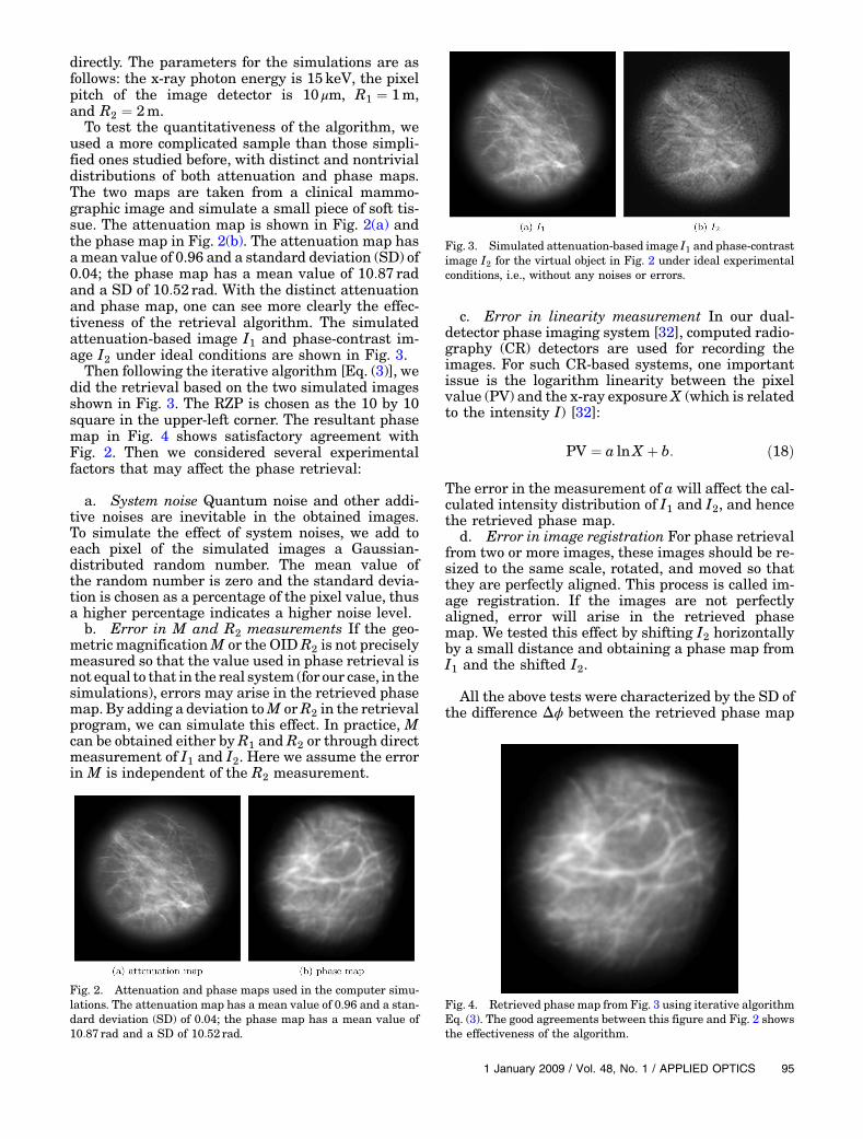
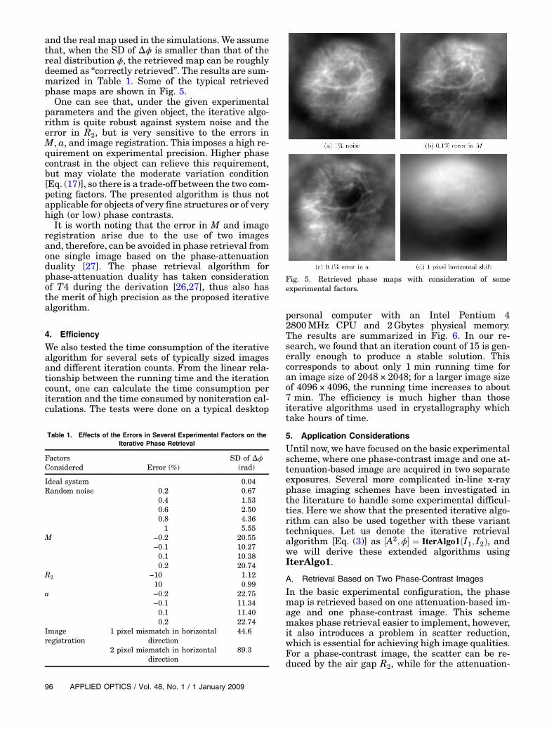
used a more complicated sample than those simpli-fied ones studied before, with distinct and nontrivialdistributions of both attenuation and phase maps.The two maps are taken from a clinical mammo-graphic image and simulate a small piece of soft tis-sue. The attenuation map is shown in Fig. 2(a) andthe phase map in Fig. 2(b). The attenuation map hasamean value of 0.96 and a standard deviation (SD) of0.04; the phase map has a mean value of 10:87 radand a SD of 10:52 rad. With the distinct attenuationand phase map, one can see more clearly the effec-tiveness of the retrieval algorithm. The simulatedattenuation-based image I1 and phase-contrast im-age I2 under ideal conditions are shown in Fig. 3.Then following the iterative algorithm [Eq. (3)], we
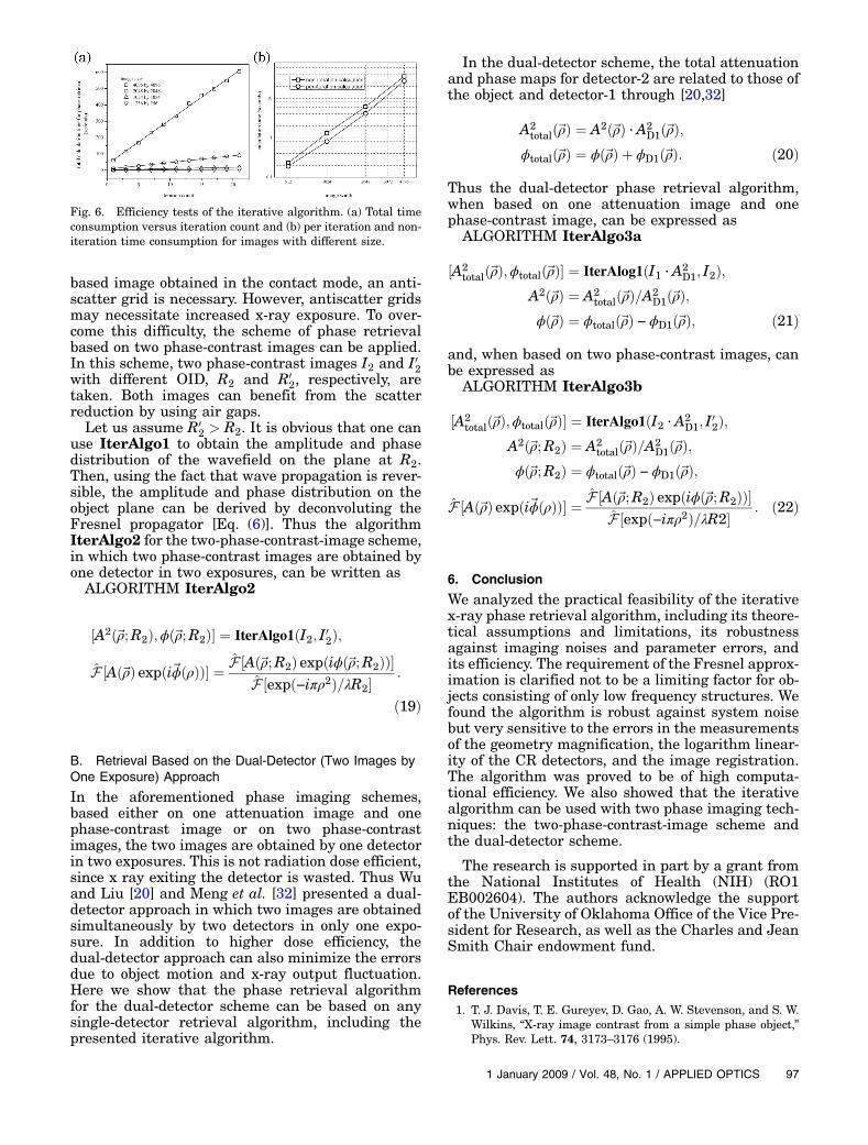
did the retrieval based on the two simulated imagesshown in Fig. 3. The RZP is chosen as the 10 by 10square in the upper-left corner. The resultant phasemap in Fig. 4 shows satisfactory agreement withFig. 2. Then we considered several experimentalfactors that may affect the phase retrieval:
a. System noise Quantum noise and other addi-tive noises are inevitable in the obtained images.To simulate the effect of system noises, we add toeach pixel of the simulated images a Gaussian-distributed random number. The mean value ofthe random number is zero and the standard devia-tion is chosen as a percentage of the pixel value, thusa higher percentage indicates a higher noise level.b. Error in M and R2 measurements If the geo-
metric magnificationM or the OIDR2 is not preciselymeasured so that the value used in phase retrieval isnot equal to that in the real system (for our case, in thesimulations), errors may arise in the retrieved phasemap. By adding a deviation toM orR2 in the retrievalprogram, we can simulate this effect. In practice, Mcan be obtained either byR1 and R2 or through directmeasurement of I1 and I2. Here we assume the errorin M is independent of the R2 measurement.
c. Error in linearity measurement In our dual-detector phase imaging system [32], computed radio-graphy (CR) detectors are used for recording theimages. For such CR-based systems, one importantissue is the logarithm linearity between the pixelvalue (PV) and the x-ray exposure X (which is relatedto the intensity I) [32]:
PV ¼ a lnX þ b: ð18Þ
The error in the measurement of a will affect the cal-culated intensity distribution of I1 and I2, and hencethe retrieved phase map.
d. Error in image registration For phase retrievalfrom two or more images, these images should be re-sized to the same scale, rotated, and moved so thatthey are perfectly aligned. This process is called im-age registration. If the images are not perfectlyaligned, error will arise in the retrieved phasemap. We tested this effect by shifting I2 horizontallyby a small distance and obtaining a phase map fromI1 and the shifted I2.
All the above tests were characterized by the SD ofthe difference Δϕ between the retrieved phase map
Fig. 2. Attenuation and phase maps used in the computer simu-lations. The attenuation map has a mean value of 0.96 and a stan-dard deviation (SD) of 0.04; the phase map has a mean value of10:87 rad and a SD of 10:52 rad.
Fig. 3. Simulated attenuation-based image I1 and phase-contrastimage I2 for the virtual object in Fig. 2 under ideal experimentalconditions, i.e., without any noises or errors.
Fig. 4. Retrieved phasemap from Fig. 3 using iterative algorithmEq. (3). The good agreements between this figure and Fig. 2 showsthe effectiveness of the algorithm.
1 January 2009 / Vol. 48, No. 1 / APPLIED OPTICS 95
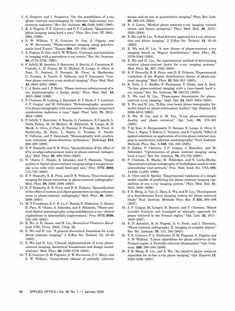
and the real map used in the simulations. We assumethat, when the SD of Δϕ is smaller than that of thereal distribution ϕ, the retrieved map can be roughlydeemed as “correctly retrieved”. The results are sum-marized in Table 1. Some of the typical retrievedphase maps are shown in Fig. 5.One can see that, under the given experimental
parameters and the given object, the iterative algo-rithm is quite robust against system noise and theerror in R2, but is very sensitive to the errors inM, a, and image registration. This imposes a high re-quirement on experimental precision. Higher phasecontrast in the object can relieve this requirement,but may violate the moderate variation condition[Eq. (17)], so there is a trade-off between the two com-peting factors. The presented algorithm is thus notapplicable for objects of very fine structures or of veryhigh (or low) phase contrasts.It is worth noting that the error in M and image
registration arise due to the use of two imagesand, therefore, can be avoided in phase retrieval fromone single image based on the phase-attenuationduality [27]. The phase retrieval algorithm forphase-attenuation duality has taken considerationof T4 during the derivation [26,27], thus also hasthe merit of high precision as the proposed iterativealgorithm.
4. Efficiency
We also tested the time consumption of the iterativealgorithm for several sets of typically sized imagesand different iteration counts. From the linear rela-tionship between the running time and the iterationcount, one can calculate the time consumption periteration and the time consumed by noniteration cal-culations. The tests were done on a typical desktop
personal computer with an Intel Pentium 42800MHz CPU and 2Gbytes physical memory.The results are summarized in Fig. 6. In our re-search, we found that an iteration count of 15 is gen-erally enough to produce a stable solution. Thiscorresponds to about only 1 min running time foran image size of 2048 × 2048; for a larger image sizeof 4096 × 4096, the running time increases to about7 min. The efficiency is much higher than thoseiterative algorithms used in crystallography whichtake hours of time.
5. Application Considerations
Until now, we have focused on the basic experimentalscheme, where one phase-contrast image and one at-tenuation-based image are acquired in two separateexposures. Several more complicated in-line x-rayphase imaging schemes have been investigated inthe literature to handle some experimental difficul-ties. Here we show that the presented iterative algo-rithm can also be used together with these varianttechniques. Let us denote the iterative retrievalalgorithm [Eq. (3)] as ½A2;ϕ� ¼ IterAlgo1ðI1; I2Þ, andwe will derive these extended algorithms usingIterAlgo1.
A. Retrieval Based on Two Phase-Contrast Images
In the basic experimental configuration, the phasemap is retrieved based on one attenuation-based im-age and one phase-contrast image. This schememakes phase retrieval easier to implement, however,it also introduces a problem in scatter reduction,which is essential for achieving high image qualities.For a phase-contrast image, the scatter can be re-duced by the air gap R2, while for the attenuation-
Table 1. Effects of the Errors in Several Experimental Factors on theIterative Phase Retrieval
FactorsConsidered Error (%)
SD of Δϕ(rad)
Ideal system 0.04Random noise 0.2 0.67
0.4 1.530.6 2.500.8 4.361 5.55
M −0:2 20.55−0:1 10.270.1 10.380.2 20.74
R2 −10 1.1210 0.99
a −0:2 22.75−0:1 11.340.1 11.400.2 22.74
Imageregistration
1 pixel mismatch in horizontaldirection
44.6
2 pixel mismatch in horizontaldirection
89.3
Fig. 5. Retrieved phase maps with consideration of someexperimental factors.
96 APPLIED OPTICS / Vol. 48, No. 1 / 1 January 2009
based image obtained in the contact mode, an anti-scatter grid is necessary. However, antiscatter gridsmay necessitate increased x-ray exposure. To over-come this difficulty, the scheme of phase retrievalbased on two phase-contrast images can be applied.In this scheme, two phase-contrast images I2 and I02with different OID, R2 and R0
2, respectively, aretaken. Both images can benefit from the scatterreduction by using air gaps.Let us assume R0
2 > R2. It is obvious that one canuse IterAlgo1 to obtain the amplitude and phasedistribution of the wavefield on the plane at R2.Then, using the fact that wave propagation is rever-sible, the amplitude and phase distribution on theobject plane can be derived by deconvoluting theFresnel propagator [Eq. (6)]. Thus the algorithmIterAlgo2 for the two-phase-contrast-image scheme,in which two phase-contrast images are obtained byone detector in two exposures, can be written asALGORITHM IterAlgo2
½A2ð~ρ;R2Þ;ϕð~ρ;R2Þ� ¼ IterAlgo1ðI2; I02Þ;
F̂ ½Að~ρÞ expði~ϕðρÞÞ� ¼ F̂ ½Að~ρ;R2Þ expðiϕð~ρ;R2ÞÞ�F̂ ½expð−iπρ2Þ=λR2�
:
ð19Þ
B. Retrieval Based on the Dual-Detector (Two Images byOne Exposure) Approach
In the aforementioned phase imaging schemes,based either on one attenuation image and onephase-contrast image or on two phase-contrastimages, the two images are obtained by one detectorin two exposures. This is not radiation dose efficient,since x ray exiting the detector is wasted. Thus Wuand Liu [20] and Meng et al. [32] presented a dual-detector approach in which two images are obtainedsimultaneously by two detectors in only one expo-sure. In addition to higher dose efficiency, thedual-detector approach can also minimize the errorsdue to object motion and x-ray output fluctuation.Here we show that the phase retrieval algorithmfor the dual-detector scheme can be based on anysingle-detector retrieval algorithm, including thepresented iterative algorithm.
In the dual-detector scheme, the total attenuationand phase maps for detector-2 are related to those ofthe object and detector-1 through [20,32]
A2totalð~ρÞ ¼ A2ð~ρÞ · A2
D1ð~ρÞ;ϕtotalð~ρÞ ¼ ϕð~ρÞ þ ϕD1ð~ρÞ: ð20Þ
Thus the dual-detector phase retrieval algorithm,when based on one attenuation image and onephase-contrast image, can be expressed as
ALGORITHM IterAlgo3a
½A2totalð~ρÞ;ϕtotalð~ρÞ� ¼ IterAlog1ðI1 · A2
D1; I2Þ;A2ð~ρÞ ¼ A2
totalð~ρÞ=A2D1ð~ρÞ;
ϕð~ρÞ ¼ ϕtotalð~ρÞ − ϕD1ð~ρÞ; ð21Þ
and, when based on two phase-contrast images, canbe expressed as
ALGORITHM IterAlgo3b
½A2totalð~ρÞ;ϕtotalð~ρÞ� ¼ IterAlgo1ðI2 · A2
D1; I02Þ;
A2ð~ρ;R2Þ ¼ A2totalð~ρÞ=A2
D1ð~ρÞ;ϕð~ρ;R2Þ ¼ ϕtotalð~ρÞ − ϕD1ð~ρÞ;
F̂ ½Að~ρÞ expði~ϕðρÞÞ� ¼ F̂ ½Að~ρ;R2Þ expðiϕð~ρ;R2ÞÞ�F̂ ½expð−iπρ2Þ=λR2� : ð22Þ
6. Conclusion
We analyzed the practical feasibility of the iterativex-ray phase retrieval algorithm, including its theore-tical assumptions and limitations, its robustnessagainst imaging noises and parameter errors, andits efficiency. The requirement of the Fresnel approx-imation is clarified not to be a limiting factor for ob-jects consisting of only low frequency structures. Wefound the algorithm is robust against system noisebut very sensitive to the errors in the measurementsof the geometry magnification, the logarithm linear-ity of the CR detectors, and the image registration.The algorithm was proved to be of high computa-tional efficiency. We also showed that the iterativealgorithm can be used with two phase imaging tech-niques: the two-phase-contrast-image scheme andthe dual-detector scheme.
The research is supported in part by a grant fromthe National Institutes of Health (NIH) (RO1EB002604). The authors acknowledge the supportof the University of Oklahoma Office of the Vice Pre-sident for Research, as well as the Charles and JeanSmith Chair endowment fund.
References1. T. J. Davis, T. E. Gureyev, D. Gao, A. W. Stevenson, and S. W.
Wilkins, “X-ray image contrast from a simple phase object,”Phys. Rev. Lett. 74, 3173–3176 (1995).
Fig. 6. Efficiency tests of the iterative algorithm. (a) Total timeconsumption versus iteration count and (b) per iteration and non-iteration time consumption for images with different size.
1 January 2009 / Vol. 48, No. 1 / APPLIED OPTICS 97
2. A. Snigirev and I. Snigireva, “On the possibilities of x-rayphase contrast microimaging by coherent high-energy syn-chrotron radiation,” Rev. Sci. Instrum. 66, 5486–5492 (1995).
3. K. A. Nugent, T. E. Gureyev, and D. F. Cookson, “Quantitativephase imaging using hard x rays,” Phys. Rev. Lett. 77, 2961–2964 (1996).
4. S. W. Wilkins, T. E. Gureyev, D. Gao, A. Pogany, andA. W. Stevenson, “Phase-contrast imaging using polychro-matic hard X-rays,” Nature 384, 335–338 (1996).
5. A. Pogany, D. Gao, and S. W. Wilkins, “Contrast and resolutionin imaging with a microfocus x-ray source,” Rev. Sci. Instrum.68, 2774–2782 (1997).
6. F. Arfelli, M. Assante, V. Bonvicini, A. Bravin, G. Cantatore, E.Castelli, L. D. Palma, M. D. Michiel, R. Longo, A. Olivo, S.Pani, D. Pontoni, P. Poropat, M. Prest, A. Rashevsky,G. Tromba, A. Vacchi, E. Vallazza, and F. Zanconati, “Low-dose phase contrast x-ray medical imaging,” Phys. Med. Biol.43, 2845–2852 (1998).
7. C. J. Kotre and I. P. Birch, “Phase contrast enhancement of x-ray mammography: a design study,” Phys. Med. Biol. 44,2853–2866 (1999).
8. P. Cloetens, W. Ludwig, J. Baruchel, D. V. Dyck, J. V. Landuyt,J. P. Guigay, and M. Schlenker, “Holotomography: quantita-tive phase tomography with micrometer resolution using hardsynchrotron radiation x rays,” Appl. Phys. Lett. 75, 2912–2914 (1999).
9. F. Arfelli, V. Bonvicini, A. Bravin, G. Cantatore, E. Castelli, L.Dalla Palma, M. Di Michiel, M. Fabrizioli, R. Longo, R. H.Menk, A. Olivo, S. Pani, D. Pontoni, P. Poropat, M. Prest, A.Rashevsky, M. Ratti, L. Rigon, G. Tromba, A. Vacchi,E. Vallazza, and F. Zanconati, “Mammography with synchro-tron radiation: phase-detection techniques,” Radiology 215,286–293 (2000).
10. E. F. Donnelly and R. R. Price, “Quantification of the effect ofkVp on edge-enhancement index in phase-contrast radiogra-phy,” Med. Phys. 29, 999–1002 (2002).
11. H. Ohara, C. Honda, A. Ishisaka, and F. Shimada, “Imagequality in digital phase contrast imaging using a tungsten an-ode x-ray tube with small focal-spot size,” Proc. SPIE 4682,713–723 (2002).
12. E. F. Donnelly, R. R. Price, and D. R. Pickens, “Dual focal-spotimaging for phase extraction in phase-contrast radiography,”Med. Phys. 30, 2292–2296 (2003).
13. E. F. Donnelly, R. R. Price, and D. R. Pickens, “Quantificationof the effect of system and object parameters on edge enhance-ment in phase-contrast radiography,” Med. Phys. 30, 2888–2896 (2003).
14. M. T. Freedman, S.-C. B. Lo, C. Honda, E. Makariou, G. Sisney,E. Pien, H. Ohara, A. Ishisaka, and F. Shimada, “Phase con-trast digital mammography using molybdenum x-ray: clinicalimplications in detectability improvement,” Proc. SPIE 5030,533–540 (2003).
15. X. Wu, A. E. Deans, and H. Liu, Biomedical Photonics Hand-book (CRC Press, 2003), Chap. 26.
16. X. Wu and H. Liu, “A general theoretical formalism for x-rayphase contrast imaging,” J. X-Ray Sci. Technol. 11, 33–42(2003).
17. X. Wu and H. Liu, “Clinical implementation of x-ray phase-contrast imaging: theoretical foundations and design consid-erations,” Med. Phys. 30, 2169–2179 (2003).
18. T. E. Gureyev, D. M. Paganin, A. W. Stevenson, S. C. Mayo, andS. W. Wilkins, “Generalized eikonal of partially coherent
beams and its use in quantitative imaging,” Phys. Rev. Lett.93, 068,103 (2004).
19. R. A. Lewis, “Medical phase contrast x-ray imaging: currentstatus and future prospects,” Phys. Med. Biol. 49, 3573–3583 (2004).
20. X. Wu and H. Liu, “A dual detector approach for x-ray attenua-tion and phase imaging,” J. X-Ray Sci. Technol. 12, 35–42(2004).
21. X. Wu and H. Liu, “A new theory of phase-contrast x-rayimaging based on Wigner distributions,” Med. Phys. 31,2378–2384 (2004).
22. X. Wu and H. Liu, “An experimental method of determiningrelative phase-contrast factor for x-ray imaging systems,”Med. Phys. 31, 997–1002 (2004).
23. E. F. Donnelly, R. R. Price, and D. R. Pickens, “Experimentalvalidation of the Wigner distributions theory of phase-con-trast imaging,” Med. Phys. 32, 928–931 (2005).
24. R. Toth, J. C. Kieffer, S. Fourmaux, T. Ozaki, and A. Krol,“In-line phase-contrast imaging with a laser-based hard x-ray source,” Rev. Sci. Instrum. 76, 083701 (2005).
25. X. Wu and H. Liu, “Phase-space formulation for phase-contrast x-ray imaging,” Appl. Opt. 44, 5847–5854 (2005).
26. X. Wu and H. Liu, “X-Ray cone-beam phase tomography for-mulas based on phase-attenuation duality,” Opt. Express 13,6000–6014 (2005).
27. X. Wu, H. Liu, and A. M. Yan, “X-ray phase-attenuationduality and phase retrieval,” Opt. Lett. 30, 379–381(2005).
28. T. Q. Xiao, A. Bergamaschi, D. Dreossi, R. Longo, A. Olivo, S.Pani, L. Rigon, T. Rokvic, C. Venanzi, and E. Castelli, “Effect ofspatial coherence on application of in-line phase contrast ima-ging to synchrotron radiation mammography,” Nucl. Instrum.Methods Phys. Res. A 548, 155–162 (2005).
29. S. Zabler, P. Cloetens, J.-P. Guigay, J. Baruchel, and M.Schlenker, “Optimization of phase contrast imaging usinghard x rays,” Rev. Sci. Instrum. 76, 073,705 (2005).
30. P. Cloetens, R. Mache, M. Schlenker, and S. Lerbs-Mache,“Quantitative phase tomography of Arabidopsis seeds revealsintercellular void network,” Proc. Natl. Acad. Sci. USA 103,14,626–14,630 (2006).
31. A. Olivo and R. Speller, “Experimental validation of a simplemodel capable of predicting the phase contrast imaging cap-abilities of any x-ray imaging system,” Phys. Med. Biol. 51,3015–3030 (2006).
32. F. B. Meng, A. Yan, G. Zhou, X. Wu, and H. Liu, “Developmentof a dual-detector X-ray imaging system for phase retrievalstudy,” Nucl. Instrum. Methods Phys. Res. B 254, 300–306(2007).
33. J. P. Guigay, M. Langer, R. Boistel, and P. Cloetens, “Mixedtransfer function and transport of intensity approach forphase retrieval in the Fresnel region.” Opt. Lett. 32, 1617–1619 (2007).
34. B. D. Arhatari, K. A. Nugent, A. G. Peele, and J. Thornton,“Phase contrast radiography. II. Imaging of complex objects,”Rev. Sci. Instrum. 76, 113, 704 (2005).
35. T. E. Gureyev, Y. L. Nesterets, D. M. Paganin, A. Pogany, andS. W. Wilkins, “Linear algorithms for phase retrieval in theFresnel region. 2. Partially coherent illumination,” Opt. Com-mun. 259, 569–580 (2006).
36. F. B. Meng, H. Liu, and X. Wu, “An iterative phase retrievalalgorithm for in-line x-ray phase imaging,” Opt. Express 15,8383–8390 (2007).
98 APPLIED OPTICS / Vol. 48, No. 1 / 1 January 2009