Embed Size (px)
Citation preview
a SpringerOpen Journal
Seno et al. SpringerPlus (2015) 4:250 DOI 10.1186/s40064-015-0998-3
CASE STUDY Open Access
FDG-PET findings of Ameloblastoma: a casereport
Satoshi Seno1, Kazuhiro Kitajima1*, Go Inokuchi2, Ken-ichi Nibu2, Tomoo Itoh3, Yasuo Ejima4, Ryohei Sasaki4,Koji Sugimoto1 and Kazuro Sugimura1Abstract
Introduction: Ameloblastoma is a benign odontogenic neoplasm of the jaw, rarely presenting as a malignanttumor. Although it is very important to discriminate ameloblastoma from ameloblastic carcinoma in order to decidethe appropriate operative procedure, this is difficult using conventional CT and MRI.
Case descriptions: We report a case of maxillar ameloblastoma in a 78-year-old man where FDG-PET/CT was usefulfor making this discrimination. CT demonstrated a 31 × 43 × 46-mm mass in the left posterior maxillary sinus withdestruction of its posterior and lateral wall and alveolar bone. MRI demonstrated a hypo- to isointenseheterogeneous pattern on T1WI, heterogeneous hyperintensity with a prominent high-signal spot on T2WI, highsignal intensity on DWI reflecting restricted diffusion, and strong heterogeneous enhancement. Because FDG-PET/CT showed mild FDG uptake (SUVmax 2.40) by the mass, ameloblastoma, rather than ameloblastic carcinoma, wasconsidered to be the correct diagnosis.
Discussion and evaluation: It appears that ameloblastic carcinoma shows intense FDG uptake, whereasameloblastoma shows mild or moderate FDG uptake, and only rarely intense FDG uptake. Our experience suggeststhat FDG-PET/CT may be effective for discriminating ameloblastoma from ameloblastic carcinoma. Especially, incases showing mild FDG uptake, benign ameloblastoma would seem the most likely diagnosis.
Conclusions: FDG-PET/CT may be useful as an adjunctive modality for diagnosis, treatment planning andsurveillance of ameloblastoma and ameloblastic carcinoma.
Keywords: 18F-fluorodeoxyglucose (FDG); PET; CT; MRI; Ameloblastoma
BackgroundAmeloblastoma is a benign odontogenic neoplasm de-rived from ameloblastic epithelial tissue. It comprisesonly 1% of all jaw tumors and 11% of all odontogenicneoplasms (being the second most common) (Nevilleet al. 2002). The 2005 WHO histological classification ofodontogenic tumors classified ameloblastoma into severaltypes: solid/multicystic, extraosseous/peripheral, desmo-plastic, and unicystic (Gardner et al. 2005). Although ame-loblastoma is a benign neoplasm, it is locally aggressiveand local recurrence is not rare (Inoue et al. 1988). Ame-loblastic carcinoma is a term given to tumors that displayhistologically malignant features at both primary and
* Correspondence: [email protected] of Radiology, Kobe University Graduate School of Medicine,7-5-2 Kusunoki-cho, Chuo-ku, Kobe 650-0017, JapanFull list of author information is available at the end of the article
© 2015 Seno et al. This is an Open Access artic(http://creativecommons.org/licenses/by/4.0), wprovided the original work is properly credited
metastatic sites with a pattern otherwise resembling ame-loblastoma (Newman et al. 1995). To plan the most ap-propriate treatment, it is very important to determinewhether a tumor is benign or malignant. However, dis-crimination between ameloblastoma and ameloblastic car-cinoma is difficult by conventional morphological imagingmodalities such as radiography, computed tomography(CT) and magnetic resonance imaging (MRI) (Devenney-Cakir et al. 2010, Dunfee et al. 2006, Matsuzaki et al.2011, Minami et al. 1992).Recently, assessment of glucose metabolism in cells
using 18F-fluorodeoxyglucose (FDG) positron emissiontomography (PET) has been used to identify aggressive tu-mors and to predict their degree of malignant potential.Here we describe a case of ameloblastoma with emphasison the imaging features revealed by FDG-PET/CT.
le distributed under the terms of the Creative Commons Attribution Licensehich permits unrestricted use, distribution, and reproduction in any medium,.
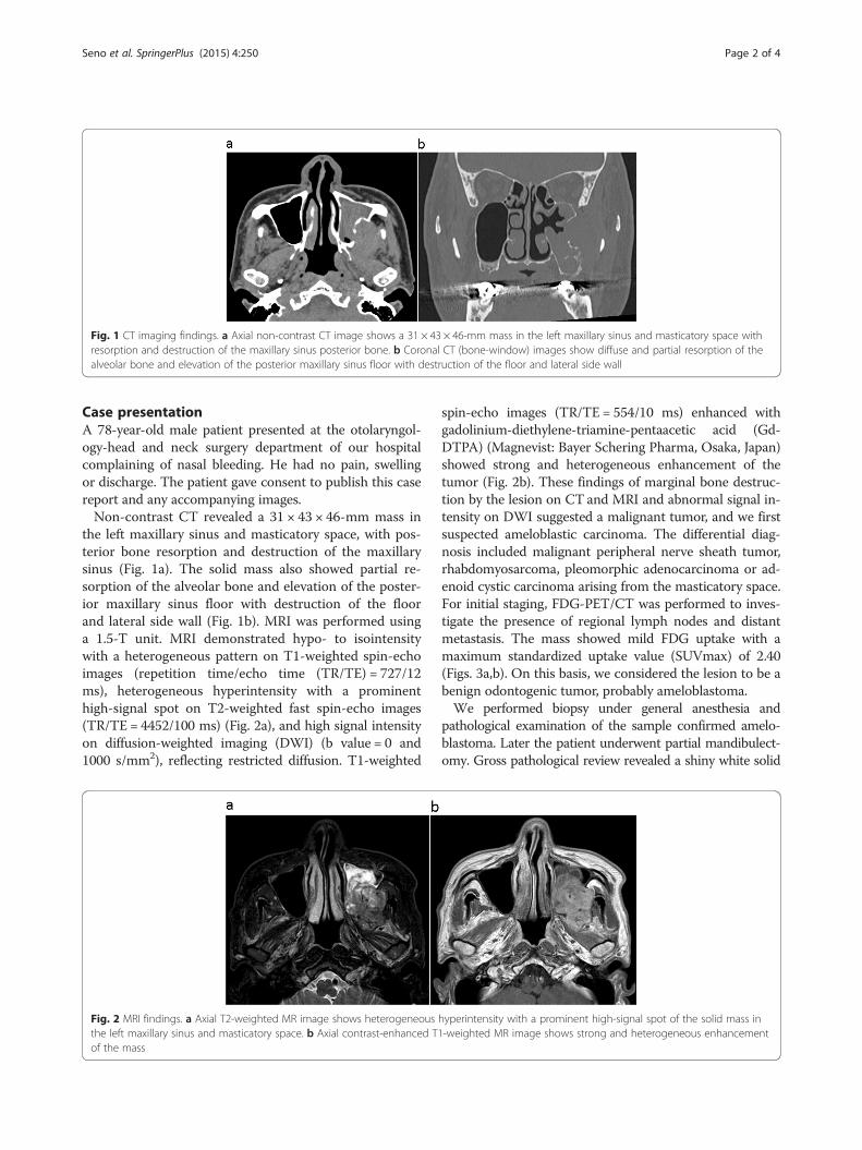
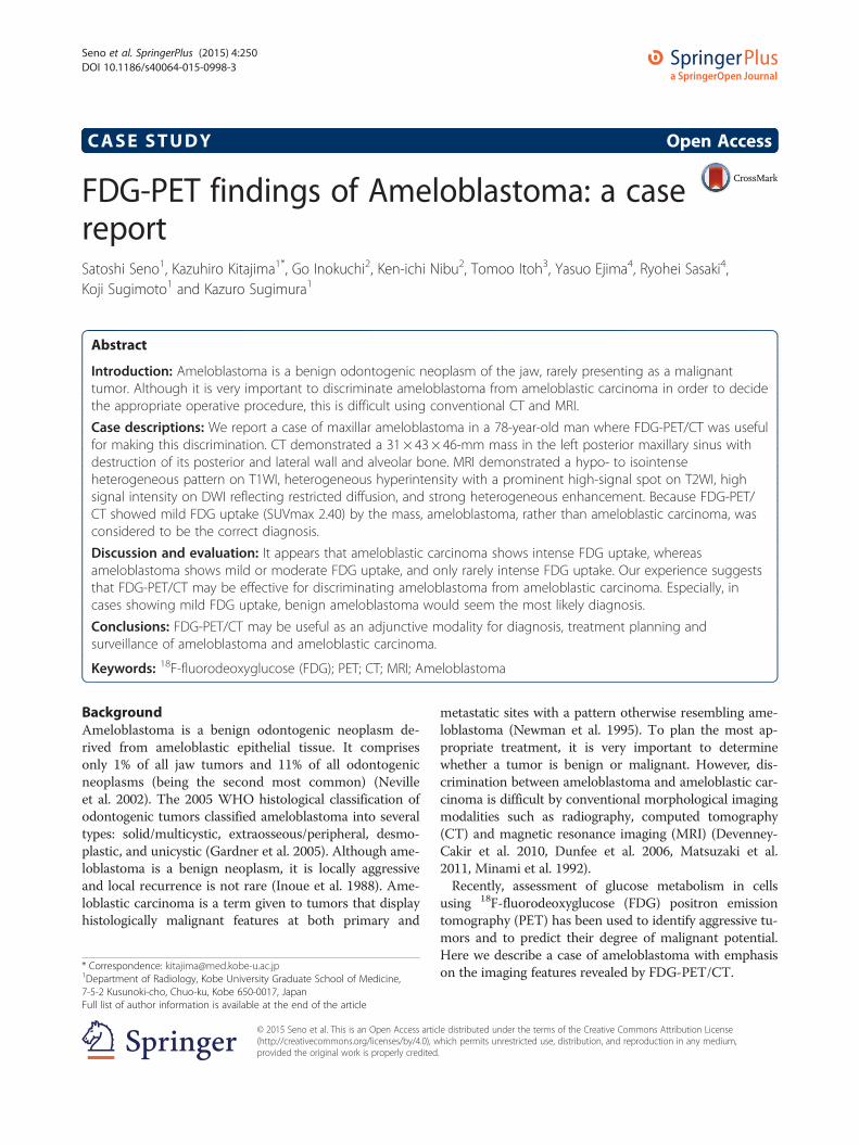
Fig. 1 CT imaging findings. a Axial non-contrast CT image shows a 31 × 43 × 46-mm mass in the left maxillary sinus and masticatory space withresorption and destruction of the maxillary sinus posterior bone. b Coronal CT (bone-window) images show diffuse and partial resorption of thealveolar bone and elevation of the posterior maxillary sinus floor with destruction of the floor and lateral side wall
Seno et al. SpringerPlus (2015) 4:250 Page 2 of 4
Case presentationA 78-year-old male patient presented at the otolaryngol-ogy-head and neck surgery department of our hospitalcomplaining of nasal bleeding. He had no pain, swellingor discharge. The patient gave consent to publish this casereport and any accompanying images.Non-contrast CT revealed a 31 × 43 × 46-mm mass in
the left maxillary sinus and masticatory space, with pos-terior bone resorption and destruction of the maxillarysinus (Fig. 1a). The solid mass also showed partial re-sorption of the alveolar bone and elevation of the poster-ior maxillary sinus floor with destruction of the floorand lateral side wall (Fig. 1b). MRI was performed usinga 1.5-T unit. MRI demonstrated hypo- to isointensitywith a heterogeneous pattern on T1-weighted spin-echoimages (repetition time/echo time (TR/TE) = 727/12ms), heterogeneous hyperintensity with a prominenthigh-signal spot on T2-weighted fast spin-echo images(TR/TE = 4452/100 ms) (Fig. 2a), and high signal intensityon diffusion-weighted imaging (DWI) (b value = 0 and1000 s/mm2), reflecting restricted diffusion. T1-weighted
Fig. 2 MRI findings. a Axial T2-weighted MR image shows heterogeneousthe left maxillary sinus and masticatory space. b Axial contrast-enhanced T1of the mass
spin-echo images (TR/TE = 554/10 ms) enhanced withgadolinium-diethylene-triamine-pentaacetic acid (Gd-DTPA) (Magnevist: Bayer Schering Pharma, Osaka, Japan)showed strong and heterogeneous enhancement of thetumor (Fig. 2b). These findings of marginal bone destruc-tion by the lesion on CT and MRI and abnormal signal in-tensity on DWI suggested a malignant tumor, and we firstsuspected ameloblastic carcinoma. The differential diag-nosis included malignant peripheral nerve sheath tumor,rhabdomyosarcoma, pleomorphic adenocarcinoma or ad-enoid cystic carcinoma arising from the masticatory space.For initial staging, FDG-PET/CT was performed to inves-tigate the presence of regional lymph nodes and distantmetastasis. The mass showed mild FDG uptake with amaximum standardized uptake value (SUVmax) of 2.40(Figs. 3a,b). On this basis, we considered the lesion to be abenign odontogenic tumor, probably ameloblastoma.We performed biopsy under general anesthesia and
pathological examination of the sample confirmed amelo-blastoma. Later the patient underwent partial mandibulect-omy. Gross pathological review revealed a shiny white solid
hyperintensity with a prominent high-signal spot of the solid mass in-weighted MR image shows strong and heterogeneous enhancement
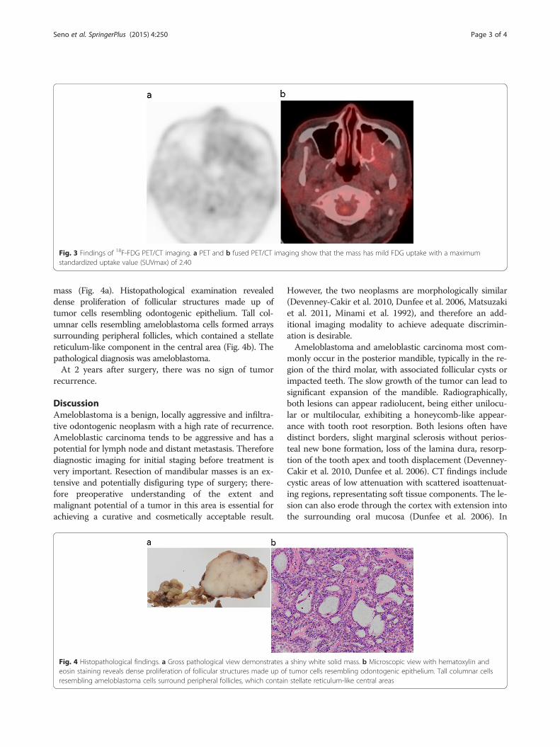
Fig. 3 Findings of 18F-FDG PET/CT imaging. a PET and b fused PET/CT imaging show that the mass has mild FDG uptake with a maximumstandardized uptake value (SUVmax) of 2.40
Seno et al. SpringerPlus (2015) 4:250 Page 3 of 4
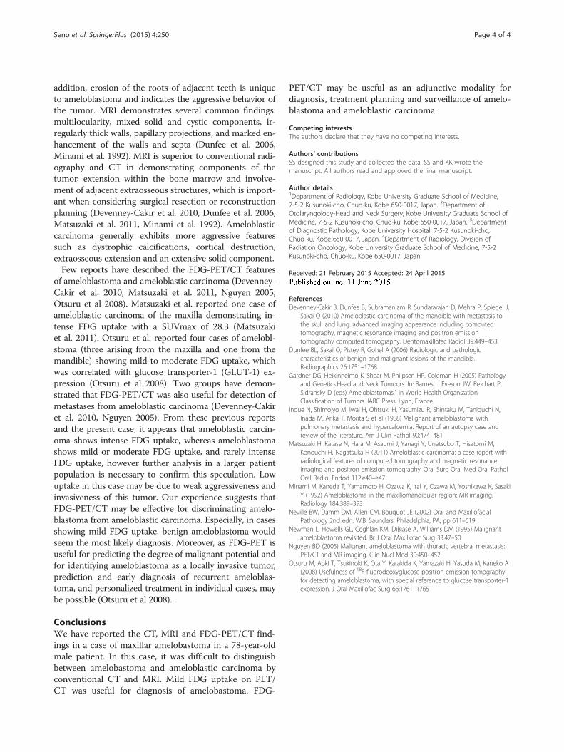
mass (Fig. 4a). Histopathological examination revealeddense proliferation of follicular structures made up oftumor cells resembling odontogenic epithelium. Tall col-umnar cells resembling ameloblastoma cells formed arrayssurrounding peripheral follicles, which contained a stellatereticulum-like component in the central area (Fig. 4b). Thepathological diagnosis was ameloblastoma.At 2 years after surgery, there was no sign of tumor
recurrence.
DiscussionAmeloblastoma is a benign, locally aggressive and infiltra-tive odontogenic neoplasm with a high rate of recurrence.Ameloblastic carcinoma tends to be aggressive and has apotential for lymph node and distant metastasis. Thereforediagnostic imaging for initial staging before treatment isvery important. Resection of mandibular masses is an ex-tensive and potentially disfiguring type of surgery; there-fore preoperative understanding of the extent andmalignant potential of a tumor in this area is essential forachieving a curative and cosmetically acceptable result.
Fig. 4 Histopathological findings. a Gross pathological view demonstrateseosin staining reveals dense proliferation of follicular structures made up oresembling ameloblastoma cells surround peripheral follicles, which contain
However, the two neoplasms are morphologically similar(Devenney-Cakir et al. 2010, Dunfee et al. 2006, Matsuzakiet al. 2011, Minami et al. 1992), and therefore an add-itional imaging modality to achieve adequate discrimin-ation is desirable.Ameloblastoma and ameloblastic carcinoma most com-
monly occur in the posterior mandible, typically in the re-gion of the third molar, with associated follicular cysts orimpacted teeth. The slow growth of the tumor can lead tosignificant expansion of the mandible. Radiographically,both lesions can appear radiolucent, being either unilocu-lar or multilocular, exhibiting a honeycomb-like appear-ance with tooth root resorption. Both lesions often havedistinct borders, slight marginal sclerosis without perios-teal new bone formation, loss of the lamina dura, resorp-tion of the tooth apex and tooth displacement (Devenney-Cakir et al. 2010, Dunfee et al. 2006). CT findings includecystic areas of low attenuation with scattered isoattenuat-ing regions, representating soft tissue components. The le-sion can also erode through the cortex with extension intothe surrounding oral mucosa (Dunfee et al. 2006). In
a shiny white solid mass. b Microscopic view with hematoxylin andf tumor cells resembling odontogenic epithelium. Tall columnar cellsstellate reticulum-like central areas
Seno et al. SpringerPlus (2015) 4:250 Page 4 of 4
addition, erosion of the roots of adjacent teeth is uniqueto ameloblastoma and indicates the aggressive behavior ofthe tumor. MRI demonstrates several common findings:multilocularity, mixed solid and cystic components, ir-regularly thick walls, papillary projections, and marked en-hancement of the walls and septa (Dunfee et al. 2006,Minami et al. 1992). MRI is superior to conventional radi-ography and CT in demonstrating components of thetumor, extension within the bone marrow and involve-ment of adjacent extraosseous structures, which is import-ant when considering surgical resection or reconstructionplanning (Devenney-Cakir et al. 2010, Dunfee et al. 2006,Matsuzaki et al. 2011, Minami et al. 1992). Ameloblasticcarcinoma generally exhibits more aggressive featuressuch as dystrophic calcifications, cortical destruction,extraosseous extension and an extensive solid component.Few reports have described the FDG-PET/CT features
of ameloblastoma and ameloblastic carcinoma (Devenney-Cakir et al. 2010, Matsuzaki et al. 2011, Nguyen 2005,Otsuru et al 2008). Matsuzaki et al. reported one case ofameloblastic carcinoma of the maxilla demonstrating in-tense FDG uptake with a SUVmax of 28.3 (Matsuzakiet al. 2011). Otsuru et al. reported four cases of amelobl-stoma (three arising from the maxilla and one from themandible) showing mild to moderate FDG uptake, whichwas correlated with glucose transporter-1 (GLUT-1) ex-pression (Otsuru et al 2008). Two groups have demon-strated that FDG-PET/CT was also useful for detection ofmetastases from ameloblastic carcinoma (Devenney-Cakiret al. 2010, Nguyen 2005). From these previous reportsand the present case, it appears that ameloblastic carcin-oma shows intense FDG uptake, whereas ameloblastomashows mild or moderate FDG uptake, and rarely intenseFDG uptake, however further analysis in a larger patientpopulation is necessary to confirm this speculation. Lowuptake in this case may be due to weak aggressiveness andinvasiveness of this tumor. Our experience suggests thatFDG-PET/CT may be effective for discriminating amelo-blastoma from ameloblastic carcinoma. Especially, in casesshowing mild FDG uptake, benign ameloblastoma wouldseem the most likely diagnosis. Moreover, as FDG-PET isuseful for predicting the degree of malignant potential andfor identifying ameloblastoma as a locally invasive tumor,prediction and early diagnosis of recurrent ameloblas-toma, and personalized treatment in individual cases, maybe possible (Otsuru et al 2008).
ConclusionsWe have reported the CT, MRI and FDG-PET/CT find-ings in a case of maxillar amelobastoma in a 78-year-oldmale patient. In this case, it was difficult to distinguishbetween amelobastoma and ameloblastic carcinoma byconventional CT and MRI. Mild FDG uptake on PET/CT was useful for diagnosis of amelobastoma. FDG-
PET/CT may be useful as an adjunctive modality fordiagnosis, treatment planning and surveillance of amelo-blastoma and ameloblastic carcinoma.
Competing interestsThe authors declare that they have no competing interests.
Authors’ contributionsSS designed this study and collected the data. SS and KK wrote themanuscript. All authors read and approved the final manuscript.
Author details1Department of Radiology, Kobe University Graduate School of Medicine,7-5-2 Kusunoki-cho, Chuo-ku, Kobe 650-0017, Japan. 2Department ofOtolaryngology-Head and Neck Surgery, Kobe University Graduate School ofMedicine, 7-5-2 Kusunoki-cho, Chuo-ku, Kobe 650-0017, Japan. 3Departmentof Diagnostic Pathology, Kobe University Hospital, 7-5-2 Kusunoki-cho,Chuo-ku, Kobe 650-0017, Japan. 4Department of Radiology, Division ofRadiation Oncology, Kobe University Graduate School of Medicine, 7-5-2Kusunoki-cho, Chuo-ku, Kobe 650-0017, Japan.
Received: 21 February 2015 Accepted: 24 April 2015
ReferencesDevenney-Cakir B, Dunfee B, Subramaniam R, Sundararajan D, Mehra P, Spiegel J,
Sakai O (2010) Ameloblastic carcinoma of the mandible with metastasis tothe skull and lung: advanced imaging appearance including computedtomography, magnetic resonance imaging and positron emissiontomography computed tomography. Dentomaxillofac Radiol 39:449–453
Dunfee BL, Sakai O, Pistey R, Gohel A (2006) Radiologic and pathologiccharacteristics of benign and malignant lesions of the mandible.Radiographics 26:1751–1768
Gardner DG, Heikinheimo K, Shear M, Philpsen HP, Coleman H (2005) Pathologyand Genetics.Head and Neck Tumours. In: Barnes L, Eveson JW, Reichart P,Sidransky D (eds) Ameloblastomas,” in World Health OrganizationClassification of Tumors. IARC Press, Lyon, France
Inoue N, Shimojyo M, Iwai H, Ohtsuki H, Yasumizu R, Shintaku M, Taniguchi N,Inada M, Arika T, Morita S et al (1988) Malignant ameloblastoma withpulmonary metastasis and hypercalcemia. Report of an autopsy case andreview of the literature. Am J Clin Pathol 90:474–481
Matsuzaki H, Katase N, Hara M, Asaumi J, Yanagi Y, Unetsubo T, Hisatomi M,Konouchi H, Nagatsuka H (2011) Ameloblastic carcinoma: a case report withradiological features of computed tomography and magnetic resonanceimaging and positron emission tomography. Oral Surg Oral Med Oral PatholOral Radiol Endod 112:e40–e47
Minami M, Kaneda T, Yamamoto H, Ozawa K, Itai Y, Ozawa M, Yoshikawa K, SasakiY (1992) Ameloblastoma in the maxillomandibular region: MR imaging.Radiology 184:389–393
Neville BW, Damm DM, Allen CM, Bouquot JE (2002) Oral and MaxillofacialPathology 2nd edn. W.B. Saunders, Philadelphia, PA, pp 611–619
Newman L, Howells GL, Coghlan KM, DiBiase A, Williams DM (1995) Malignantameloblastoma revisited. Br J Oral Maxillofac Surg 33:47–50
Nguyen BD (2005) Malignant ameloblastoma with thoracic vertebral metastasis:PET/CT and MR imaging. Clin Nucl Med 30:450–452
Otsuru M, Aoki T, Tsukinoki K, Ota Y, Karakida K, Yamazaki H, Yasuda M, Kaneko A(2008) Usefulness of 18F-fluorodeoxyglucose positron emission tomographyfor detecting ameloblastoma, with special reference to glucose transporter-1expression. J Oral Maxillofac Surg 66:1761–1765


![FDG-PET in Large Vessel Vasculitis...FDG-PET in Large Vessel Vasculitis 61 5. [18 F]FDG-PET and [18 F]FDG-PET/CT [18 F]FDG-PET is an operator-independent, non- invasive imaging modality](https://img.pdfslide.us/doc/110x75/5f6c13132f0609183b646bce/fdg-pet-in-large-vessel-vasculitis-fdg-pet-in-large-vessel-vasculitis-61-5.jpg)