Embed Size (px)
Citation preview
IMMUNOBIOLOGY
Fc-dependent expression of CD137 on human NK cells: insights into “agonistic”effects of anti-CD137 monoclonal antibodies*Wei Lin,1 *Caroline J. Voskens,2 Xiaoyu Zhang,1 Daniel G. Schindler,3 Aaron Wood,1 Erin Burch,1 Yadong Wei,4
Lieping Chen,5 Guoliang Tian,6 Koji Tamada,1 Lai-Xi Wang,4 Dan H. Schulze,1,7 Dean Mann,2 and Scott E. Strome1,7
1Department of Otorhinolaryngology-Head and Neck Surgery, University of Maryland, Baltimore; 2Department of Pathology, University of Maryland School ofMedicine, Baltimore; 3GTC Biotherapeutics, Framingham, MA; 4Institute of Human Virology, University of Maryland, Baltimore; 5Department of Dermatology,Johns Hopkins University, Baltimore, MD; 6Division of Biostatistics, University of Maryland Greenebaum Cancer Center, Baltimore; and 7Department ofMicrobiology and Immunology, University of Maryland School of Medicine, Baltimore
CD137 (4-1BB) is a costimulatory mol-ecule that can be manipulated for thetreatment of cancer and autoimmune dis-ease. Although it is known that agonisticantibodies (mAbs) against CD137 en-hance the rejection of murine tumors ina natural killer (NK) cell– and T cell–dependent fashion, the mechanism forNK dependence is poorly understood. Inthis study, we evaluated the ability of 2different glycoforms of a chimerized anti-human CD137 mAb, an aglycosylated (GA)
and a low fucose form (GG), to react withhuman NK cells. Both mAbs bound simi-larly to CD137 and partially blocked theinteraction between CD137 and CD137ligand. However, unlike GA mAb, immobi-lized GG mAb activated NK cells andenhanced CD137 expression. These ef-fects were seemingly dependent on Fcinteraction with putative Fc receptors onthe NK-cell surface, as only the immobi-lized Fc-fragment of GG was required forCD137 expression. Furthermore, CD137
expression could be enhanced with anti-bodies directed against non-CD137epitopes, and the expression levels di-rectly correlated with patterns of Fc-glycosylation recognized to improve Fcinteraction with Fc� receptors. Our datasuggest that CD137 can be enhanced onNK cells in an Fc-dependent fashion andthat expression correlates with pheno-typic and functional parameters of activa-tion. (Blood. 2008;112:699-707)
Introduction
CD137 (4-1BB), a member of the TNF receptor superfamily, isincreasingly recognized as an important target for the treatmentof both cancer and autoimmunity.1-5 Specifically, in murinemodels, it is clear that ligation of CD137 on the surface ofactivated T cells, through either CD137 ligand (CD137L) oragonistic monoclonal antibodies (mAbs), potentiates the im-mune response to weakly immunogenic tumors in a naturalkiller (NK)–dependent fashion.3,6 Unlike other costimulatory-based antitumor immunotherapies (eg, CTLA-4 blockade),CD137 ligation does not induce self-reactivity, but rather hastherapeutic benefit in multiple murine models of autoimmunedisease such as rheumatoid arthritis,7 systemic lupus erythema-tosus,8 and inflammatory bowel disease.9 In many studies,functional conclusions regarding CD137 have been based onmAb or fusion protein manipulation of receptor/ligand path-ways, with the assumption being that observed effects weresecondary to Fab region or ligand interaction with CD137.Importantly, little attention has been paid to the potential linkbetween the Fc region of these molecules and alternate pathwaysof CD137 regulation through Fc receptors (Fc�Rs).
It is now evident that Fc cross-linking of Fc�RIII (CD16) onhuman NK cells induces cellular activation defined by bothphenotypic change and release of proinflammatory cytokines.10
The affinity and functional consequences of the interactionbetween Fc and Fc�RIII is dependent on the presence ofoligosaccharides (N-glycan) covalently attached at asparagine
297 (Asn297) of the Fc heavy chain.11 For example, Fcfragments with low fucose content at Asn297 have enhancedbinding affinity for Fc�RIII.12-14 In addition, aglycosylated Fcfragments are unable to efficiently bind the Fc�RIII.15,16 Theinteraction between Fc-Fc�Rs also has clinical implications, asit is now evident that polymorphisms within the Fc�RIII (eg,V/F at amino acid position 158), which impact Fc-Fc�RIIIinteractions, can be used to define the therapeutic efficacy oftargeted anticancer therapeutics such as rituximab.17,18
Based on the therapeutic potential of anti-CD137 mAbs andthe recognized importance of Fc-Fc�R interactions on mAbfunction, in collaboration with GTC Biotherapeutics, 2 chimericanti-CD137 mAbs were developed. The first, a glycosylatedform (GG) is likely to cross-link the Fc�R and the second, anaglycosylated form (GA), is unlikely to efficiently cross-link theFc�R. Because of the recognized role of NK cells in theantitumor function of anti-CD137 mAbs in murine models, weinitially hypothesized that interleukin-2 (IL-2)–stimulated hu-man NK cells would express CD137 and that ligation withchimeric anti-CD137 mAb would result in cytokine releaseand degranulation.
Surprisingly, we observed that, after IL-2 stimulation andsubsequent culture, human NK cells do not express high levels ofCD137. However, CD137 is enhanced on IL-2–stimulated humanNK cells after culture in the presence of immobilized glycosylatedmAbs or papain-cleaved Fc fragments. The ability to enhance
Submitted November 6, 2007; accepted May 12, 2008. Prepublished online asBlood First Edition paper, June 2, 2008; DOI 10.1182/blood-2007-11-122465.
*W.L. and C.J.V. contributed equally to this study.
The publication costs of this article were defrayed in part by page chargepayment. Therefore, and solely to indicate this fact, this article is herebymarked ‘‘advertisement’’ in accordance with 18 USC section 1734.
© 2008 by The American Society of Hematology
699BLOOD, 1 AUGUST 2008 � VOLUME 112, NUMBER 3
For personal use only.on April 10, 2019. by guest www.bloodjournal.orgFrom
CD137 expression is independent of the antigen specificity of theFab region, and the magnitude of CD137 expression is associatedwith patterns of glycosylation known to enhance the interactionbetween Fc and Fc�Rs. These data suggest that “agonistic effects”of select antibodies on costimulatory molecules may be in partsecondary to Fc-Fc�R interactions and provide important insightinto the design of antibodies intended to manipulate costimulatorypathways for clinical benefit.
Methods
All experimental work related to human materials was approved by theUniversity of Maryland’s institutional review board (IRB), under IRBnumber H-27785.
Chimeric monoclonal antibodies
Mouse anti–human CD137 mAb (G6) was generated in the laboratory ofL. C. A glycosylated chimeric version of G6, hereafter called GG, wasdeveloped by replacing the mouse IgG2a Fc region with a human IgG1 Fcregion. Likewise, an aglycosylated (GA) chimeric anti-CD137 mAb wascreated by mutating Asn to glutamine (Gln) at amino acid position 297 inthe Fc region. Both GA and GG mAbs were produced in the milk oftransgenic goats. Cetuximab (hereafter called CTM) was obtained throughthe Greenebaum Cancer Center (University of Maryland Medical System,Baltimore). For flow cytometric experiments, all chimeric mAbs weredirectly labeled to allophycocyanin (APC) through the custom conjugationservice of Invitrogen (Invitrogen, Carlsbad, CA).
Cells and transfectants
Human blood samples were purchased (Biological Specialty Corporation,Colmar, PA) and whole peripheral blood mononuclear cells (PBMCs) wereisolated using density-gradient centrifugation. Freshly purified resting NKcells (CD3�CD56�CD16� as defined by flow cytometry) were obtained bynegative magnetic cell sorting using NK-cell isolation beads (MiltenyiBiotec, Auburn, CA) according to the manufacturer’s instructions. Chinesehamster ovary (CHO) cells stably transfected with human CD137 receptorand nontransfected wild-type controls were established in the laboratory ofL.C. CHO cells were maintained in Dulbecco modified Eagle medium(DMEM) supplemented with 10% fetal bovine serum (FBS) (AtlantaBiological, Lawrenceville, GA), 1% penicillin/streptomycin (P/S), 1%Glutamax-1 (both Invitrogen), and 1% HEPES (Mediatech, Herndon, VA).K562 cells were cultured in RPMI 1640 supplemented with 10% FBS, 1%P/S, and 1% Glutamax-1.
In vitro NK experiments
Fresh purified resting NK cells (CD3�CD56�CD16� as defined by flowcytometry; � 90% purity) were stimulated for 72 hours in RPMI 1640supplemented with 10% FBS, 1% P/S, 1% HEPES, 1% Glutamax-1, and200 IU/mL recombinant human IL-2 (rhIL-2, aldesleukin; Chiron, Em-eryville, CA). After 72 hours, NK cells were washed 3 times withphosphate-buffered saline and resuspended in RPMI 1640 supplementedwith 10% FBS, 1% P/S, 1% HEPES, and 1% Glutamax-1. IL-2–stimulatedNK cells were plated at a concentration of 2 � 105 cells/well in a 96-wellflat-bottom plate in the presence of either precoated (immobilized) orsoluble chimeric mAb or polyclonal human IgG1 (huIgG1; Sigma-Aldrich,St Louis, MO) at a concentration of 20 �g/mL. Control wells wereprecoated overnight with RPMI 1640 supplemented with 10% FBS tominimize cross-linking of soluble positively charged immunoglobulinspresent in the culture media. At the indicated time points, NK cells andcell-free supernatants were harvested and analyzed for cell surface expres-sion of defined proteins and secreted cytokines.
Flow cytometry
NK-cell phenotype was determined by staining with directly conjugatedmouse antihuman mAbs against CD3, CD56, CD16, CD69, CD54, andCD137 (BD Biosciences, San Diego, CA). Unless indicated otherwise,CD137 expression was measured using commercial mouse anti–humanCD137 mAb (clone 4B4-1). In some experiments, NK cells were stainedwith annexinV/7-AAD (annexin V apoptosis detection kit I; BD Bio-sciences) to distinguish live cells from apoptotic and/or necrotic cells. Thenumber of degranulating NK cells was determined by staining with CD107amAb (BD Biosciences). In brief, NK-cell cultures were incubated for 1 hourat 37°C in 5% CO2 after which brefeldin A and monensin (both BDBiosciences) were added. NK cells were cultured for an additional 5 hoursand subsequently stained for CD56 and CD16. Directly conjugated mouseIgGs were used as isotype controls. A minimum of 10 000 events wereacquired using a BD LSR II flow cytometer and analyzed with BD FACSDIVA Software (BD Biosciences).
Analysis of collected cell-free supernatants
Cell-free culture supernatants were collected at indicated time points duringincubation. Concentrations of IFN-� and TNF-� were determined byenzyme-linked immunosorbent assay (ELISA) according to the manufactur-er’s instructions (BD Biosciences).
Flow cytometry–based competitive binding assays
CD137-expressing CHO cells were incubated with different concentra-tions (0.8 �g/mL-100 �g/mL) of G6, GA, and GG mAb at 4°C for1 hour. Purified mouse IgG2a (muIgG2a) and huIgG1 (BD Biosciences)were used as control antibodies at a concentration of 100 �g/mL. Next,cells were incubated with 5 �g/mL human CD137L-muCD8 (Ancell,Bayport, MN) for 45 minutes at 4°C. Cells were further stained withPE-conjugated anti-muCD8 mAb (BD Biosciences) and acquired usinga BD LSR II flow cytometer.
Generation of Fab fragments and Fc fragments
Fab and Fc fragments from GG, CTM, and huIgG1 were generated bypapain digestion followed by protein A and protein L chromatography usingan ImmunoPure Fab Preparation kit (Pierce, Rockford, IL) according to themanufacturer’s instructions. Briefly, all mAbs were digested into Fc andFab fragments using papain. The digests were dialyzed against protein A orprotein L binding solutions and subsequently passed through protein A orprotein L columns, and the flow through, containing Fab or the Fcfragments, respectively, was collected. Purity, determined by both immuno-blotting and ELISA, was greater than 75% and 95% for the Fc and Fabfractions, respectively.
Compositional monosaccharide analysis of IgG1-Fc
Purified Fc fragments (100-200 �g) were diluted to 200 �L using deionizedwater and mixed with an equal volume of 4N TFA (trifluoroacetic acid;Sigma-Aldrich). Samples were hydrolyzed at 100°C for 5 hours. Afterlyophilization, the residues were reconstituted with water prior to high-performance anion-exchange chromatography (HPAEC) analysis. Thechromatography was performed on a Dionex DX600 chromatographysystem (Dionex, Sunnyvale, CA) equipped with an electrochemical detec-tor (ED50). Monosaccharides were separated on a CarboPac PA10 (Dionex)column (4 � 250 mm) with an isocratic 18 mM NaOH as the eluent.Monosaccharides were detected by the pulsed amperometric electrochemi-cal detector with the following waveforms (potentials and durations):E1 � �0.05 V (T1 � 0 to 0.4 seconds), E2 � �0.75 V (T2 � 0.41 to0.6 seconds), and E3 � �0.15 V (T3 � 0.61 to 1 seconds). Typically,10 �L of sample containing 0.1 to 1 nmol of monosaccharides was injected.For quantification, the peaks corresponding to the monosaccharides wereintegrated and peak areas were normalized to molar quantity by comparisonwith standard solution of monosaccharide standards.
700 LIN et al BLOOD, 1 AUGUST 2008 � VOLUME 112, NUMBER 3
For personal use only.on April 10, 2019. by guest www.bloodjournal.orgFrom
Cytotoxicity assays
NK cytotoxicity was measured by 4-hour chromium-51 (51Cr)–releaseassays. K562 tumor targets were labeled with 150 �Ci (5.55 MBq) 51Cr(Na2
51CrO4; Perkin Elmer, Shelton, CT) for 1 hour at 37°C in 5% CO2.After incubation, cells were washed 2 times and incubated for an additional30 minutes to reduce spontaneous release. Finally, tumor targets wereplated at 5000 targets/well in triplicate wells in a 96-well V-bottom plate.Effector cells consisted of CD3�CD56� NK cells that were IL-2 stimulatedfor 72 hours and subsequently cultured for 24 hours in the presence ofimmobilized GA or GG mAb. NK effectors (100 �L) were added atindicated effector-target ratios to give a final volume of 200 �L per well.After 4 hours of incubation, 100 �L supernatant was harvested and mixedwith 100 �L scintillation cocktail (Perkin Elmer) in a 96-well sample plate(Perkin Elmer). Its radioactive content was measured using a gamma-counter and the percentage of specific lysis was calculated using theformula: 100 � (experimental release � spontaneous release)/(maximumrelease � spontaneous release).
Statistical analysis
A Q-Q plot was used to assess whether normality could be assumed forthe flow cytometric parameters. Statistical significance, as measured bya 2-sided paired Student t test (or the nonparametric sign test if thenormality was not plausible) was calculated using Excel v2003 (Mi-crosoft, Redmond, WA) and Splus (Insightful, Seattle, WA), based onthe number of experiments as indicated in the figure legends. NK-cellcultures with immobilized GA mAb were used as control cultures tocalculate statistical differences. Differences were considered to besignificant at P value less than .05.
Results
Immobilized glycosylated mAbs enhance CD137 expression onhuman NK cells
Several groups, including ours, have demonstrated in murinemodels that the antitumor activity of anti-CD137 agonistic mAbs isNK dependent.2,3,6 Based on the crucial role of NK cells inantitumor activity and the recognized expression of CD137 onIL-2–stimulated murine NK cells, we initially hypothesized thathuman NK cells cultured in IL-2 would express high levels ofCD137 and that chimeric anti-CD137 mAbs would react withCD137 expressed on IL-2–stimulated human NK cells, inducingdegranulation and cytokine release.
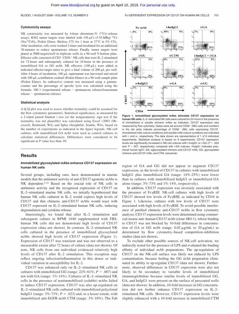
Interestingly, we found that after IL-2 stimulation andsubsequent culture in RPMI 1640 supplemented with FBS,human NK cells did not demonstrate high levels of CD137expression (data not shown). In contrast, IL-2–stimulated NKcells cultured in the presence of immobilized glycosylatedmAbs showed high levels of CD137 expression (Figure 1).Expression of CD137 was transient and was not observed to ameasurable extent after 72 hours of culture (data not shown). Ofnote, NK cells from one individual evidenced relatively highlevels of CD137 after IL-2 stimulation. This exception mayreflect ongoing infection/inflammation in this donor or indi-vidual variation in susceptibility for IL-2.
CD137 was enhanced only on IL-2–stimulated NK cells incultures with immobilized GG (range: 22%-61%; P � .007) andnot with GA (range: 1%-14%). Cultures of IL-2–stimulated NKcells in the presence of nonimmobilized (soluble) mAbs failedto induce CD137 expression. CD137 was also up-regulated onIL-2–stimulated NK cells cultured with immobilized polyclonalhuIgG1 (range: 3%-73%; P � .023) and, to a lesser extent, withimmobilized anti-EGFR mAb CTM (range: 3%-10%). The Fab
region of GA and GG did not appear to augment CD137expression, as the levels of CD137 in cultures with immobilizedhuIgG1 plus immobilized GA (range: 14%-25%) were lowerthan in cultures with immobilized huIgG1 or immobilized GAalone (range: 3%-73% and 1%-14%, respectively).
In addition, CD137 expression was inversely associated withthe presence of Fc�RIII. NK-cell cultures with high levels ofCD137 showed low levels of Fc�RIII, as indicated by CD16 inFigure 1. Likewise, cultures with low levels of CD137 wereassociated with high levels of Fc�RIII. To avoid possible interfer-ence of purified chimeric anti-CD137 mAbs in flow cytometryanalysis, CD137 expression levels were determined using commer-cial mouse anti–human CD137 mAb (clone 4B4-1), whose bindingto CD137 was not blocked by 10-fold differences in concentra-tion of GA or GG mAb (range: 0.02 �g/mL to 20 �g/mL) asdetermined by flow cytometry–based competition-inhibitionstudies (data not shown).
To exclude other possible sources of NK-cell activation, weindirectly tested for the presence of LPS and evaluated the bindingaffinity of individual mAb preparations. The up-regulation ofCD137 on the NK-cell surface was likely not induced by LPScontamination, because boiling the GG mAb preparation elimi-nated its ability to up-regulate CD137 (data not shown). Further-more, observed differences in CD137 expression were also notlikely to be secondary to variable levels of immobilizedimmunoglobulins because similar levels of immobilized GG,GA, and huIgG1 were present on the surface of precoated wells(data not shown). In addition, 10-fold increases in GG concentra-tion did not further enhance CD137 expression on IL-2–stimulated NK cells. However, CD137 expression levels wereslightly enhanced with a 10-fold increase in immobilized CTM
Figure 1. Immobilized glycosylated mAbs stimulate CD137 expression onhuman NK cells. IL-2–stimulated NK cells were cultured for 24 hours in the presenceof immobilized or soluble chimeric mAbs as indicated. CD137 expression wasmeasured by flow cytometry. Gates were set around CD56� (NK) cells and numbersin the dot plots indicate percentage of CD56� (NK) cells expressing CD137.Immobilized mAb culture conditions and soluble mAb culture conditions are indicatedwith i- and s-, respectively. The data shown are representative of 1 of 6 individualexperiments. Statistical analysis is based on 6 experiments; CD137 expressionlevels are significantly increased in NK-cell cultures with i-huIgG1 or i-GG (P � .023and P � .007, respectively) compared with i-GA cultures. HuIgG1 indicates poly-clonal human IgG1; GA, aglycosylated chimeric anti-CD137 mAb; GG, glycosylatedchimeric anti-CD137 mAb; and CTM, cetuximab.
Fc-DEPENDENT EXPRESSION OF CD137 ON HUMAN NK CELLS 701BLOOD, 1 AUGUST 2008 � VOLUME 112, NUMBER 3
For personal use only.on April 10, 2019. by guest www.bloodjournal.orgFrom
concentration (10 �g/mL to 100 �g/mL) and CD137 started toreach expression levels as observed in cultures with 10 �g/mLGG (data not shown). These experiments support the conclusionthat CD137 expression on NK cells is Fc dependent and thatdifferent Fc regions have variable ability to induce CD137 onhuman NK cells.
CD137 expression on NK cells is induced in an Fc-dependentfashion
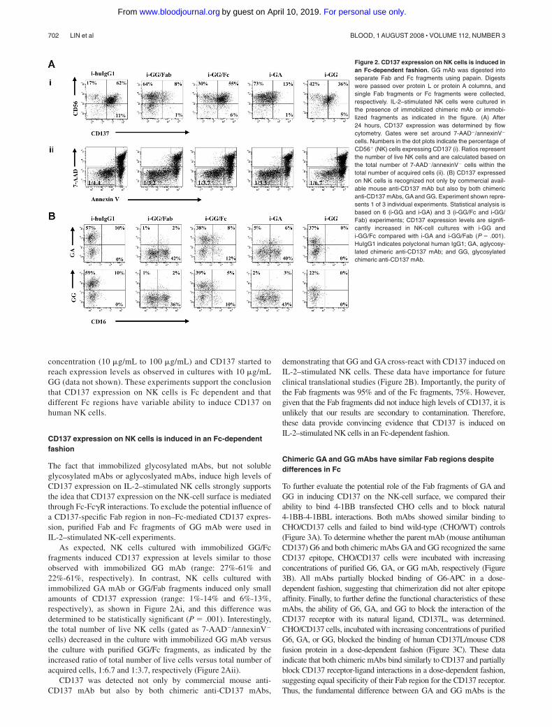
The fact that immobilized glycosylated mAbs, but not solubleglycosylated mAbs or aglycoslyated mAbs, induce high levels ofCD137 expression on IL-2–stimulated NK cells strongly supportsthe idea that CD137 expression on the NK-cell surface is mediatedthrough Fc-Fc�R interactions. To exclude the potential influence ofa CD137-specific Fab region in non–Fc-mediated CD137 expres-sion, purified Fab and Fc fragments of GG mAb were used inIL-2–stimulated NK-cell experiments.
As expected, NK cells cultured with immobilized GG/Fcfragments induced CD137 expression at levels similar to thoseobserved with immobilized GG mAb (range: 27%-61% and22%-61%, respectively). In contrast, NK cells cultured withimmobilized GA mAb or GG/Fab fragments induced only smallamounts of CD137 expression (range: 1%-14% and 6%-13%,respectively), as shown in Figure 2Ai, and this difference wasdetermined to be statistically significant (P � .001). Interestingly,the total number of live NK cells (gated as 7-AAD�/annexinV�
cells) decreased in the culture with immobilized GG mAb versusthe culture with purified GG/Fc fragments, as indicated by theincreased ratio of total number of live cells versus total number ofacquired cells, 1:6.7 and 1:3.7, respectively (Figure 2Aii).
CD137 was detected not only by commercial mouse anti-CD137 mAb but also by both chimeric anti-CD137 mAbs,
demonstrating that GG and GA cross-react with CD137 induced onIL-2–stimulated NK cells. These data have importance for futureclinical translational studies (Figure 2B). Importantly, the purity ofthe Fab fragments was 95% and of the Fc fragments, 75%. However,given that the Fab fragments did not induce high levels of CD137, it isunlikely that our results are secondary to contamination. Therefore,these data provide convincing evidence that CD137 is induced onIL-2–stimulated NK cells in an Fc-dependent fashion.
Chimeric GA and GG mAbs have similar Fab regions despitedifferences in Fc
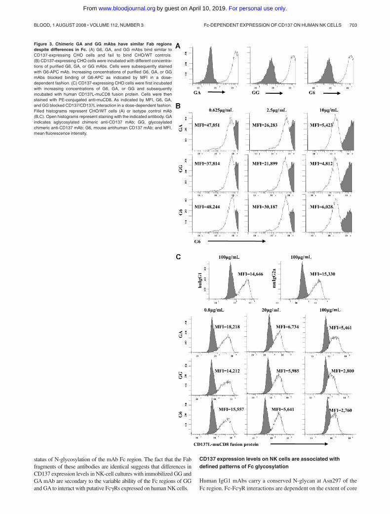
To further evaluate the potential role of the Fab fragments of GA andGG in inducing CD137 on the NK-cell surface, we compared theirability to bind 4-1BB transfected CHO cells and to block natural4-1BB-4-1BBL interactions. Both mAbs showed similar binding toCHO/CD137 cells and failed to bind wild-type (CHO/WT) controls(Figure 3A). To determine whether the parent mAb (mouse antihumanCD137) G6 and both chimeric mAbs GA and GG recognized the sameCD137 epitope, CHO/CD137 cells were incubated with increasingconcentrations of purified G6, GA, or GG mAb, respectively (Figure3B). All mAbs partially blocked binding of G6-APC in a dose-dependent fashion, suggesting that chimerization did not alter epitopeaffinity. Finally, to further define the functional characteristics of thesemAbs, the ability of G6, GA, and GG to block the interaction of theCD137 receptor with its natural ligand, CD137L, was determined.CHO/CD137 cells, incubated with increasing concentrations of purifiedG6, GA, or GG, blocked the binding of human CD137L/mouse CD8fusion protein in a dose-dependent fashion (Figure 3C). These dataindicate that both chimeric mAbs bind similarly to CD137 and partiallyblock CD137 receptor-ligand interactions in a dose-dependent fashion,suggesting equal specificity of their Fab region for the CD137 receptor.Thus, the fundamental difference between GA and GG mAbs is the
Figure 2. CD137 expression on NK cells is induced inan Fc-dependent fashion. GG mAb was digested intoseparate Fab and Fc fragments using papain. Digestswere passed over protein L or protein A columns, andsingle Fab fragments or Fc fragments were collected,respectively. IL-2–stimulated NK cells were cultured inthe presence of immobilized chimeric mAb or immobi-lized fragments as indicated in the figure. (A) After24 hours, CD137 expression was determined by flowcytometry. Gates were set around 7-AAD�/annexinV�
cells. Numbers in the dot plots indicate the percentage ofCD56� (NK) cells expressing CD137 (i). Ratios representthe number of live NK cells and are calculated based onthe total number of 7-AAD�/annexinV� cells within thetotal number of acquired cells (ii). (B) CD137 expressedon NK cells is recognized not only by commercial avail-able mouse anti-CD137 mAb but also by both chimericanti-CD137 mAbs, GA and GG. Experiment shown repre-sents 1 of 3 individual experiments. Statistical analysis isbased on 6 (i-GG and i-GA) and 3 (i-GG/Fc and i-GG/Fab) experiments; CD137 expression levels are signifi-cantly increased in NK-cell cultures with i-GG andi-GG/Fc compared with i-GA and i-GG/Fab (P � .001).HuIgG1 indicates polyclonal human IgG1; GA, aglycosy-lated chimeric anti-CD137 mAb; and GG, glycosylatedchimeric anti-CD137 mAb.
702 LIN et al BLOOD, 1 AUGUST 2008 � VOLUME 112, NUMBER 3
For personal use only.on April 10, 2019. by guest www.bloodjournal.orgFrom
status of N-glycosylation of the mAb Fc region. The fact that the Fabfragments of these antibodies are identical suggests that differences inCD137 expression levels in NK-cell cultures with immobilized GG andGA mAb are secondary to the variable ability of the Fc regions of GGand GA to interact with putative Fc�Rs expressed on human NK cells.
CD137 expression levels on NK cells are associated withdefined patterns of Fc glycosylation
Human IgG1 mAbs carry a conserved N-glycan at Asn297 of theFc region. Fc-Fc�R interactions are dependent on the extent of core
Figure 3. Chimeric GA and GG mAbs have similar Fab regionsdespite differences in Fc. (A) G6, GA, and GG mAbs bind similar toCD137-expressing CHO cells and fail to bind CHO/WT controls.(B) CD137-expressing CHO cells were incubated with different concentra-tions of purified G6, GA, or GG mAbs. Cells were subsequently stainedwith G6-APC mAb. Increasing concentrations of purified G6, GA, or GGmAbs blocked binding of G6-APC as indicated by MFI in a dose-dependent fashion. (C) CD137-expressing CHO cells were first incubatedwith increasing concentrations of G6, GA, or GG and subsequentlyincubated with human CD137L-muCD8 fusion protein. Cells were thenstained with PE-conjugated anti-muCD8. As indicated by MFI, G6, GA,and GG blocked CD137/CD137L interaction in a dose-dependent fashion.Filled histograms represent CHO/WT cells (A) or isotype control mAb(B,C). Open histograms represent staining with the indicated antibody. GAindicates aglycosylated chimeric anti-CD137 mAb; GG, glycosylatedchimeric anti-CD137 mAb; G6, mouse antihuman CD137 mAb; and MFI,mean fluorescence intensity.
Fc-DEPENDENT EXPRESSION OF CD137 ON HUMAN NK CELLS 703BLOOD, 1 AUGUST 2008 � VOLUME 112, NUMBER 3
For personal use only.on April 10, 2019. by guest www.bloodjournal.orgFrom
fucosylation of the Fc N-glycan, with lower fucosylated IgG1having significantly enhanced affinity for the Fc�III receptor incomparison with those with high fucose content. The level offucosylation has functional implications, as lower fucosylatedmAbs induce more potent antibody-dependent cellular cytotoxicity(ADCC) at lower antigen densities.13 Based on our observationsthat mAbs from different sources stimulated variable levels ofCD137 on the NK-cell surface, we sought to investigate whetherthe difference in fucose composition of N-glycans was associatedwith CD137 expression.
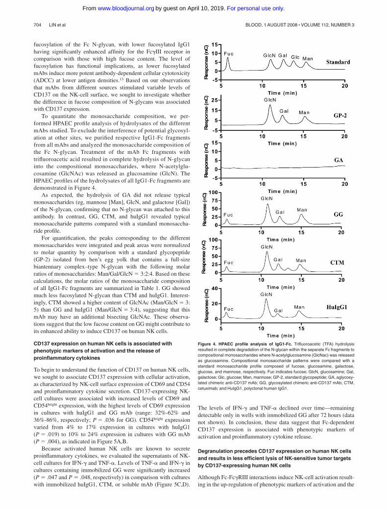
To quantitate the monosaccharide composition, we per-formed HPAEC profile analysis of hydrolysates of the differentmAbs studied. To exclude the interference of potential glycosyl-ation at other sites, we purified respective IgG1-Fc fragmentsfrom all mAbs and analyzed the monosaccharide composition ofthe Fc N-glycan. Treatment of the mAb Fc fragments withtrifluoroacetic acid resulted in complete hydrolysis of N-glycaninto the compositional monosaccharides, where N-acetylglu-cosamine (GlcNAc) was released as glucosamine (GlcN). TheHPAEC profiles of the hydrolysates of all IgG1-Fc fragments aredemonstrated in Figure 4.
As expected, the hydrolysis of GA did not release typicalmonosaccharides (eg, mannose [Man], GlcN, and galactose [Gal])of the N-glycan, confirming that no N-glycan was attached to thisantibody. In contrast, GG, CTM, and huIgG1 revealed typicalmonosaccharide patterns compared with a standard monosaccha-ride profile.
For quantification, the peaks corresponding to the differentmonosaccharides were integrated and peak areas were normalizedto molar quantity by comparison with a standard glycopeptide(GP-2) isolated from hen’s egg yolk that contains a full-sizebiantennary complex–type N-glycan with the following molarratios of monosaccharides: Man/Gal/GlcN � 3:2:4. Based on thesecalculations, the molar ratios of the monosaccharide compositionof all IgG1-Fc fragments are summarized in Table 1. GG showedmuch less fucosylated N-glycan than CTM and huIgG1. Interest-ingly, CTM showed a higher content of GlcNAc (Man/GlcN � 3:5) than GG and huIgG1 (Man/GlcN � 3:4), suggesting that thismAb may have an additional bisecting GlcNAc. These observa-tions suggest that the low fucose content on GG might contribute toits enhanced ability to induce CD137 on human NK cells.
CD137 expression on human NK cells is associated withphenotypic markers of activation and the release ofproinflammatory cytokines
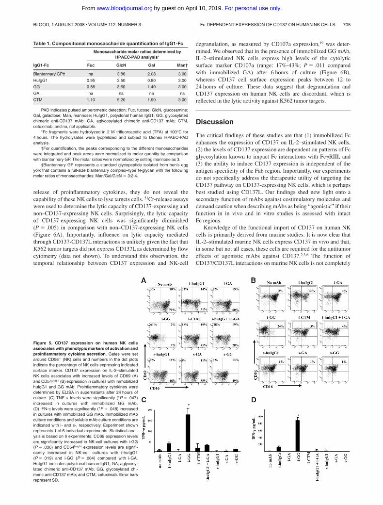
To begin to understand the function of CD137 on human NK cells,we sought to associate CD137 expression with cellular activation,as characterized by NK-cell surface expression of CD69 and CD54and proinflammatory cytokine secretion. CD137-expressing NK-cell cultures were associated with increased levels of CD69 andCD54bright expression, with the highest levels of CD69 expressionin cultures with huIgG1 and GG mAb (range: 32%-62% and36%-86%, respectively; P � .036 for GG). CD54bright expressionvaried from 4% to 17% expression in cultures with huIgG1(P � .019) to 10% to 24% expression in cultures with GG mAb(P � .004), as indicated in Figure 5A,B.
Because activated human NK cells are known to secreteproinflammatory cytokines, we evaluated the supernatants of NK-cell cultures for IFN-� and TNF-�. Levels of TNF-� and IFN-� incultures containing immobilized GG were significantly increased(P � .047 and P � .048, respectively) in comparison with cultureswith immobilized huIgG1, CTM, or soluble mAb (Figure 5C,D).
The levels of IFN-� and TNF-� declined over time—remainingdetectable only in wells with immobilized GG after 72 hours (datanot shown). In conclusion, these data suggest that Fc-dependentCD137 expression is associated with phenotypic markers ofactivation and proinflammatory cytokine release.
Degranulation precedes CD137 expression on human NK cellsand results in less efficient lysis of NK-sensitive tumor targetsby CD137-expressing human NK cells
Although Fc-Fc�RIII interactions induce NK-cell activation result-ing in the up-regulation of phenotypic markers of activation and the
Figure 4. HPAEC profile analysis of IgG1-Fc. Trifluoroacetic (TFA) hydrolysisresulted in complete degradation of the N-glycan within the separate Fc fragments tocompositional monosaccharides where N-acetylglucosamine (GlcNac) was releasedas glucosamine. Compositional monosaccharide patterns were compared with astandard monosaccharide profile composed of fucose, glucosamine, galactose,glucose, and mannose, respectively. Fuc indicates fucose; GlcN, glucosamine; Gal,galactose; Glc, glucose; Man, mannose; GP-2, standard glycopeptide; GA, aglycosy-lated chimeric anti-CD137 mAb; GG, glycosylated chimeric anti-CD137 mAb; CTM,cetuximab; and HuIgG1, polyclonal human IgG1.
704 LIN et al BLOOD, 1 AUGUST 2008 � VOLUME 112, NUMBER 3
For personal use only.on April 10, 2019. by guest www.bloodjournal.orgFrom
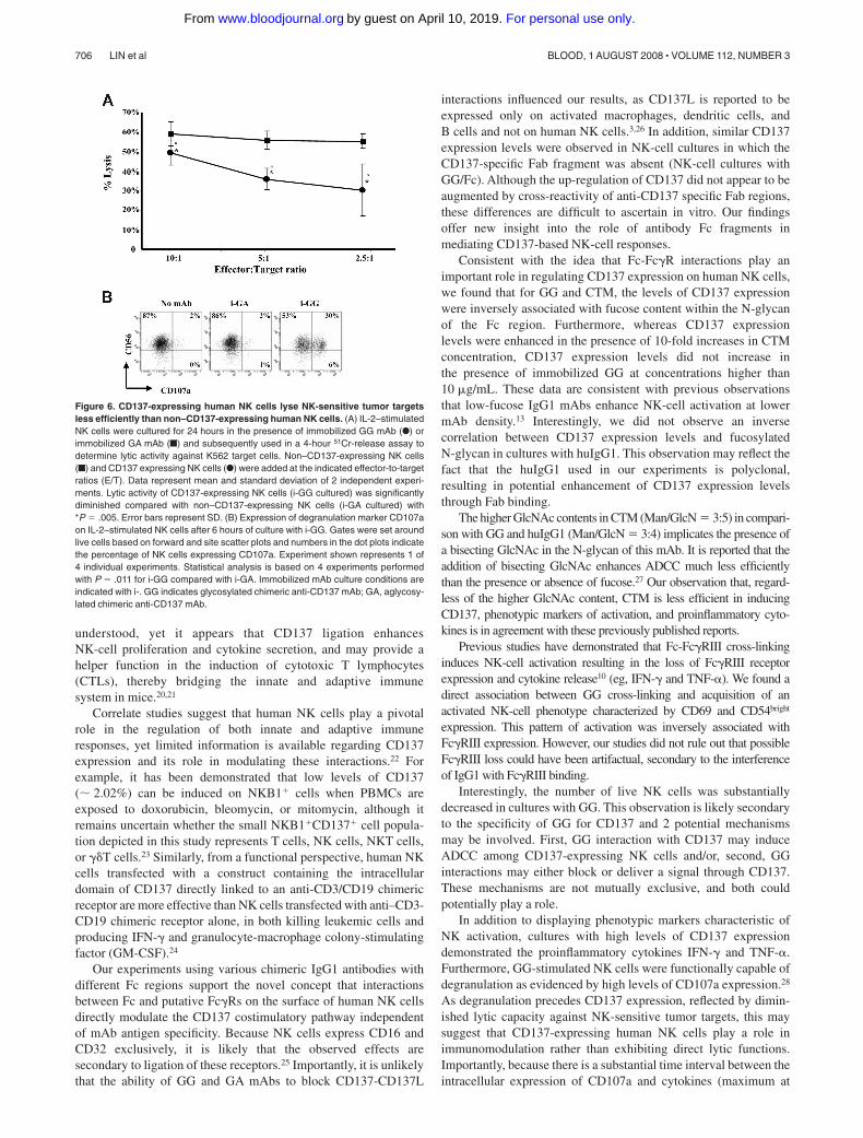
release of proinflammatory cytokines, they do not reveal thecapability of these NK cells to lyse targets cells. 51Cr-release assayswere used to determine the lytic capacity of CD137-expressing andnon–CD137-expressing NK cells. Surprisingly, the lytic capacityof CD137-expressing NK cells was significantly diminished(P � .005) in comparison with non–CD137-expressing NK cells(Figure 6A). Importantly, influence on lytic capacity mediatedthrough CD137-CD137L interactions is unlikely given the fact thatK562 tumor targets did not express CD137L as determined by flowcytometry (data not shown). To understand this observation, thetemporal relationship between CD137 expression and NK-cell
degranulation, as measured by CD107a expression,19 was deter-mined. We observed that in the presence of immobilized GG mAb,IL-2–stimulated NK cells express high levels of the cytolyticsurface marker CD107a (range: 17%-43%; P � .011 comparedwith immobilized GA) after 6 hours of culture (Figure 6B),whereas CD137 cell surface expression peaks between 12 to24 hours of culture. These data suggest that degranulation andCD137 expression on human NK cells are discordant, which isreflected in the lytic activity against K562 tumor targets.
Discussion
The critical findings of these studies are that (1) immobilized Fcenhances the expression of CD137 on IL-2–stimulated NK cells,(2) the levels of CD137 expression are dependent on patterns of Fcglycosylation known to impact Fc interactions with Fc�RIII, and(3) the ability to induce CD137 expression is independent of theantigen specificity of the Fab region. Importantly, our experimentsdo not specifically address the therapeutic utility of targeting theCD137 pathway on CD137-expressing NK cells, which is perhapsbest studied using CD137L. Our findings shed new light onto asecondary function of mAbs against costimulatory molecules anddemand caution when describing mAbs as being “agonistic” if theirfunction in in vivo and in vitro studies is assessed with intactFc regions.
Knowledge of the functional import of CD137 on human NKcells is primarily derived from murine studies. It is now clear thatIL-2–stimulated murine NK cells express CD137 in vivo and that,in some but not all cases, these cells are required for the antitumoreffects of agonistic mAbs against CD137.2,3,6 The function ofCD137/CD137L interactions on murine NK cells is not completely
Figure 5. CD137 expression on human NK cellsassociates with phenotypic markers of activation andproinflammatory cytokine secretion. Gates were setaround CD56� (NK) cells and numbers in the dot plotsindicate the percentage of NK cells expressing indicatedsurface marker. CD137 expression on IL-2–stimulatedNK cells associates with increased levels of CD69 (A)and CD54bright (B) expression in cultures with immobilizedhuIgG1 and GG mAb. Proinflammatory cytokines weredetermined by ELISA in supernatants after 24 hours ofculture. (C) TNF-� levels were significantly (*P � .047)increased in cultures with immobilized GG mAb.(D) IFN-� levels were significantly (*P � .048) increasedin cultures with immobilized GG mAb. Immobilized mAbculture conditions and soluble mAb culture conditions areindicated with i- and s-, respectively. Experiment shownrepresents 1 of 6 individual experiments. Statistical anal-ysis is based on 6 experiments; CD69 expression levelsare significantly increased in NK-cell cultures with i-GG(P � .036) and CD54bright expression levels are signifi-cantly increased in NK-cell cultures with i-huIgG1(P � .019) and i-GG (P � .004) compared with i-GA.HuIgG1 indicates polyclonal human IgG1; GA, aglycosy-lated chimeric anti-CD137 mAb; GG, glycosylated chi-meric anti-CD137 mAb; and CTM, cetuximab. Error barsrepresent SD.
Table 1. Compositional monosaccharide quantification of IgG1-Fc
IgG1-Fc
Monosaccharide molar ratios determined byHPAEC-PAD analysis*
Fuc GlcN Gal Man†
Biantennary GP‡ na 3.86 2.08 3.00
HuIgG1 0.95 3.50 0.80 3.00
GG 0.56 3.60 1.40 3.00
GA na na na na
CTM 1.10 5.20 1.90 3.00
PAD indicates pulsed amperometric detection; Fuc, fucose; GlcN, glucosamine;Gal, galactose; Man, mannose; HuIgG1, polyclonal human IgG1; GG, glycosylatedchimeric anti-CD137 mAb; GA, aglycosylated chimeric anti-CD137 mAb; CTM,cetuximab; and na, not applicable.
*Fc fragments were hydrolyzed in 2 M trifluoroacetic acid (TFA) at 100°C for4 hours. The hydrolysates were lyophilized and subject to Dionex HPAEC-PADanalysis.
†For quantification, the peaks corresponding to the different monosaccharideswere integrated and peak areas were normalized to molar quantity by comparisonwith biantennary GP. The molar ratios were normalized by setting mannose as 3.
‡Biantennary GP represents a standard glycopeptide isolated from hen’s eggyolk that contains a full-size biantennary complex–type N-glycan with the followingmolar ratios of monosaccharides: Man/Gal/GlcN � 3:2:4.
Fc-DEPENDENT EXPRESSION OF CD137 ON HUMAN NK CELLS 705BLOOD, 1 AUGUST 2008 � VOLUME 112, NUMBER 3
For personal use only.on April 10, 2019. by guest www.bloodjournal.orgFrom
understood, yet it appears that CD137 ligation enhancesNK-cell proliferation and cytokine secretion, and may provide ahelper function in the induction of cytotoxic T lymphocytes(CTLs), thereby bridging the innate and adaptive immunesystem in mice.20,21
Correlate studies suggest that human NK cells play a pivotalrole in the regulation of both innate and adaptive immuneresponses, yet limited information is available regarding CD137expression and its role in modulating these interactions.22 Forexample, it has been demonstrated that low levels of CD137( 2.02%) can be induced on NKB1� cells when PBMCs areexposed to doxorubicin, bleomycin, or mitomycin, although itremains uncertain whether the small NKB1�CD137� cell popula-tion depicted in this study represents T cells, NK cells, NKT cells,or �T cells.23 Similarly, from a functional perspective, human NKcells transfected with a construct containing the intracellulardomain of CD137 directly linked to an anti-CD3/CD19 chimericreceptor are more effective than NK cells transfected with anti–CD3-CD19 chimeric receptor alone, in both killing leukemic cells andproducing IFN-� and granulocyte-macrophage colony-stimulatingfactor (GM-CSF).24
Our experiments using various chimeric IgG1 antibodies withdifferent Fc regions support the novel concept that interactionsbetween Fc and putative Fc�Rs on the surface of human NK cellsdirectly modulate the CD137 costimulatory pathway independentof mAb antigen specificity. Because NK cells express CD16 andCD32 exclusively, it is likely that the observed effects aresecondary to ligation of these receptors.25 Importantly, it is unlikelythat the ability of GG and GA mAbs to block CD137-CD137L
interactions influenced our results, as CD137L is reported to beexpressed only on activated macrophages, dendritic cells, andB cells and not on human NK cells.3,26 In addition, similar CD137expression levels were observed in NK-cell cultures in which theCD137-specific Fab fragment was absent (NK-cell cultures withGG/Fc). Although the up-regulation of CD137 did not appear to beaugmented by cross-reactivity of anti-CD137 specific Fab regions,these differences are difficult to ascertain in vitro. Our findingsoffer new insight into the role of antibody Fc fragments inmediating CD137-based NK-cell responses.
Consistent with the idea that Fc-Fc�R interactions play animportant role in regulating CD137 expression on human NK cells,we found that for GG and CTM, the levels of CD137 expressionwere inversely associated with fucose content within the N-glycanof the Fc region. Furthermore, whereas CD137 expressionlevels were enhanced in the presence of 10-fold increases in CTMconcentration, CD137 expression levels did not increase inthe presence of immobilized GG at concentrations higher than10 �g/mL. These data are consistent with previous observationsthat low-fucose IgG1 mAbs enhance NK-cell activation at lowermAb density.13 Interestingly, we did not observe an inversecorrelation between CD137 expression levels and fucosylatedN-glycan in cultures with huIgG1. This observation may reflect thefact that the huIgG1 used in our experiments is polyclonal,resulting in potential enhancement of CD137 expression levelsthrough Fab binding.
The higher GlcNAc contents in CTM (Man/GlcN � 3:5) in compari-son with GG and huIgG1 (Man/GlcN � 3:4) implicates the presence ofa bisecting GlcNAc in the N-glycan of this mAb. It is reported that theaddition of bisecting GlcNAc enhances ADCC much less efficientlythan the presence or absence of fucose.27 Our observation that, regard-less of the higher GlcNAc content, CTM is less efficient in inducingCD137, phenotypic markers of activation, and proinflammatory cyto-kines is in agreement with these previously published reports.
Previous studies have demonstrated that Fc-Fc�RIII cross-linkinginduces NK-cell activation resulting in the loss of Fc�RIII receptorexpression and cytokine release10 (eg, IFN-� and TNF-�). We found adirect association between GG cross-linking and acquisition of anactivated NK-cell phenotype characterized by CD69 and CD54bright
expression. This pattern of activation was inversely associated withFc�RIII expression. However, our studies did not rule out that possibleFc�RIII loss could have been artifactual, secondary to the interferenceof IgG1 with Fc�RIII binding.
Interestingly, the number of live NK cells was substantiallydecreased in cultures with GG. This observation is likely secondaryto the specificity of GG for CD137 and 2 potential mechanismsmay be involved. First, GG interaction with CD137 may induceADCC among CD137-expressing NK cells and/or, second, GGinteractions may either block or deliver a signal through CD137.These mechanisms are not mutually exclusive, and both couldpotentially play a role.
In addition to displaying phenotypic markers characteristic ofNK activation, cultures with high levels of CD137 expressiondemonstrated the proinflammatory cytokines IFN-� and TNF-�.Furthermore, GG-stimulated NK cells were functionally capable ofdegranulation as evidenced by high levels of CD107a expression.28
As degranulation precedes CD137 expression, reflected by dimin-ished lytic capacity against NK-sensitive tumor targets, this maysuggest that CD137-expressing human NK cells play a role inimmunomodulation rather than exhibiting direct lytic functions.Importantly, because there is a substantial time interval between theintracellular expression of CD107a and cytokines (maximum at
Figure 6. CD137-expressing human NK cells lyse NK-sensitive tumor targetsless efficiently than non–CD137-expressing human NK cells. (A) IL-2–stimulatedNK cells were cultured for 24 hours in the presence of immobilized GG mAb (F) orimmobilized GA mAb (f) and subsequently used in a 4-hour 51Cr-release assay todetermine lytic activity against K562 target cells. Non–CD137-expressing NK cells(f) and CD137 expressing NK cells (F) were added at the indicated effector-to-targetratios (E/T). Data represent mean and standard deviation of 2 independent experi-ments. Lytic activity of CD137-expressing NK cells (i-GG cultured) was significantlydiminished compared with non–CD137-expressing NK cells (i-GA cultured) with*P � .005. Error bars represent SD. (B) Expression of degranulation marker CD107aon IL-2–stimulated NK cells after 6 hours of culture with i-GG. Gates were set aroundlive cells based on forward and site scatter plots and numbers in the dot plots indicatethe percentage of NK cells expressing CD107a. Experiment shown represents 1 of4 individual experiments. Statistical analysis is based on 4 experiments performedwith P � .011 for i-GG compared with i-GA. Immobilized mAb culture conditions areindicated with i-. GG indicates glycosylated chimeric anti-CD137 mAb; GA, aglycosy-lated chimeric anti-CD137 mAb.
706 LIN et al BLOOD, 1 AUGUST 2008 � VOLUME 112, NUMBER 3
For personal use only.on April 10, 2019. by guest www.bloodjournal.orgFrom

6 hours) and the surface expression of CD137 (maximum at� 12 hours), we cannot ascertain whether CD137-expressing cellsare directly responsible for the observed differences.
In summary, our findings that immobilized Fc induces CD137expression on IL-2–stimulated NK cells are pivotal to the interpre-tation of studies using anti-CD137 mAbs with an Fc region capableof binding Fc�Rs. Specifically, under these circumstances, effectspreviously attributed to CD137 interactions with the Fab region,must now be reevaluated as potentially related to Fc-inducedexpression of CD137 on NK cells with subsequent effects due to(1) direct interaction of CD137L with Fc-induced CD137, (2) directstimulation of Fc-induced CD137 by agonistic anti-CD137 mAbs,or (3) antibody blockade of interactions between Fc-inducedCD137 and CD137L.
Acknowledgment
This study was partially supported by National Institutes of Health(Bethesda, MD) grant 5R44CA107608-04 and by an Small Busi-ness Innovation Research grant from the National Cancer Institute,Bethesda, MD, on which S.E.S. and L.C. are subcontractors.
Authorship
Contribution: W.L., X.Z., A.W., E.B., and Y.W. performed experi-ments and analyzed data; C.J.V. designed and performed experi-ments, analyzed data, and wrote the paper; L.C. provided transfec-tants and the mouse antihuman anti-CD137 monoclonal antibody;D.G.S. provided the chimeric anti-CD137 monoclonal antibodies;G.T. performed statistical analysis; L.-X.W., K.T, D.H.S., D.M.,and S.E.S. designed experiments, and reviewed data and the paper.
Conflict-of-interest disclosure: S.E.S. and L.C. receive royaltiesfrom GTC Biotherapeutics through the Mayo Clinic College ofMedicine for licensure of intellectual property related to CD137.S.E.S. is a cofounder and major stockholder in Gliknik, a biotech-nology company. D.G.S. is employed by and owns stock in GTCBiotherapeutics. All other authors declare no competing financialinterests.
Correspondence: Scott E. Strome, Department of Otorhinolaryn-gology-Head and Neck Surgery, University of Maryland, 16 SouthEutaw Street, Suite 500, Baltimore, MD 21201-168; e-mail:[email protected].
References
1. Wilcox RA, Flies DB, Zhu G, et al. Provision ofantigen and CD137 signaling breaks immunologi-cal ignorance, promoting regression of poorly im-munogenic tumors. J Clin Invest. 2002;109:651-659.
2. Wilcox RA, Tamada K, Strome SE, Chen L. Sig-naling through NK cell-associated CD 137 pro-motes both helper function for CD8� cytolyticT cells and responsiveness to IL-2 but not cyto-lytic activity. J Immunol. 2002;169:4230-4236.
3. Melero I, Shuford W, Newby S, et al. Monoclonalantibodies against the 4-1BB T-cell activationmolecule eradicate established tumors. Nat Med.1997;3:682-685.
4. Martinet O, Ermekova V, Qiao JQ, et al. Immuno-modulatory gene therapy with interleukin 12 and4-1BB ligand: long-term remission of liver metas-tases in a mouse model. J Natl Cancer Inst.2000;92:931-936.
5. Melero I, Hervas-Stubbs S, Glennie M, PardollDM, Chen L. Immunostimulatory monoclonal anti-bodies for cancer therapy. Nat Rev Cancer. 2007;7:95-106.
6. Melero I, Johnston J, Shufford W, Mittler R, ChenL. NK1.1 cells express 4-1BB (CDw137) costimu-latory molecule and are required for tumor immu-nity elicited by anti-4-1-BB monoclonal antibod-ies. Cell Immunol. 1998;190:167-172.
7. Seo SK, Choi JH, Kim YH, et al. 4-1BB-mediatedimmunotherapy of rheumatoid arthritis. Nat Med.2004;10:1088-1094.
8. Foell J, Strahotin S, O’Neil SP, et al. CD137 co-stimulatory T cell receptor engagement reversesacute disease in lupus-prone NZB x NZW F1mice. J Clin Invest. 2003;111:1505-1518.
9. Lee J, Lee E-N, Kim E-Y, et al. Administration ofagonistic anti-4-1BB monoclonal antibody leadsto the amelioration of inflammatory bowel dis-ease. Immunol Lett. 2005;101:210-216.
10. Bowles JA, Weiner GJ. CD16 polymorphisms andNK activation induced by monoclonal antibody-coated target cells. J Immunol Methods. 2005;304:88-99.
11. Shields RL, Namenuk AK, Hong K, et al. High
resolution mapping of the binding site on humanIgG1 for Fcgamma RI, Fcgamma RII, FcgammaRIII, and FcRn and design of IgG1 variants withimproved binding to the Fcgamma R. J BiolChem. 2001;276:6591-6604.
12. Shields RL, Lai J, Keck R, et al. Lack of fucose onhuman IgG1 N-linked oligosaccharide improvesbinding to human Fcgamma RIII and antibody-dependent cellular toxicity. J Biol Chem. 2002;277:26733-26740.
13. Niwa R, Sakurada M, Kobayashi Y, et al. En-hanced natural killer cell binding and activation bylow-fucose IgG1 antibody results in potent anti-body-dependent cellular cytotoxicity induction atlower antigen density. Clin Cancer Res. 2005;11:2327-2336.
14. Okazaki A, Shoji-Hosaka E, Nakamura K, et al.Fucose depletion from human IgG1 oligosaccha-ride enhances binding enthalpy and associationrate between IgG1 and Fc[gamma]RIIIa. J MolBiol. 2004;336:1239-1249.
15. Nose M, Wigzell H. Biological significance of car-bohydrate chains on monoclonal antibodies. ProcNatl Acad Sci U S A. 1983;80:6632-6636.
16. Tao MH, Morrison SL. Studies of aglycosylatedchimeric mouse-human IgG: role of carbohydratein the structure and effector functions mediatedby the human IgG constant region. J Immunol.1989;143:2595-2601.
17. Weng W-K, Czerwinski D, Timmerman J, Hsu FJ,Levy R. Clinical outcome of lymphoma patientsafter idiotype vaccination is correlated with hu-moral immune response and immunoglobulin GFc receptor genotype. J Clin Oncol. 2004;22:4717-4724.
18. Weng W-K, Levy R. Two immunoglobulin G frag-ment C receptor polymorphisms independentlypredict response to rituximab in patients with fol-licular lymphoma. J Clin Oncol. 2003;21:3940-3947.
19. Fischer L, Penack O, Gentilini C, et al. The anti-lymphoma effect of antibody-mediated immuno-therapy is based on an increased degranulationof peripheral blood natural killer (NK) cells. ExpHematol. 2006;34:753-759.
20. Pan P-Y, Gu P, Li Q, Xu D, Weber K, Chen S-H.Regulation of dendritic cell function by NK cells:mechanisms underlying the synergism in thecombination therapy of IL-12 and 4-1BB activa-tion. J Immunol. 2004;172:4779-4789.
21. Vinay DS, Choi BK, Bae JS, Kim WY, GebhardtBM, Kwon BS. CD137-deficient mice have re-duced NK/NKT cell numbers and function, areresistant to lipopolysaccharide-induced shocksyndromes, and have lower IL-4 responses. J Im-munol. 2004;173:4218-4229.
22. Cooper MA, Fehniger TA, Caligiuri MA. The biol-ogy of human natural killer-cell subsets. TrendsImmunol. 2001;22:633-640.
23. Kim KM, Kim HW, Kim JO, Baek KM, Kim JG,Kang CY. Induction of 4-1BB (CD137) expressionby DNA damaging agents in human T lympho-cytes. Immunology. 2002;107:472-479.
24. Imai C, Iwamoto S, Campana D. Genetic modifi-cation of primary natural killer cells overcomesinhibitory signals and induces specific killing ofleukemic cells. Blood. 2005;106:376-383.
25. Metes D, Ernst LK, Chambers WH, Sulica A, Her-berman RB, Morel PA. Expression of functionalCD32 molecules on human NK cells is deter-mined by an allelic polymorphism of the Fc-gamma RIIC gene. Blood. 1998;91:2369-2380.
26. Houtenbos I, Westers TM, Dijkhuis A, de GruijlTD, Ossenkoppele GJ, van de Loosdrecht AA.Leukemia-specific T-cell reactivity induced by leu-kemic dendritic cells is augmented by 4-1BB tar-geting. Clin Cancer Res. 2007;13:307-315.
27. Shinkawa T, Nakamura K, Yamane N, et al. Theabsence of fucose but not the presence of galac-tose or bisecting N-acetylglucosamine of humanIgG1 complex-type oligosaccharides shows thecritical role of enhancing antibody-dependent cel-lular cytotoxicity. J Biol Chem. 2003;278:3466-3473.
28. Betts MR, Brenchley JM, Price DA, et al. Sensi-tive and viable identification of antigen-specificCD8� T cells by a flow cytometric assay for de-granulation. J Immunol Methods. 2003;281:65-78.
Fc-DEPENDENT EXPRESSION OF CD137 ON HUMAN NK CELLS 707BLOOD, 1 AUGUST 2008 � VOLUME 112, NUMBER 3
For personal use only.on April 10, 2019. by guest www.bloodjournal.orgFrom
online June 2, 2008 originally publisheddoi:10.1182/blood-2007-11-122465
2008 112: 699-707
E. StromeScottWei, Lieping Chen, Guoliang Tian, Koji Tamada, Lai-Xi Wang, Dan H. Schulze, Dean Mann and
Wei Lin, Caroline J. Voskens, Xiaoyu Zhang, Daniel G. Schindler, Aaron Wood, Erin Burch, Yadong ''agonistic'' effects of anti-CD137 monoclonal antibodiesFc-dependent expression of CD137 on human NK cells: insights into
http://www.bloodjournal.org/content/112/3/699.full.htmlUpdated information and services can be found at:
(5663 articles)Immunobiology and Immunotherapy Articles on similar topics can be found in the following Blood collections
http://www.bloodjournal.org/site/misc/rights.xhtml#repub_requestsInformation about reproducing this article in parts or in its entirety may be found online at:
http://www.bloodjournal.org/site/misc/rights.xhtml#reprintsInformation about ordering reprints may be found online at:
http://www.bloodjournal.org/site/subscriptions/index.xhtmlInformation about subscriptions and ASH membership may be found online at:
Copyright 2011 by The American Society of Hematology; all rights reserved.of Hematology, 2021 L St, NW, Suite 900, Washington DC 20036.Blood (print ISSN 0006-4971, online ISSN 1528-0020), is published weekly by the American Society
For personal use only.on April 10, 2019. by guest www.bloodjournal.orgFrom
![NK Cell-Fc Receptors Advance Tumor Immunotherapy · The Fc receptors bind the Fc portion of the antibody and transduce activating or inhibitory signals into the cells [20]. The Fc](https://img.pdfslide.us/doc/110x75/5fe70adeb441d3706c1900ec/nk-cell-fc-receptors-advance-tumor-immunotherapy-the-fc-receptors-bind-the-fc-portion.jpg)