Embed Size (px)
Citation preview
Clinico-pathological assessment of common medical liver diseases-
Fatty Liver Disease
Judy Wyatt
And
Mervyn Davies

Fat and the liver
• Hepatocytes use lipid (membranes, bile, energy metabolism) but do not normally store it.
• In fatty liver disease, stored triglyceride = steatosis
• A small amount of fatty change is very common -
not considered pathological if <5% hepatocytes
• Fatty liver disease = includes steatosis and steatohepatitis and cirrhosis
• Alcoholic liver disease (ALD) or non-alcoholic fatty liver disease (NAFLD)
• Steatohepatitis = metabolic injury, leading to fibrosis and cirrhosis
– Alcoholic steatohepatitis (ASH) and non-alcoholic steatohepatitis (NASH)
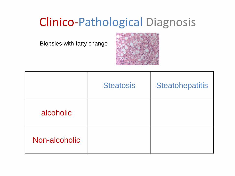
Clinico-Pathological Diagnosis
Steatosis Steatohepatitis
alcoholic
Non-alcoholic
Biopsies with fatty change

Fatty liver disease is very common –who should have a biopsy?
Estimated prevalence up to 44% in Europe*
Clinical diagnosis based on:
Abnormal liver enzymes – can be normal
Fatty liver on ultrasound
Aetiology from clinical history and examination
Why would you do a biopsy??
Suspect severe/advanced fatty liver disease
– non-invasive liver screen
Exclude an additional / alternative diagnosis
Medication such as methotrexate
*Blachier M et al. J Hepatol 2013;58;593-608

The spectrum of fatty liver disease
Case 1. Steatohepatitis - diagnosis and severity
Case 2. Cirrhosis, alcoholic steatohepatitis
Case 3. Steatosis

• Referred for abnormal LFTs to St. James’s Hospital in November 2014
• Background
- Obesity (BMI 32)
- Moderate Alcohol consumption (14 units/week)
- Diet controlled type 2 diabetes, no complications
- Married with 3 kids
- Takes lansoprazole
Case 1. JSF b 1961 Age 53, F

1. JSF – Aged 53
• ALT 152 – 186 (<40), AST 110 (<40), AST:ALT ratio 0.6
• Ferritin – 441 (30 – 300) – transferrin sat 28%
• Viral Serology – HBV/HCV negative
• Auto-antibodies –ve: ANA/SMA/LKM/AMA
• Ig’s Normal, ttg – Negative. TFT normal
• Glu - DM HBA1c good control.
• A-1 AT slightly low, Pi MS heterozygote
• U/S – Bright fatty liver
1. JSF – Aged 53
On examination, occasional spider naevi
Over weight, BMI 32
Lost some weight on a low carbohydrate diet (South Beach Diet)
• LFTs continued to fluctuate therefore whether to biopsy (to assess pathological process and stage)
• Use non invasive fibrosis scores

Fibroscan and other non invasive measures of fibrosis
in NAFLD
In cases where the only question is the extent of fibrosis, then a
fibroscan is often used to replace a liver biopsy – eg HCV

Fibroscan
– Non-invasive liver stiffness measurement
– Measures velocity of a low frequency shear wave
– Velocity is directly related to liver stiffness
– Measures a volume of liver 1x4cm at 2.5-6.5cm below skin
– LSM ranges from 2.5-75kPa

Fibroscan and biopsy evaluation of fibrosis

Fibroscan ROC curves

BARD score
Points 1 or 2 ZERO
DIABETES Yes = 1 No = 0
BMI >28 Yes = 1 No = 0
AST/ALT ratio >0.8 Yes = 2 No = o
TOTAL
A score >2 potential for fibrosispositive and negative predictive values of the BARD score
for advanced fibrosis were 69% and 96%

NAFLD score
• Age
• Platelets
• Albumin
• Diabetes/impaired glucose tolerance
• ALT
• AST
On line calculator – low risk/intermediate/high
High score (>0.676) positive predictive value for advanced fibrosis (F3-F4) of 82% and a low score (<-1.455) negative predictive value of 88%

ELF Score European liver fibrosis panel
• Proprietary algorithm
Age
Hyaluronic acid level
PIIIP
TIMP-1
Threshold score of 0.102 sensitivity of 87 to 90% and specificity of 41 to 51% for moderate or severe fibrosis
Threshold >0.457 specificity 95 %.

Scores
• NAFLD score low
• BARD score 2 = intermediate
Therefore proceed to a fibroscan:
Median stiffness 15kpa, with satisfactory quality measures
High ALT and high fibroscan, plan liver biopsy to assess process and stage
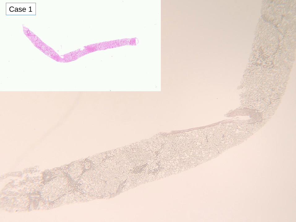
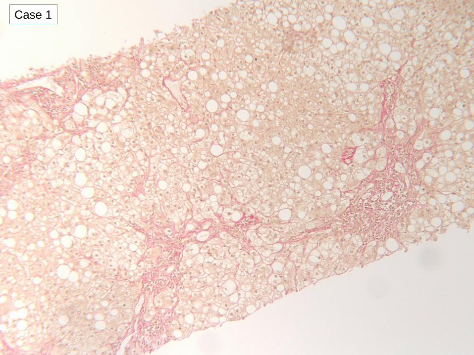
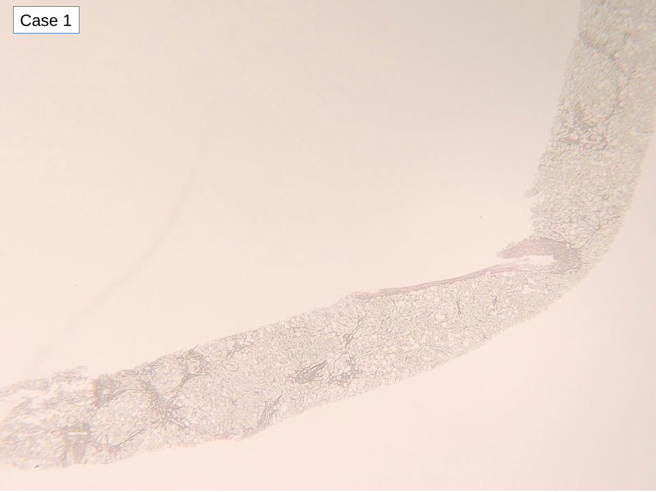
Case 1

Case 1

Case 1

Case 1

Case 1
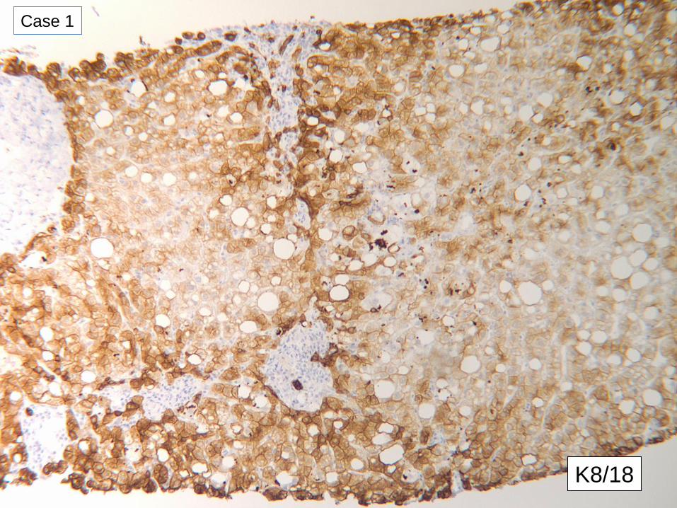
K8/18
Case 1

K8/18
Case 1

K8/18
Case 1
Case 1

Case 1

Case 1. JSF – Aged 53
• Diagnosis – steatohepatitis, consistent with non-alcoholic steatohepatitis,
• Bridging fibrosis (Kleiner stage 3)
• No evidence of other liver disease

Fibroscan result v. Biopsy stage, NAFLD, 16 recent patients
8/16 patients with
fibroscan >10.5 had
stage 0-2 fibrosis
F2 F4

• Necessary components – must see– Steatosis, Macro>micro, mainly zone 3– Mixed mild lobular inflammation– Hepatocyte ballooning, most apparent near steatotic cells
• Usually present, not necessary for diagnosis – often see– Mallory’s hyaline, usually zone 3– Perisinusoidal fibrosis (zone 3)– Glycogenated nuclei (zone 1)– Lipogranulomas (usually small)– Occasional apoptotic hepatocytes/PASD+ve Kupffer cells
• May be present, not necessary for diagnosis – may see– Mild siderosis,– Megamitochondria
• Not present in NASH – don’t see = something else as well
consider other causes of liver disease Next slide
Hepatology. 2003 May;37(5):1202-19.
Necessary features to diagnose steatohepatitisAASLD single topic conference NASH Atlanta, 2002

Unusual for NASH, consider other causes of liver disease instead/as well
– Sclerosing hyaline necrosis
= severe pattern of steatohepatitis seen in alcoholic liver disease
– Portal inflammation > lobular inflammation in early stage disease
– Lymphoid aggregates, plasma cells
– Significant eosinophils, granulomas
– Portal/periportal fibrosis much greater than zone 3 fibrosis
– Lobular disarray
– Acute cholestasis, bile plugs
– Chronic cholestasis, copper associated protein
– Significant iron, evidence of alpha 1 antitrypsin deficiency
Hepatology. 2003 May;37(5):1202-19.

Staining for keratins 8/18 improves detection of
hepatocyte injury in NAFLD
• More sensitive and specific for fibrogenic hepatocellular injury than H&E staining
• 40 biopsies from NASH CRN database study for no steatohepatitis (18): suspicious(7): definite (15) steatohepatitis
Results:
• 2 NASH weren’t
• 5/7 suspicious were no-NASH
• Improved inter-observer agreement on NASH
• Correlates with insulin resistanceGuy CD et al. Human Pathology 2012;43;790-800

How much portal inflammation/fibrosis in steatohepatitis?
When to suspect an additional cause of chronic liver disease?
• Clinicians suspect –
– another cause of liver disease in liver screen,
e.g. Autoantibodies, high ferritin, cholestatic LFTs
? Look for features of these in the biopsy
Up to 30% NAFLD have liver autoantibodies, usually low titre
• Pathologists suspects –
– Disproportionate portal tract inflammation, definite interface hepatitis
? Ask about autoantibodies, Ig level, hepatitis C
– Any copper associated protein in non-cirrhotic biopsy
? Ask about autoantibodies , cholestatic liver function tests
• Portal inflammation in NASH is predictor of fibrosis progression•

Clinico-Pathological Diagnosis
Steatosis Steatohepatitis
either
>5% hepatocytes with steatosis, without
features of steatohepatitis
SteatosisInflammation
Ballooned hepatocytes+/- Mallory-Denk bodies,
pericellular fibrosis
alcoholic
More fibrosisMore Mallory-Denk bodies
PolymorphsCholestasis
Non-alcoholicGlycogenated nuclei
More steatosis v. other features
or
Can the pathologist distinguish ASH from NASH?

How bad is it?
Stage:
1 perisinusoidal (1a, 1b) or periportal (1c)
2 peri-sinusoidal and portal/periportal
3 bridging fibrosis
4 cirrhosis
Kleiner, Hepatology. 2005 Jun;41(6):1313-21
Case 1
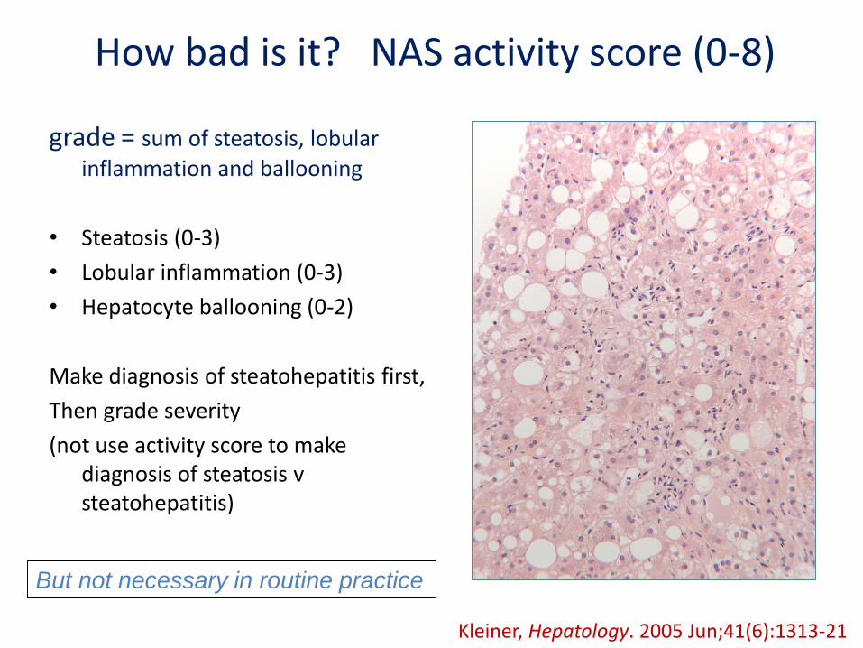
How bad is it? NAS activity score (0-8)
grade = sum of steatosis, lobular
inflammation and ballooning
• Steatosis (0-3)
• Lobular inflammation (0-3)
• Hepatocyte ballooning (0-2)
Make diagnosis of steatohepatitis first,
Then grade severity
(not use activity score to make diagnosis of steatosis v steatohepatitis)
But not necessary in routine practice
Kleiner, Hepatology. 2005 Jun;41(6):1313-21

Scoring system for evaluation of liver lesions in morbidly obese patients
Steatosis, Activity, Fibrosis (SAF) score
• Steatosis (0-3), Fibrosis (0-4), scored as NASH-CRN (Kleiner 2005)
• Activity score = combined scores for ballooning (0-2) and inflammation (0-2)
• Activity score >2 closely correlated with original histological diagnosis of NASH, and with serum AST and ALT and fibrosis
Bedossa P et al Hepatology 2012;56;1751-1759

Scoring system for evaluation of liver lesions in morbidly obese patients
Steatosis, Activity, Fibrosis (SAF) score
• Steatosis (0-3), Fibrosis (0-4), scored as NASH-CRN (Kleiner 2005)
• Activity score = combined scores for ballooning (0-2) and inflammation (0-2)
• Activity score >2 closely correlated with original histological diagnosis of NASH, and with serum AST and ALT and fibrosis
Natural history of NASH:
Initially steatosis – protective, non-progressive
May Metabolic cell injury – hepatocyte damage and senescence
• Increasing fibrosis and nodular regeneration, decreasing steatosis
Bedossa P et al Hepatology 2012;56;1751-1759
Genes, toxins
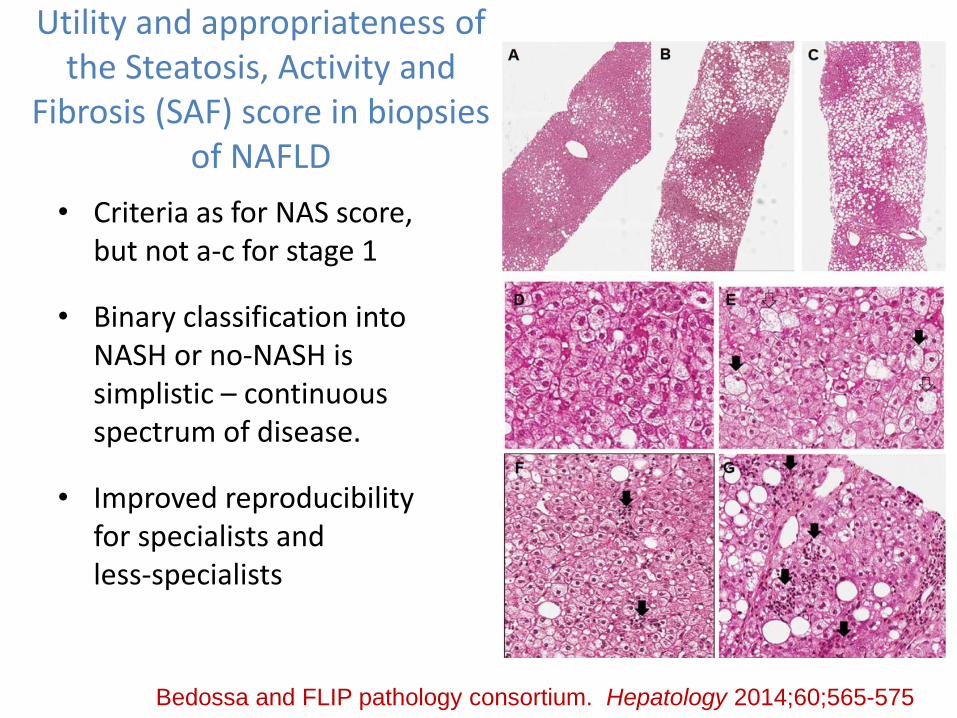
Utility and appropriateness of the Steatosis, Activity and
Fibrosis (SAF) score in biopsies of NAFLD
• Criteria as for NAS score, but not a-c for stage 1
• Binary classification into NASH or no-NASH is simplistic – continuous spectrum of disease.
• Improved reproducibility for specialists and less-specialists
Bedossa and FLIP pathology consortium. Hepatology 2014;60;565-575
The spectrum of fatty liver disease
Case 1. Steatohepatitis - diagnosis and severity
Case 2. Cirrhosis, alcoholic steatohepatitis
Case 3. Steatosis
Case 2: AA – aged 33
• Male, born in Slovenia 1981
• Healthy childhood
• Elite athlete – joined the military development squad
IV anabolic steroids
Continued to compete at a high level

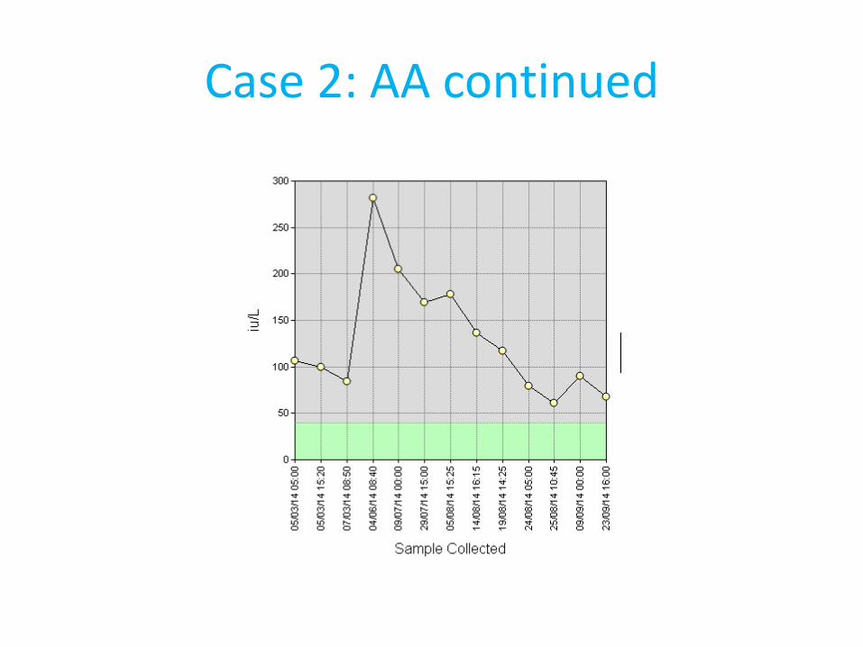
Case 2: AA continued
• Commenced heroin and amphetamine 1990s
• Migrated to UK 2009
• Clean of illicit drugs since 1999
• August 2011 admission with acute alcoholic hepatitis
• Screening positive for HCV PCR + Genotype 3a
• Previous hepatitis B – immune (sAg –ve, cAb+) and HIV -ve

Case 2: AA continued
• Too high risk currently for anti viral therapy
• Appearing dishevelled, but confident he can stop all alcohol
• July 2012 – “not drinking” ALT 121, bilirubin 46, plan liver biopsy pre-treatment
• Biopsy – steatohepatitis, presumed to be alcohol.
Not for anti viral therapy MUST STOP alcohol

Case 2: AA continued
• Therapy deferred
Referral to alcohol team
Good progress and rapidly achieved abstinence
Patient requested review at viral hepatitis clinic

Case 2: AA continued
• Attended follow up,
Keen again on anti viral therapy – patient denies drinking alcohol
Liver function stable
Plan to proceed with anti viral therapy:
Commenced PEG IFN/Ribavirin
Week 6 became jaundiced – biopsy ? Toxic drug reaction or relapse of alcohol

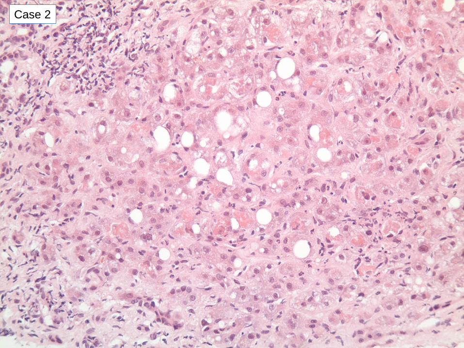
Case 2

Case 2

Case 2

Case 2
Case 2

Case 2

Case 2
Case 2
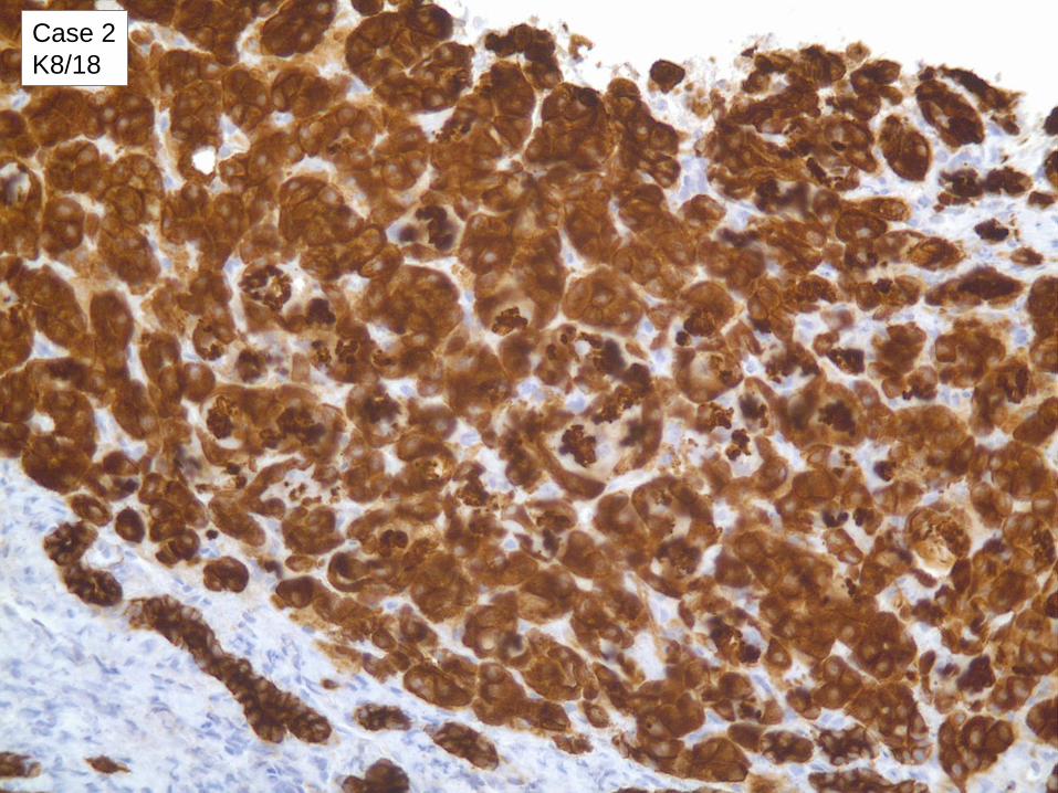
K8/18

Case 2
K8/18
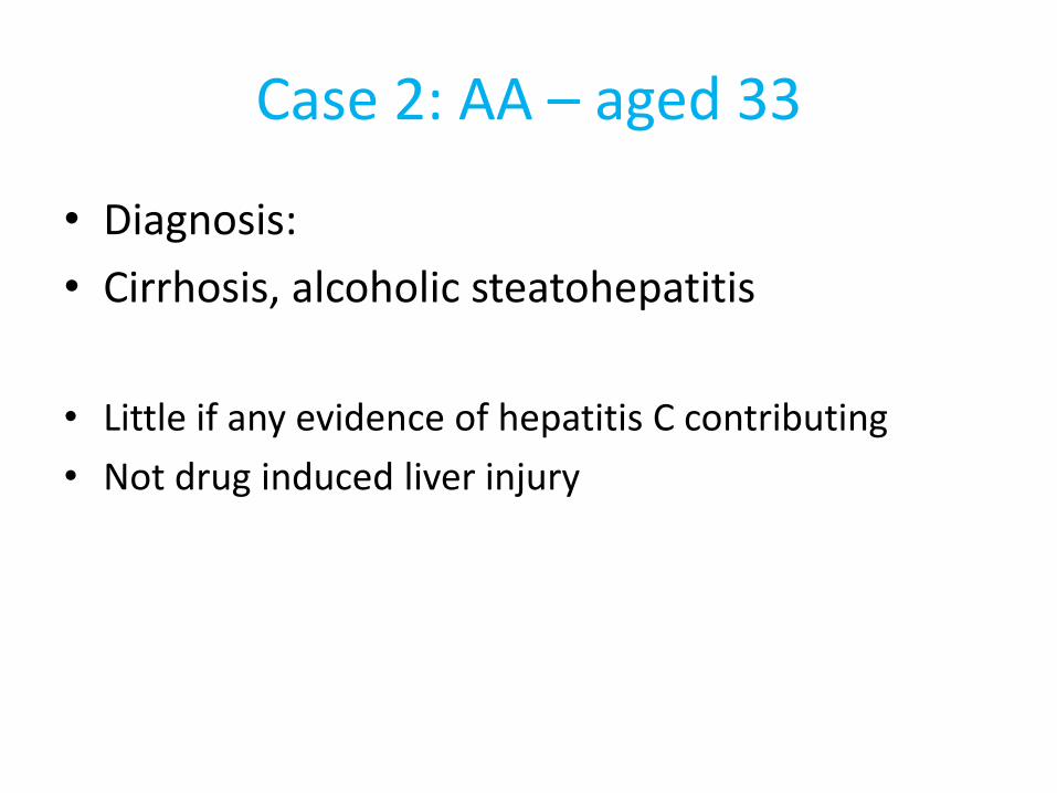
Case 2: AA – aged 33
• Diagnosis:
• Cirrhosis, alcoholic steatohepatitis
• Little if any evidence of hepatitis C contributing
• Not drug induced liver injury

Cirrhosis, Acute alcoholic hepatitis
Biopsy to confirm the diagnosis,
Distinguish from other causes of acute liver failure
Can it tell us anything else?
Histology reflects acute reversible deterioration from inexorable decline in late stage of alcoholic cirrhosis.
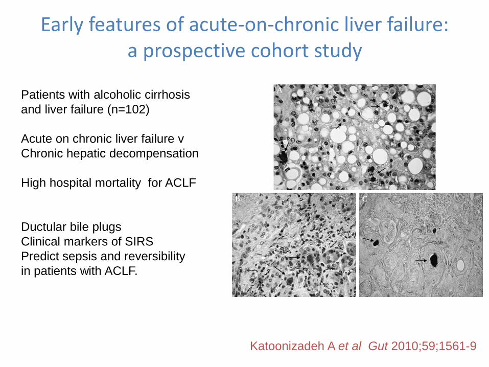
Early features of acute-on-chronic liver failure: a prospective cohort study
Katoonizadeh A et al Gut 2010;59;1561-9
Patients with alcoholic cirrhosis
and liver failure (n=102)
Acute on chronic liver failure v
Chronic hepatic decompensation
High hospital mortality for ACLF
Ductular bile plugs
Clinical markers of SIRS
Predict sepsis and reversibility
in patients with ACLF.
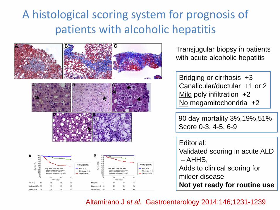
A histological scoring system for prognosis of patients with alcoholic hepatitis
Altamirano J et al. Gastroenterology 2014;146;1231-1239
Bridging or cirrhosis +3
Canalicular/ductular +1 or 2
Mild poly infiltration +2
No megamitochondria +2
Editorial:
Validated scoring in acute ALD
– AHHS,
Adds to clinical scoring for
milder disease
Not yet ready for routine use
Transjugular biopsy in patients
with acute alcoholic hepatitis
90 day mortality 3%,19%,51%
Score 0-3, 4-5, 6-9

Variation in liver biopsies on my desk…..

Bradford = 18G, usually 2 passes
Leeds = 16G, single pass
same laboratory – how good are the sections?
Illustrating median area
of tissue sections
Bradford 18G Leeds 16G
Improved tissue sections for medical liver biopsies:
a comparison of 16G v 18G biopsy needles.
18G (n=49) 16G (n=41)
% intact 29% 76%
Max diameter Median
0.74mm 1.04mm
Average diameter Median
0.53mm 0.87mm
Average area per passmedian
8.3mm2 11.4mm2
P<0.001 for all
Palmer T et al. J Clin Pathol 2014

Size of biopsy
RCPath Tissue Pathways – 16G needle, 1 core >15mm long is sufficient, >6 portal tracts
.
Portal tracts
9/111 (8%) of cores had <6 portal
tracts per section
All biopsies > 16mm in length
when taken contained 6 or more
portal tracts
The number of portal tracts per cm
of biopsy is very variable and
therefore cannot be predicted from
the length of core at biopsy R=0.23
Halas R et al, ESP meeting September 2014

Case 2: AA continued
• Rx with prednisolone for acute alcoholic hepatitis
Good response
Anti viral therapy discontinued
Monitor for complications of cirrhosis
Attends alcohol nurse
Case 2: AA continued

The spectrum of fatty liver disease
Case 1. Steatohepatitis - diagnosis and severity
Case 2. Cirrhosis, alcoholic steatohepatitis
Case 3. Steatosis

Case 3 JES 25.08.1958 age 54
• September 2012 – dermatology clinic, aged 54
- Severe psoriasis
Previous good response to ciclosporin, which was then weaned
Now psoriasis breaking through
Multiple topical agents unhelpful
Restart ciclosporin

Case 3 JES - continued
• Developing side effects from ciclosporin
Psoriasis troublesome
Consider methotrexate – but abnormal liver function tests, SMA + and elevation of PIIINP
• Referral to hepatology
Case 3 JES - continued
• Non invasive scores:
BARD
NAFLD
ELF
PIIINP
Fibroscan
• This lady has a mix of risk factors for chronic liver disease

Case 3 JES - continued
• Obese BMI 33.2
• No risk factors otherwise for chronic liver disease – alcohol never heavy 7 units per week
On examination no signs of liver disease, liver feels unremarkable
USS - fatty liver and gallstone, slightly wide calibre CBD
MRCP – no stones

Case 3 JES - continued
• BARD, NAFLD & Fibroscan scores low
HBV HCV negative
SMA positive, LKM, ANA and AMA and Ig’s normal, tTg –ve,
Ferritin normal. A-1 AT normal, Glu and HBA1c normal. TFT normal
But, team want to use methotrexate, LFT elevated, SMA +, high PIIINP
Case 3 JES - continued
• If Steatohepatitis present, then Methotrexate best avoided
• Also issue of SMA positivity
Biopsy:
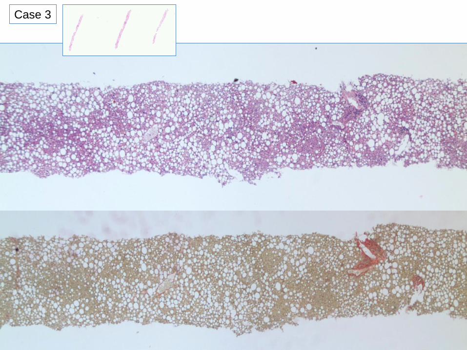
Case 3
Case 3

Case 3
Van Gieson

CK8/18
Case 3
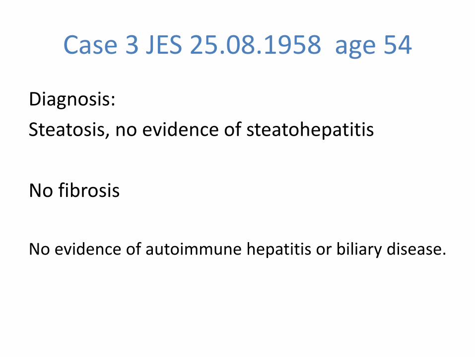
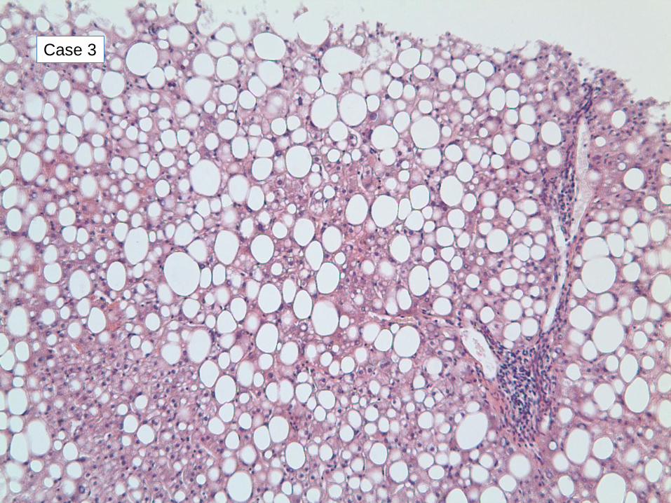
Case 3 JES 25.08.1958 age 54
Diagnosis:
Steatosis, no evidence of steatohepatitis
No fibrosis
No evidence of autoimmune hepatitis or biliary disease.
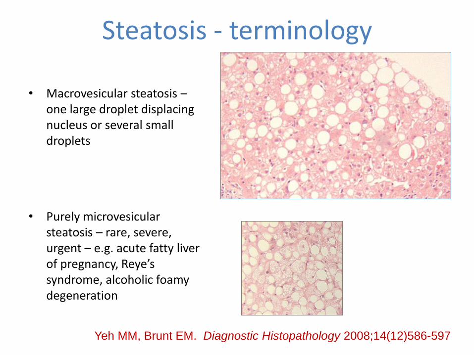
Steatosis - terminology
• Macrovesicular steatosis –one large droplet displacing nucleus or several small droplets
• Purely microvesicular steatosis – rare, severe, urgent – e.g. acute fatty liver of pregnancy, Reye’s syndrome, alcoholic foamy degeneration
Yeh MM, Brunt EM. Diagnostic Histopathology 2008;14(12)586-597

Fatty liver disease: alcohol, non-alcoholic,
also drugs causing fatty liver disease
• Cause steatohepatitis directly
– Amiodarone, irinotecan, ?methotrexate
• Promote steatohepatitis in patients with other risk factors
– Tamoxifen, methotrexate, steroids,
• Association with steatosis
– Steroids, 5FU, Brufen, anti-TB drugs, spironolactone.......
Ramachandran R & Kakar S. J Clin Pathol 2009;62;481-492

SummaryCan’t biopsy everyone with ? Fatty liver disease……
Not every obese patient has fatty liver disease
Purpose of biopsy: guide clinical management when non-invasive tests are insufficient:
- clinical question must be on the request form
1. Establish diagnosis – necessary features present
+/- features of other disease
2. and assess severity – stage fibrosis (not = Ishak)
- grade - steatosis
- inflammation/ballooning (K8/18)
3. Consider aetiology – needs clinical information
especially alcohol history

The challenge of obesity
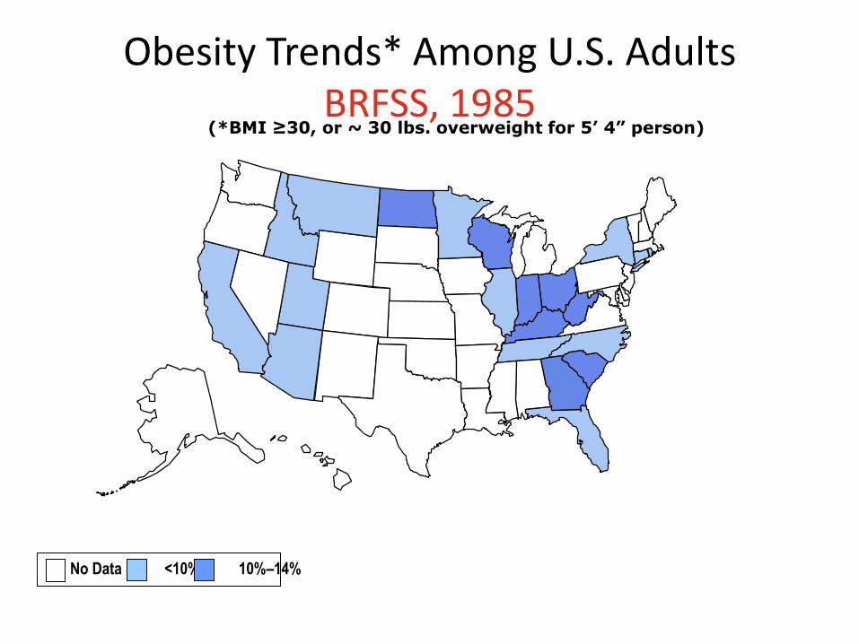
Obesity Trends* Among U.S. AdultsBRFSS, 1985
(*BMI ≥30, or ~ 30 lbs. overweight for 5’ 4” person)
No Data <10% 10%–14%

Obesity Trends* Among U.S. AdultsBRFSS, 1990
(*BMI ≥30, or ~ 30 lbs. overweight for 5’ 4” person)
No Data <10% 10%–14%

Obesity Trends* Among U.S. AdultsBRFSS, 1995
(*BMI ≥30, or ~ 30 lbs. overweight for 5’ 4” person)
No Data <10% 10%–14% 15%–19%

Obesity Trends* Among U.S. AdultsBRFSS, 2000
(*BMI ≥30, or ~ 30 lbs. overweight for 5’ 4” person)
No Data <10% 10%–14% 15%–19% ≥20%

Obesity Trends* Among U.S. AdultsBRFSS, 2005
(*BMI ≥30, or ~ 30 lbs. overweight for 5’ 4” person)
No Data <10% 10%–14% 15%–19% 20%–24% 25%–29% ≥30%

Obesity Trends* Among U.S. AdultsBRFSS, 2010
(*BMI ≥30, or ~ 30 lbs. overweight for 5’ 4” person)
No Data <10% 10%–14% 15%–19% 20%–24% 25%–29% ≥30%

2000
Obesity Trends* Among U.S. AdultsBRFSS, 1990, 2000, 2010
(*BMI 30, or about 30 lbs. overweight for 5’4” person)
2010
1990
No Data <10% 10%–14% 15%–19% 20%–24% 25%–29% ≥30%

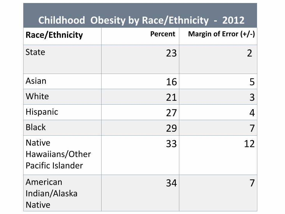
Childhood Obesity by Race/Ethnicity - 2012
Race/Ethnicity Percent Margin of Error (+/-)
State 23 2
Asian 16 5
White 21 3
Hispanic 27 4
Black 29 7
Native Hawaiians/Other Pacific Islander
33 12
American Indian/Alaska Native
34 7
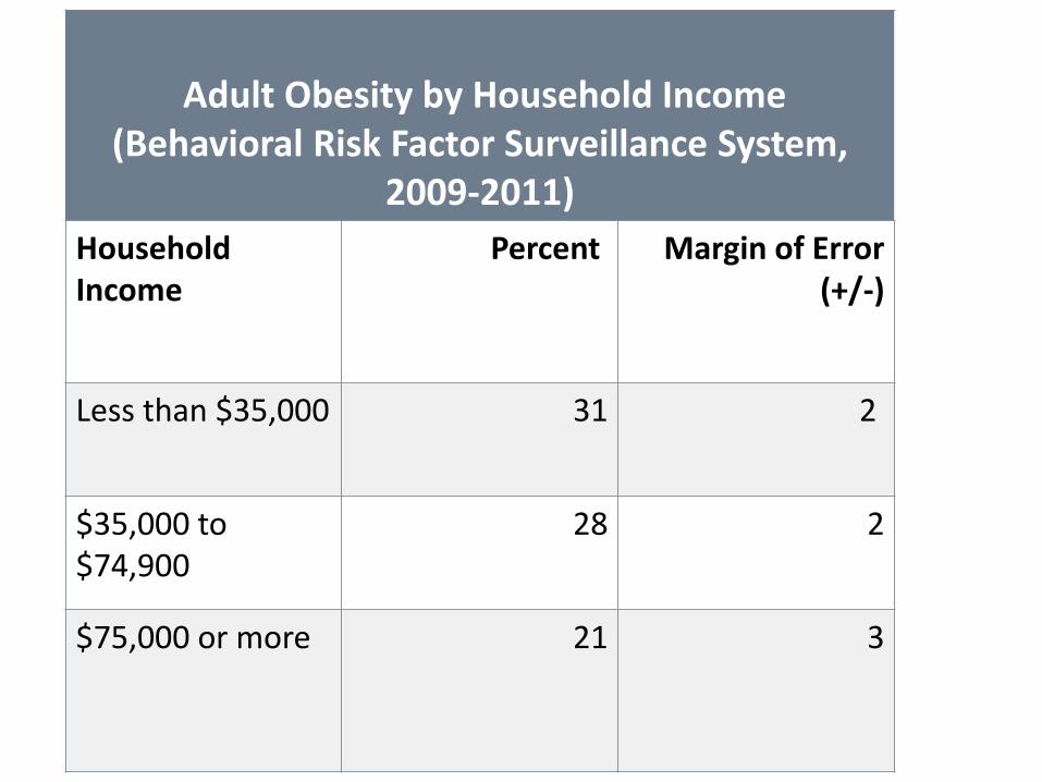
Adult Obesity by Household Income(Behavioral Risk Factor Surveillance System,
2009-2011)
Household Income
Percent Margin of Error (+/-)
Less than $35,000 31 2
$35,000 to $74,900
28 2
$75,000 or more 21 3
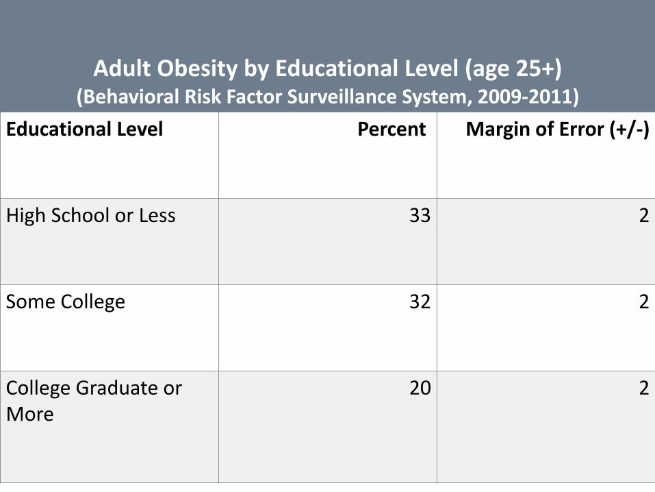
Adult Obesity by Educational Level (age 25+)(Behavioral Risk Factor Surveillance System, 2009-2011)
Educational Level Percent Margin of Error (+/-)
High School or Less 33 2
Some College 32 2
College Graduate or More
20 2

Prevalence of Self-Reported Obesity Among
Non-Hispanic White Adults, by State and
Territory, BRFSS, 2012-2014
*Sample size <50 or the relative standard error (dividing the standard error by the prevalence) ≥ 30%.

Prevalence of Self-Reported Obesity Among
Hispanic Adults, by State and Territory,
BRFSS, 2012-2014
*Sample size <50 or the relative standard error (dividing the standard error by the prevalence) ≥ 30%.

Prevalence of Self-Reported Obesity Among
Non-Hispanic Black Adults, by State and
Territory, BRFSS, 2012-2014
*Sample size <50 or the relative standard error (dividing the standard error by the prevalence) ≥ 30%.

Trends for Obesity (BMI>30) rates in UK and Ireland

Trends for Obesity (BMI>30) rates in UK
and Ireland

International comparison of obesity rates

UK changing obesity rates with age

UK Childhood obesity and deprivation index
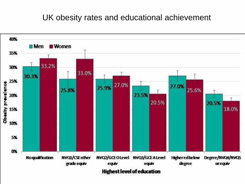
UK obesity rates and educational achievement

Trends for smoking rates in UK