Embed Size (px)
Citation preview
Fast delayed rectifier potassium current is requiredfor circadian neural activity
Jason N Itri1, Stephan Michel1,2, Mariska J Vansteensel2, Johanna H Meijer2 & Christopher S Colwell1
In mammals, the precise circadian timing of many biological processes depends on the generation of oscillations in neural
activity of pacemaker cells in the suprachiasmatic nucleus (SCN). The ionic mechanisms that underlie these rhythms are largely
unknown. Using the mouse brain slice preparation, we show that the magnitude of fast delayed rectifier (FDR) potassium
currents has a diurnal rhythm that peaks during the day. Notably, this rhythm continues in constant darkness, providing the first
demonstration of the circadian regulation of an intrinsic voltage-gated current in mammalian cells. Blocking this current
prevented the daily rhythm in firing rate in SCN neurons. Kv3.1b and Kv3.2 potassium channels were widely distributed within
the SCN, with higher expression during the day. We conclude that the FDR is necessary for the circadian modulation of electrical
activity in SCN neurons and represents an important part of the ionic basis for the generation of rhythmic output.
Almost all organisms, including humans, show daily rhythms in theirbehavior and physiology. In most cases, endogenous cellular networkscomposed of multiple circadian oscillators generate these rhythms.These oscillators provide temporal structure to an organism’s physio-logical systems. Nearly all behavioral processes show significant dailyvariations. This temporal variation is important in the body’s homeo-static mechanisms and has a major impact on the function of thenervous system. In mammals, the part of the nervous system that isresponsible for most circadian behavior can be localized to a bilaterallypaired structure in the hypothalamus known as the SCN1. Neurons inthe SCN are intrinsic oscillators that continue to generate near 24-hrhythms in electrical activity, secretion and gene expression whenisolated from the rest of the organism2. A key component that isresponsible for the generation of these rhythms may be a molecularfeedback loop that occurs in individual SCN neurons3,4. There is alsoevidence, however, that membrane excitability and/or synaptic trans-mission may be required for generation of the molecular oscillations5,6.Thus, clarifying the ionic mechanisms that interact with the molecularfeedback loop is critical to understanding the generation of circadianoscillations at both cellular and molecular levels of organization7.
It is well accepted that voltage-dependent potassium (K+) currentsare primary regulators of membrane excitability8. Given their role inother neuronal systems, K+ currents are likely candidates to coupleclock-related gene expression to membrane excitability and sponta-neous firing rate in the SCN. K+ currents are a large and diverse familyof voltage regulators, and previous studies have characterized a numberof intrinsic voltage-gated K+ currents in SCN neurons that are likely tobe important in regulating the firing rate of SCN neurons9–12. Thepossibility of diurnal or circadian modulation of these K+ currents hasnot, however, been explored in any detail, nor do we understand how
selective currents regulate the daily rhythm in the frequency of actionpotentials in SCN neurons. We became interested in examining thepossible circadian regulation of a subtype of K+ currents known as theFDR for two reasons. First, in molluskan retinal neurons, tetraethyl-ammonium (TEA)-sensitive K+ currents are thought to underlie thedaily rhythm in electrical activity of these circadian pacemaker cells13,and the slow delayed rectifier (SDR) current undergoes a circadianmodulation14. Second, during the subjective day, SCN neurons showsustained discharge for hours without spike adaptation, and the FDRcurrent may allow for this type of discharge in other neurons15,16. In thepresent study, we find that the FDR currents are under circadianregulation and that these currents are critical for controlling the rhythmin firing rate that is characteristic of SCN neurons.
RESULTS
Characterization of SDR and FDR currents
We used the whole-cell voltage clamp technique to isolate and recordK+ currents from neurons in the mouse SCN. Each of these cells wasdetermined to be within the SCN by directly visualizing the cell’slocation with infrared–differential interference contrast videomicro-scopy. In most cases, these images were sufficient to label a cell as beingin either ventral or dorsal subregions17. Although these currents arepresent throughout the SCN, we focused on the dorsal subregion(dSCN) because this region shows more-robust circadian rhythms inthe transcription of clock-related genes and electrical activity18,19.
Every SCN neuron had SDR currents (n ¼ 76). The SDR currentswere isolated by subtraction (I1 mM TEA – I20 mM TEA; Fig. 1a) using avoltage step protocol20,21 with a prepulse potential of �90 mV and testpulse potentials ranging from �80 to +50 mV (10-mV increments;Fig. 1a). The control artificial cerebrospinal fluid (ACSF) perfusion
Published online 24 April 2005; doi:10.1038/nn1448
1Department of Psychiatry and Biobehavioral Sciences, University of California Los Angeles, 760 Westwood Plaza, Los Angeles, California 90024-1759, USA.2Department of Neurophysiology, Leiden University Medical Center, P.O. Box 9604, 2300 RC Leiden, The Netherlands. Correspondence should be addressed to C.S.C.([email protected]).
65 0 VOLUME 8 [ NUMBER 5 [ MAY 2005 NATURE NEUROSCIENCE
A R T I C L E S©
2005
Nat
ure
Pub
lishi
ng G
roup
ht
tp://
ww
w.n
atur
e.co
m/n
atur
eneu
rosc
ienc
e
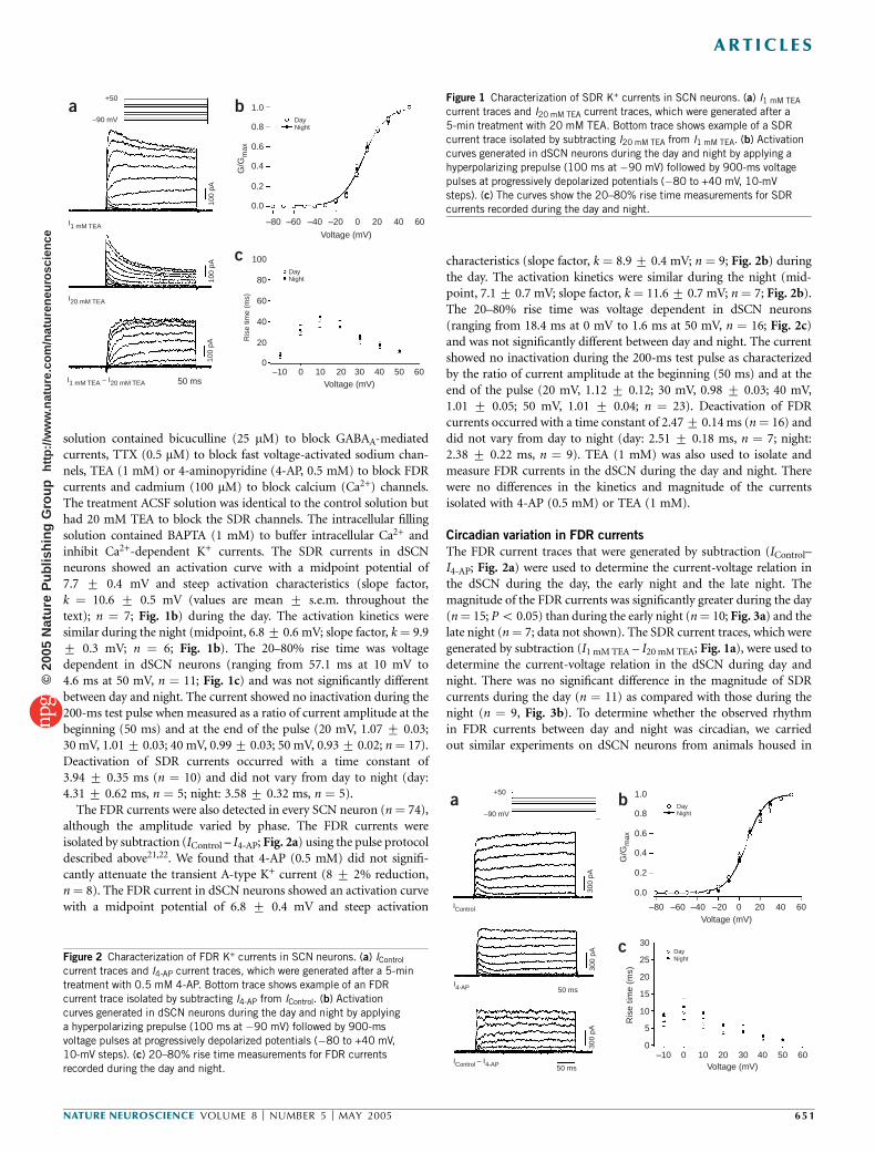
solution contained bicuculline (25 mM) to block GABAA-mediatedcurrents, TTX (0.5 mM) to block fast voltage-activated sodium chan-nels, TEA (1 mM) or 4-aminopyridine (4-AP, 0.5 mM) to block FDRcurrents and cadmium (100 mM) to block calcium (Ca2+) channels.The treatment ACSF solution was identical to the control solution buthad 20 mM TEA to block the SDR channels. The intracellular fillingsolution contained BAPTA (1 mM) to buffer intracellular Ca2+ andinhibit Ca2+-dependent K+ currents. The SDR currents in dSCNneurons showed an activation curve with a midpoint potential of7.7 7 0.4 mV and steep activation characteristics (slope factor,k ¼ 10.6 7 0.5 mV (values are mean 7 s.e.m. throughout thetext); n ¼ 7; Fig. 1b) during the day. The activation kinetics weresimilar during the night (midpoint, 6.8 7 0.6 mV; slope factor, k ¼ 9.97 0.3 mV; n ¼ 6; Fig. 1b). The 20–80% rise time was voltagedependent in dSCN neurons (ranging from 57.1 ms at 10 mV to4.6 ms at 50 mV, n ¼ 11; Fig. 1c) and was not significantly differentbetween day and night. The current showed no inactivation during the200-ms test pulse when measured as a ratio of current amplitude at thebeginning (50 ms) and at the end of the pulse (20 mV, 1.07 7 0.03;30 mV, 1.01 7 0.03; 40 mV, 0.99 7 0.03; 50 mV, 0.93 7 0.02; n ¼ 17).Deactivation of SDR currents occurred with a time constant of3.94 7 0.35 ms (n ¼ 10) and did not vary from day to night (day:4.31 7 0.62 ms, n ¼ 5; night: 3.58 7 0.32 ms, n ¼ 5).
The FDR currents were also detected in every SCN neuron (n ¼ 74),although the amplitude varied by phase. The FDR currents wereisolated by subtraction (IControl – I4-AP; Fig. 2a) using the pulse protocoldescribed above21,22. We found that 4-AP (0.5 mM) did not signifi-cantly attenuate the transient A-type K+ current (8 7 2% reduction,n ¼ 8). The FDR current in dSCN neurons showed an activation curvewith a midpoint potential of 6.8 7 0.4 mV and steep activation
characteristics (slope factor, k ¼ 8.9 7 0.4 mV; n ¼ 9; Fig. 2b) duringthe day. The activation kinetics were similar during the night (mid-point, 7.1 7 0.7 mV; slope factor, k ¼ 11.6 7 0.7 mV; n ¼ 7; Fig. 2b).The 20–80% rise time was voltage dependent in dSCN neurons(ranging from 18.4 ms at 0 mV to 1.6 ms at 50 mV, n ¼ 16; Fig. 2c)and was not significantly different between day and night. The currentshowed no inactivation during the 200-ms test pulse as characterizedby the ratio of current amplitude at the beginning (50 ms) and at theend of the pulse (20 mV, 1.12 7 0.12; 30 mV, 0.98 7 0.03; 40 mV,1.01 7 0.05; 50 mV, 1.01 7 0.04; n ¼ 23). Deactivation of FDRcurrents occurred with a time constant of 2.47 7 0.14 ms (n ¼ 16) anddid not vary from day to night (day: 2.51 7 0.18 ms, n ¼ 7; night:2.38 7 0.22 ms, n ¼ 9). TEA (1 mM) was also used to isolate andmeasure FDR currents in the dSCN during the day and night. Therewere no differences in the kinetics and magnitude of the currentsisolated with 4-AP (0.5 mM) or TEA (1 mM).
Circadian variation in FDR currents
The FDR current traces that were generated by subtraction (IControl–I4-AP; Fig. 2a) were used to determine the current-voltage relation inthe dSCN during the day, the early night and the late night. Themagnitude of the FDR currents was significantly greater during the day(n ¼ 15; P o 0.05) than during the early night (n ¼ 10; Fig. 3a) and thelate night (n ¼ 7; data not shown). The SDR current traces, which weregenerated by subtraction (I1 mM TEA – I20 mM TEA; Fig. 1a), were used todetermine the current-voltage relation in the dSCN during day andnight. There was no significant difference in the magnitude of SDRcurrents during the day (n ¼ 11) as compared with those during thenight (n ¼ 9, Fig. 3b). To determine whether the observed rhythmin FDR currents between day and night was circadian, we carriedout similar experiments on dSCN neurons from animals housed in
6050403020100–10Voltage (mV)50 ms
100
pA10
0 pA
100
pA
0
20
40
60
80
100
Ris
e tim
e (m
s)
DayNight
I1 mM TEA – I20 mM TEA
I20 mM TEA
I1 mM TEA
–90 mV
+50
Voltage (mV)
6040200–20–40–60–80
0.0
0.2
0.4
0.6
0.8
1.0
G/G
max
DayNight
a b
c
6050403020100–10Voltage (mV)
Voltage (mV)
0
5
10
15
20
25
30
Ris
e tim
e (m
s)
DayNight
50 ms
50 ms
300
pA30
0 pA
300
pA
IControl – I4-AP
IControl
I4-AP
+50
–90 mV
6040200–20–40–60–80
0.0
0.2
0.4
0.6
0.8
1.0
G/G
max
DayNight
a b
c
Figure 1 Characterization of SDR K+ currents in SCN neurons. (a) I1 mM TEA
current traces and I20 mM TEA current traces, which were generated after a
5-min treatment with 20 mM TEA. Bottom trace shows example of a SDR
current trace isolated by subtracting I20 mM TEA from I1 mM TEA. (b) Activation
curves generated in dSCN neurons during the day and night by applying a
hyperpolarizing prepulse (100 ms at �90 mV) followed by 900-ms voltage
pulses at progressively depolarized potentials (�80 to +40 mV, 10-mV
steps). (c) The curves show the 20–80% rise time measurements for SDRcurrents recorded during the day and night.
Figure 2 Characterization of FDR K+ currents in SCN neurons. (a) IControl
current traces and I4-AP current traces, which were generated after a 5-min
treatment with 0.5 mM 4-AP. Bottom trace shows example of an FDR
current trace isolated by subtracting I4-AP from IControl. (b) Activation
curves generated in dSCN neurons during the day and night by applying
a hyperpolarizing prepulse (100 ms at �90 mV) followed by 900-ms
voltage pulses at progressively depolarized potentials (�80 to +40 mV,
10-mV steps). (c) 20–80% rise time measurements for FDR currents
recorded during the day and night.
NATURE NEUROSCIENCE VOLUME 8 [ NUMBER 5 [ MAY 2005 6 5 1
A R T I C L E S©
2005
Nat
ure
Pub
lishi
ng G
roup
ht
tp://
ww
w.n
atur
e.co
m/n
atur
eneu
rosc
ienc
e

constant darkness (Fig. 3c). We found the same relationship in themagnitude of FDR currents in the dSCN between subjective day (n ¼ 6;P o 0.02) and subjective night (n ¼ 6), confirming that the rhythm issustained in constant darkness and is endogenously generated. Incontrast, there was no significant rhythm in SDR currents betweensubjective day (n ¼ 6) and subjective night (n ¼ 6; Fig. 3d).
Kv3.1b and Kv3.2 are expressed throughout the SCN
The Kv3.1 and Kv3.2 channels are responsible for the FDR currents15.To examine the pattern of expression of these channels in the SCN, weused antibodies against Kv3.1b and Kv3.2 channels (anti-Kv3.1b andanti-Kv3.2, respectively; Fig. 4). Mice were perfused during the day(zeitgeber time 4–6) and at night (zeitgeber time 14–16). We groupedtissue sections from each time point, and we ran immunohistochemicalprocedures in parallel to avoid procedural artifacts and to ensureconsistency. Kv3.1b immunoreactivity was evident throughout theSCN, and most cell bodies were labeled. Staining was robust in boththe dorsal and ventral regions of the SCN. This general pattern was seenthroughout the rostral to caudal extent of the SCN. The mean numberof immunopositive neurons per SCN section was significantly higherduring the day than during the night (day: 1,941 7 11 cells, n ¼ 5;night: 59 7 24 cells, n ¼ 5; P o 0.001). Optical analyses of digitalimages of these sections indicated that the immunopositive neuronswere significantly darker during the day than during the night (day,0.427 0.01 OD (optical density; see Methods); night, 0.277 0.01 OD;P o 0.001). Anti-Kv3.2 also labeled cell bodies throughout thedorsal and ventral SCN. The staining was most robust in the rostraland central SCN regions with relatively less staining present in thecaudal aspects of the nucleus. The mean number of immunopositiveneurons per SCN section was also significantly higher during the day(day: 103 7 13 cells, n ¼ 6; night: 40 7 19 cells, n ¼ 5; P o 0.02).Again, optical density measurements indicate that the immunopositiveneurons were significantly darker in the day (day, 0.37 7 0.01 OD;night, 0.25 7 0.01 OD; P o 0.001). These differences between day
and night were not seen in the number (day, 80 7 9 cells; night,73 7 7 cells) or optical density (day, 0.46 7 0.02 OD; night,0.43 7 0.01 OD) of immunoreactive cells in the piriform cortex regionof the same sections. Overall, the immunocytochemical analysisindicates that Kv3.1b and Kv3.2 channels are expressed within broadregions of the SCN and that expression of these channels is significantlyhigher in the day.
Regulation of spontaneous firing rate by FDR currents
Finally, we determined the contribution of FDR currents to thespontaneous firing rate (SFR) in SCN neurons with two sets ofexperiments. Using the current clamp recording technique in theperforated-patch configuration, we applied either 0.5 mM 4-AP or1 mM TEA to dSCN neurons and found that blocking FDR currentsreduced the mean SFR by 417 4% (5.32 to 3.13 Hz; n¼ 14; Po 0.001;Fig. 5a) during the day in the presence of bicuculline (25 mM) andcadmium (100 mM). This treatment prolonged repolarization andreduced the amplitude and duration of the after-hyperpolarization ofthe action potential in nearly every cell treated (11 of 14; Fig. 5b). Thereduction in SFR was long lasting and was not relieved by up to 30 minof washout (n ¼ 5). Three of fourteen dSCN neurons that wererecorded from during the day did not respond to treatment.
Application of 0.5 mM 4-AP or 1 mM TEA reduced the SFR of dSCNneurons during the early night by 20 7 5% (2.80 to 2.08 Hz; n ¼ 13;Fig. 5c). These treatments significantly lengthened repolarization andreduced the amplitude and duration of the after-hyperpolarization inmost cells treated (7 of 11; Fig. 5d). Three of eleven dSCN neurons thatwere recorded from during the early night did not respond to treatmentwith either 4-AP or TEA. During the late night, treatment of dSCNneurons with 0.5 mM 4-AP or 1 mM TEA reduced the SFR by 9 7 2%(2.25 to 2.08 Hz; n ¼ 7; data not shown). Treatment resulted in changesto the action potential waveform that were identical to those observedduring early night in nearly every cell treated (5 of 7; Fig. 5e,f). One ofseven dSCN neurons that were recorded from during the late night didnot respond to treatment. Overall, a comparison of all current clamprecordings from dSCN showed a significantly higher SFR during theday (5.32 Hz) than during the early night (2.80 Hz; P o 0.001) andduring the late night (2.25 Hz; P o 0.002; Fig. 6a). After applicationof 0.5 mM 4-AP to eliminate the FDR current, the SFR was not
604020–20–40–60–80–100Voltage (mV)
604020–20–40–60–80–100Voltage (mV)
604020–20–40–60–80–100Voltage (mV)
604020–20–40–60–80–100Voltage (mV)
Subjective nightSubjective day
Subjective nightSubjective day
200
400
600
800
Am
plitu
de (
pA)
200
400
600
800
Am
plitu
de (
pA)
200
400
600
800
Am
plitu
de (
pA)
200
400
600
800
Am
plitu
de (
pA)
NightDay
NightDay
Fast delayed rectifier current Slow delayed rectifier currenta b
c d
Day Night
Kv3.1b
Kv3.2
OC
3V 3V
OC
OC
3V3V
OCFigure 4 Photomicrographs showing immunoreactivity for Kv3.1b and Kv3.2
in the SCN during the day and night. Top panels: Kv3.1b immunoreactivitywas robust throughout the SCN including the ventral lateral portions as well
as a dorsal region of the SCN near the third ventricle (3V). Positive staining
was seen throughout the rostral-to-caudal extent of the SCN. Bottom panels:
Kv3.2 immunoreactivity was also seen on cell bodies throughout the SCN.
The staining was most robust in the rostral and central aspects of the SCN.
OC, optic chiasm. Scale bars, 50 mm.
Figure 3 Current-voltage relationship of delayed rectifier K+ currents in the
mouse dSCN. (a) The FDR currents recorded during the day were significantly
greater than those recorded during the night. (b) In contrast, the SDR
currents did not vary between day and night. (c) The FDR currents recorded
during the subjective day were significantly greater than those recorded
during the subjective night. (d) The SDR currents did not vary between
subjective day and night.
65 2 VOLUME 8 [ NUMBER 5 [ MAY 2005 NATURE NEUROSCIENCE
A R T I C L E S©
2005
Nat
ure
Pub
lishi
ng G
roup
ht
tp://
ww
w.n
atur
e.co
m/n
atur
eneu
rosc
ienc
e
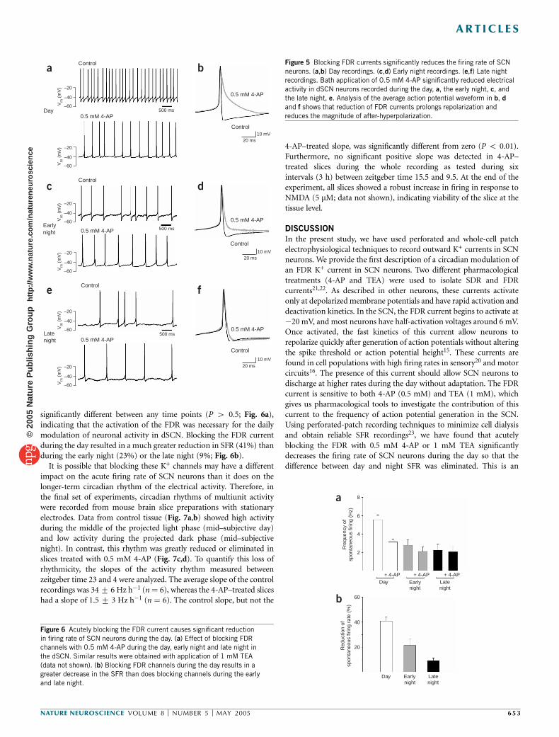
significantly different between any time points (P 4 0.5; Fig. 6a),indicating that the activation of the FDR was necessary for the dailymodulation of neuronal activity in dSCN. Blocking the FDR currentduring the day resulted in a much greater reduction in SFR (41%) thanduring the early night (23%) or the late night (9%; Fig. 6b).
It is possible that blocking these K+ channels may have a differentimpact on the acute firing rate of SCN neurons than it does on thelonger-term circadian rhythm of the electrical activity. Therefore, inthe final set of experiments, circadian rhythms of multiunit activitywere recorded from mouse brain slice preparations with stationaryelectrodes. Data from control tissue (Fig. 7a,b) showed high activityduring the middle of the projected light phase (mid–subjective day)and low activity during the projected dark phase (mid–subjectivenight). In contrast, this rhythm was greatly reduced or eliminated inslices treated with 0.5 mM 4-AP (Fig. 7c,d). To quantify this loss ofrhythmicity, the slopes of the activity rhythm measured betweenzeitgeber time 23 and 4 were analyzed. The average slope of the controlrecordings was 34 7 6 Hz h�1 (n ¼ 6), whereas the 4-AP–treated sliceshad a slope of 1.5 7 3 Hz h�1 (n ¼ 6). The control slope, but not the
4-AP–treated slope, was significantly different from zero (P o 0.01).Furthermore, no significant positive slope was detected in 4-AP–treated slices during the whole recording as tested during sixintervals (3 h) between zeitgeber time 15.5 and 9.5. At the end of theexperiment, all slices showed a robust increase in firing in response toNMDA (5 mM; data not shown), indicating viability of the slice at thetissue level.
DISCUSSION
In the present study, we have used perforated and whole-cell patchelectrophysiological techniques to record outward K+ currents in SCNneurons. We provide the first description of a circadian modulation ofan FDR K+ current in SCN neurons. Two different pharmacologicaltreatments (4-AP and TEA) were used to isolate SDR and FDRcurrents21,22. As described in other neurons, these currents activateonly at depolarized membrane potentials and have rapid activation anddeactivation kinetics. In the SCN, the FDR current begins to activate at�20 mV, and most neurons have half-activation voltages around 6 mV.Once activated, the fast kinetics of this current allow neurons torepolarize quickly after generation of action potentials without alteringthe spike threshold or action potential height15. These currents arefound in cell populations with high firing rates in sensory20 and motorcircuits16. The presence of this current should allow SCN neurons todischarge at higher rates during the day without adaptation. The FDRcurrent is sensitive to both 4-AP (0.5 mM) and TEA (1 mM), whichgives us pharmacological tools to investigate the contribution of thiscurrent to the frequency of action potential generation in the SCN.Using perforated-patch recording techniques to minimize cell dialysisand obtain reliable SFR recordings23, we have found that acutelyblocking the FDR with 0.5 mM 4-AP or 1 mM TEA significantlydecreases the firing rate of SCN neurons during the day so that thedifference between day and night SFR was eliminated. This is an
Vm
(m
V) –20
–40
–60
Vm
(m
V) –20
–40
–60
Vm
(m
V) –20
–40
–60
Vm
(m
V) –20
–40
–60
Vm
(m
V) –20
–40
–60
Vm
(m
V) –20
–40
–60
0.5 mM 4-AP
0.5 mM 4-AP
0.5 mM 4-AP
0.5 mM 4-AP
0.5 mM 4-AP
0.5 mM 4-APLatenight
night
500 ms
500 ms
500 ms
Control
Control
Control
Early
Day
Control
Control
Control
20 ms
20 ms
20 ms
10 mV
10 mV
10 mV
a b
c d
e f
Latenightnight
EarlyDay
20
40
60
Red
uctio
n of
spon
tane
ous
firin
g ra
te (
%)
Latenightnight
Early+ 4-AP+ 4-AP+ 4-AP
Day
8
6
4
2
Freq
uenc
y of
spon
tane
ous
firin
g (H
z)
a
b
Figure 5 Blocking FDR currents significantly reduces the firing rate of SCN
neurons. (a,b) Day recordings. (c,d) Early night recordings. (e,f) Late night
recordings. Bath application of 0.5 mM 4-AP significantly reduced electrical
activity in dSCN neurons recorded during the day, a, the early night, c, and
the late night, e. Analysis of the average action potential waveform in b, d
and f shows that reduction of FDR currents prolongs repolarization and
reduces the magnitude of after-hyperpolarization.
Figure 6 Acutely blocking the FDR current causes significant reduction
in firing rate of SCN neurons during the day. (a) Effect of blocking FDRchannels with 0.5 mM 4-AP during the day, early night and late night in
the dSCN. Similar results were obtained with application of 1 mM TEA
(data not shown). (b) Blocking FDR channels during the day results in a
greater decrease in the SFR than does blocking channels during the early
and late night.
NATURE NEUROSCIENCE VOLUME 8 [ NUMBER 5 [ MAY 2005 6 5 3
A R T I C L E S©
2005
Nat
ure
Pub
lishi
ng G
roup
ht
tp://
ww
w.n
atur
e.co
m/n
atur
eneu
rosc
ienc
e

unusual feature of the FDR, because blockage of most K+ currentswould be expected to increase firing rate. Finally, using extracellularrecordings of rhythms in multiunit activity, we found that longer-termapplication of 4-AP (0.5 mM) prevented expression of diurnal rhythmsin electrical activity recorded from SCN tissue.
Based on our observations, we believe that the FDR K+ current isnecessary for the expression of the circadian rhythm in the frequency ofaction potentials. We have found that the rhythm in the magnitude ofthis current is correlated with the rhythm in electrical discharge: bothpeak during the day and are low during the night. This rhythm inamplitude continues in constant darkness and seems to be a circadianrhythm that is expressed at the level of individual SCN neurons. Otherproperties of the current such as voltage dependence and activation anddeactivation kinetics do not change with the daily cycle. The SDRcurrent does not show a diurnal or circadian rhythm in amplitude orkinetic parameters in the dSCN. In addition, when the FDR is acutelyblocked with 4-AP, the frequency of firing in the SCN is significantlyreduced, with the largest effects occurring in dSCN neurons recordedduring the day. Longer treatments of 4-AP prevented the daily rhythmof firing in the SCN. Although the evidence presented points to acrucial role of the FDR in diurnal and circadian SFR modulation, thiscurrent cannot be responsible for the initial membrane depolarizationat dawn. The FDR acts only across a range of voltages that aredepolarized relative to the resting membrane potential of SCN neurons.Another class of current must be responsible for driving actionpotential generation to activate FDR channels.
SCN neurons have a slowly inactivating sodium current, whichactivates around �65 mV24,25. Although it is not known if this currentshows a circadian rhythm, blocking this sodium current inhibitsspontaneous firing25. In addition, previous work has found evidencefor a daily rhythm in an L-type calcium current26. These sodiumand calcium currents are probably critical for moving the SCN neuroninto a voltage range in which the FDR would be activated. Finally,electrophysiological measurements from SCN neurons indicate thatinput resistance also peaks during the day23,27,28. These observations
indicate that when SCN neurons are at their resting membranepotential, the net current flow through ion channels is lower duringthe day. Because the membrane is also depolarized during the day23, theclosed channels are likely to be K+ channels. Important support for thismodel comes from the observation that a TEA-sensitive K+ current iscritical for the daily change in input resistance28. The identity of thesechannels is not yet known, and these studies raise the possibility that awhole set of currents change rhythmically in the SCN as the cell movesfrom an ‘inactive/down’ state during the night to an ‘active/up’ stateduring the day. Not all currents within the SCN are rhythmicallyregulated, however, and the SDR current (this study), a barium-sensitive K+ current10 and H-currents29 all seem to be constant fromday to night.
The mechanisms that underlie the daily rhythm in FDR currents isnot known. Our immunocytochemical evidence clearly indicates thepresence of the Kv3.1b and Kv3.2 channels in the SCN and indicatesthat expression of these channel proteins may be rhythmically regu-lated. It is possible that the genes Kcnc1 (also known as Kv3.1) andKcnc2 (also known as Kv3.2) are rhythmically regulated, as many genesin this region show transcriptional rhythms30 and the promoterregion of the Kcnc1 contains both CRE and AP-1 sites31. Cells in theSCN exhibit a prominent circadian oscillation in CRE-mediatedgene expression32, and we expect to see a rhythm in the expressionof the genes coding for the fDR. In Drosophila melanogaster, circadianrhythms in levels of mRNA coding for a regulatory protein asso-ciated with Ca2+-sensitive K+ channels have been described33,34.Furthermore, in mammalian cardiac tissue, diurnal variation occursin the expression of genes that encode two K+ channels (Kv1.5 andKv4.2)35. The regulation need not be transcriptional, however, andpost-translational modifications could also be responsible for the dailyrhythms. Outside of the SCN, kinase and/or phosphatase activity isimportant for mediating short-term changes in channel function thatalter electrical excitability8. In chick photoreceptors, circadian oscilla-tions in cone cGMP-gated channels have been well described36. Withinthe SCN, circadian patterns of phosphorylation seem to be critical forthe basic molecular feedback loop that drives circadian rhythms, withthe involvement of casein kinases being particularly important37.Regardless of the underlying mechanism, the work described in thepresent study identifies a specific K+ current that is under theregulatory control of the molecular circadian feedback loop anddemonstrates that this current is necessary for the daily rhythm infrequency of action potential generation that lies at the heart of theSCN oscillator.
METHODSIn all studies, recommendations for animal use and welfare dictated by the
University of California Los Angeles Division of Laboratory Animals and guide-
lines from the U.S. National Institutes of Health or the Animal Experiments
Ethical Committee of the Leiden University Medical Center were followed.
Behavioral measurements. Male mice of at least 21 d of age were housed
individually, and their wheel-running activity was recorded (Mini Mitter).
Zeitgeber time is used to describe the projected time based on the previous light
cycle, with lights on defined as zeitgeber time 0. Circadian time was used when
mice were in constant darkness and the onset of activity was defined as
circadian time 12. When necessary, mice were killed in darkness while using an
infrared viewer. In all cases, mice were killed 1 h before recording.
Whole-cell patch clamp electrophysiology. Brain slices were prepared using
standard techniques from mice (C57Bl/6) of 30–50 d old. Methods, including
solutions, were identical to those described previously17. For perforated-patch
recordings, a standard internal solution was used to fill the tip of the patch
pipette. Amphotericin was dissolved in DMSO and was mixed with standard
108642242220181614
108642242220181614108642242220181614
108642242220181614Time (ZT)Time (ZT)
4-AP4-AP500
400
300
200
100
0
MU
A (
Hz)
500
400
300
200
100
0
MU
A (
Hz)
300
250
200
150
100
50
0
Ave
rage
MU
A (
Hz)
300
250
200
150
100
50
0
Ave
rage
MU
A (
Hz)
ControlControla b
c d
Figure 7 The daily rhythm in the SFR is lost when the FDR current is
blocked. Rhythms in multiunit activity (MUA) were recorded in mouse SCN.
(a) Control slices (n ¼ 6) had an average circadian rhythm in spontaneous
electrical activity with peak activity during the middle of the projected light
phase (mid–subjective day) and low activity during the projected dark phase
(mid–subjective night). (b) Representative examples of electrical activity
recorded from three control SCN slices. (c) In contrast, this rhythm was
greatly reduced or eliminated in those slices treated with 4-AP (0.5 mM;
n ¼ 6). (d) Representative examples of electrical activity recorded from
three SCN slices treated with 4-AP. ZT, zeitgeber time.
65 4 VOLUME 8 [ NUMBER 5 [ MAY 2005 NATURE NEUROSCIENCE
A R T I C L E S©
2005
Nat
ure
Pub
lishi
ng G
roup
ht
tp://
ww
w.n
atur
e.co
m/n
atur
eneu
rosc
ienc
e
internal solution at 0.2 mM for backfilling the patch pipette. Recordings were
obtained with an Axon Instruments 200B amplifier and were monitored on-
line with pCLAMP (Axon Instruments). To minimize changes in offset
potentials with changing ionic conditions, the ground path used a KCl agar
bridge. Whole-cell capacitance and electrode resistance were neutralized and
compensated (50–80%). Series and input resistances were monitored repeatedly
by checking the response to small pulses in a passive potential range. Series
resistance was not compensated, and the maximal voltage error that was due to
this resistance was calculated to be 6 mV. The access resistance of these cells was
15–35 MO in the whole-cell voltage clamp configuration, whereas the cell
capacitance was typically 6–18 pF.
Current traces were recorded with pClamp using the whole-cell voltage
clamp recording configuration and then were analyzed using ClampFit.
Delayed rectifier K+ currents were isolated pharmacologically using a voltage
step protocol in the whole-cell voltage clamp configuration. The protocol
consisted of a 100-ms prepulse at �90 mV followed by a 250-ms step at
progressively depolarized potentials. Leak subtraction was carried out during
acquisition using a p/4 protocol, which uses four subpulses with one-quarter of
the test pulse amplitude and reversed polarity given from a holding potential of
�70 mV. Current traces from treatments were subtracted from controls to
isolate delayed rectifier currents. Both 4-AP (0.5 mM) and TEA (1 mM) were
used to isolate FDR currents, whereas high-concentration TEA (20 mM) was
used to isolate SDR currents. Current measurements were done in the control
solution and after 5–7 min of drug treatment in each cell. Activation curves
were generated using data collected from the voltage step protocol outlined
above and were fit with a Boltzmann function f(Vm) ¼ 1/(1 + exp[�(Vm �Vhalf)/k]), where Vm is the membrane potential, Vhalf is the membrane potential
at which the value of the Boltzmann function equals 0.5 and k is the slope
factor. Inactivation curves were generated by using the following protocol in the
whole-cell voltage clamp configuration: 100-ms prepulses of varying potentials
(�100 mV to �30 mV, 5-mV steps) followed by a 900-ms step at +50 mV to
elicit maximal current. Data were fit with the Boltzmann function described for
activation curves, and 20–80% rise time measurements were made on current
traces. The SFR and action potential waveforms were recorded with pClamp
using current clamp in the whole-cell perforated-patch configuration. No
current was injected during recording. After a baseline SFR was established,
drug treatment began within 5 min of obtaining the perforated-patch config-
uration (access resistance o100 MO).
Extracellular recording. The multiunit activity rhythms of SCN neurons were
measured as described38. Coronal slices (500 mm) were prepared from male
C57Bl/6 mice at the beginning of the subjective day. The slices were kept
submerged with a thin fork in a laminar flow chamber (35.5 1C). Slices
contained at least 50% of the rostrocaudal extent of the SCN and all of the
ventrodorsal extent. Extracellular electrical activity of SCN neurons was
measured by platinum/iridium electrodes and was subsequently amplified
and bandwidth filtered39. Action potentials with a signal-to-noise ratio of 2:1
(noise o5 mV from baseline) were selected by spike triggers and were counted
electronically every 10 s for about one circadian cycle. The positions of the
electrodes and spike trigger settings were not changed during the experiment.
Linear fits were done on the multiunit activity data between zeitgeber time 23
and 4, and the slopes of the resulting lines were determined. A significant
positive slope at this circadian time is indicative of a typical rhythm in electrical
activity. The obtained slopes from control and experimental slices were tested
against zero using a one-sided t-test (P o 0.05). The individual examples in the
figures were smoothed with a box filter for clearer presentation of the data.
Immunocytochemistry. The methods were similar to those previously
described40. Polyclonal anti-Kv3.1b and anti-Kv3.2, raised in rabbit, were used
(Alomone Labs). A dilution series was done for both antibodies, with optimal
dilution found to be 1:150 for anti-Kv3.2 and 1:400 for anti-Kv3.1b. Anti-Kv3.1b
was raised to amino acids 567–585, which is a sequence that is unique to the
Kv3.1b splice variant, whereas anti-Kv3.2 recognizes a sequence that is common
to all known splice variants of Kv3.2, amino acids 184–204. Tissue sections from
each time point were grouped and processed in parallel to avoid procedural
artifacts and to ensure consistency. Immunocytochemical controls included the
omission of primary and secondary antisera and preabsorption of antibodies
with appropriate peptide epitopes. We found that that the omission of primary
antibodies and preabsorption with appropriate epitopes blocked all staining.
For each mouse, images were taken from one section from each of three
regions (rostral, middle and caudal) using a SPOT camera system (Diagnostic
Instruments). All immunopositive cells within the SCN of these three sections
were counted manually at 400� with the aid of a grid. All immunopositive
cells within the grid were counted without regard to the intensity of the
staining. Counts were done by two observers who were blind to the treat-
ment protocol, and the results were averaged. To have some measure of the
intensity of staining, optical density measurements were also undertaken using
SigmaScan Pro software (SPSS). For this analysis, digital images were converted
to an 8-bit gray scale in which each pixel would register a gray level value
between 0 (dark) and 255 (white). SCN neurons were manually outlined so
that the program could determine the average gray level per neuron. The
optical density was then calculated as OD ¼ �log(gray level of the neuron/
maximum gray level). The microscope, lighting and software parameters were
held constant to allow comparisons to be made in the optical density
measurements between tissue sections. Cell counts and optical density mea-
surements were also made in the piriform cortical region of the same sections
that contained the SCN.
Statistical analyses. Between-group differences were first evaluated using an
analysis of variance to determine whether there were any significant differences
among means of all groups. Post hoc pairwise comparisons were then done
using t-tests or Mann-Whitney rank sum tests when appropriate. Values were
considered significant if P o 0.05. All tests were carried out using SigmaStat
(SPSS). In the text, values are shown as mean 7 s.e.m.
ACKNOWLEDGMENTSWe would like to thank H. Duindam for technical assistance. We would also liketo thank E. Herzog and N. Wayne for comments on a draft of the manuscript.Supported by National Institutes of Health grants HL64582, NS043169and MH68087.
COMPETING INTERESTS STATEMENTThe authors declare that they have no competing financial interests.
Received 18 January; accepted 29 March 2005
Published online at http://www.nature.com/natureneuroscience/
1. van Esseveldt, K.E., Lehman, M.N. & Boer, G.J. The suprachiasmatic nucleus andthe circadian time-keeping system revisited. Brain Res. Brain Res. Rev. 33, 34–77(2000).
2. Gillette, M.U. Cellular and biochemical mechanisms underlying circadian rhythms invertebrates. Curr. Opin. Neurobiol. 7, 797–804 (1997).
3. King, D.P. & Takahashi, J.S. Molecular genetics of circadian rhythms in mammals. Annu.Rev. Neurosci. 23, 713–742 (2000).
4. Reppert, S.M. & Weaver, D.R. Molecular analysis of mammalian circadian rhythms.Annu. Rev. Physiol. 63, 647–676 (2001).
5. Nitabach, M.N., Blau, J. & Holmes, T.C. Electrical silencing of Drosophila pacemakerneurons stops the free-running circadian clock. Cell 109, 485–495 (2002).
6. Harmar, A.J. et al. The VPAC2 Receptor is essential for circadian function in the mousesuprachiasmatic nuclei. Cell 109, 497–508 (2002).
7. Schaap, J., Pennartz, C.M. & Meijer, J.H. Electrophysiology of the circadian pacemakerin mammals. Chronobiol. Int. 20, 171–188 (2003).
8. Hille, B. Ion Channels of Excitable Membranes (Sinauer Associates, Sunderland,Massachusetts, USA, 2001).
9. Bouskila, Y. & Dudek, F.E. A rapidly activating type of outward rectifier K+ current andA-current in rat suprachiasmatic nucleus neurones. J. Physiol. (Lond.) 488, 339–350(1995).
10. De Jeu, M., Geurtsen, A. & Pennartz, C.M.A. Ba2+-sensitive K+ current contributes to theresting membrane potential of neurons in rat suprachiasmatic nucleus. J. Neurophysiol.88, 869–878 (2002).
11. Cloues, R.K. & Sather, W.A. Afterhyperpolarization regulates firing rate in neurons ofthe suprachiasmatic nucleus. J. Neurosci. 23, 1593–1604 (2003).
12. Teshima, K., Kim, S.H. & Allen, C.N. Characterization of an apamin-sensitive potas-sium current in suprachiasmatic nucleus neurons. Neuroscience 120, 65–73(2003).
13. Michel, S., Geusz, M.E., Zaritsky, J.J. & Block, G.D. Circadian rhythm in membraneconductance expressed in isolated neurons. Science 259, 239–241 (1993).
14. Michel, S., Manivannan, K., Zaritsky, J.J. & Block, G.D. A delayed rectifier currentis modulated by the circadian pacemaker in Bulla. J. Biol. Rhythms 14, 141–150(1999).
15. Rudy, B. & McBain, C.J. Kv3 channels: voltage-gated K+ channels designed for high-frequency repetitive firing. Trends Neurosci. 24, 517–526 (2001).
NATURE NEUROSCIENCE VOLUME 8 [ NUMBER 5 [ MAY 2005 6 5 5
A R T I C L E S©
2005
Nat
ure
Pub
lishi
ng G
roup
ht
tp://
ww
w.n
atur
e.co
m/n
atur
eneu
rosc
ienc
e
16. Baranauskas, G., Tkatch, T., Nagata, K., Yeh, J.Z. & Surmeier, D.J. Kv3.4 subunitsenhance the repolarizing efficiency of Kv3.1 channels in fast-spiking neurons. Nat.Neurosci. 6, 258–266 (2003).
17. Itri, J. & Colwell, C.S. Regulation of inhibitory synaptic transmission by vasoactiveintestinal peptide (VIP) in the mouse suprachiasmatic nucleus. J. Neurophysiol. 90,1589–1597 (2003).
18. Hamada, T., Antle, M.C. & Silver, R. Temporal and spatial expression patterns ofcanonical clock genes and clock-controlled genes in the suprachiasmatic nucleus.Eur. J. Neurosci. 19, 1741–1748 (2004).
19. Yamaguchi, S. et al. Synchronization of cellular clocks in the suprachiasmatic nucleus.Science 302, 1408–1412 (2003).
20. Wang, L.Y., Gan, L., Forsythe, I.D. & Kaczmarek, L.K. Contribution of the Kv3.1potassium channel to high-frequency firing in mouse auditory neurones. J. Physiol.(Lond.) 509, 183–194 (1998).
21. Martina, M., Schultz, J.H., Ehmke, H., Monyer, H. & Jonas, P. Functional and moleculardifferences between voltage-gated K+ channels of fast-spiking interneurons andpyramidal neurons of rat hippocampus. J. Neurosci. 18, 8111–8125 (1998).
22. Kirsch, G.E. & Drewe, J.A. Gating-dependent mechanism of 4-aminopyridine block intwo related potassium channels. J. Gen. Physiol. 102, 797–816 (1993).
23. Schaap, J. et al. Neurons of the rat suprachiasmatic nucleus show a circadian rhythmin membrane properties that is lost during prolonged whole-cell recording. Brain Res.815, 154–166 (1999).
24. Pennartz, C.M., Bierlaagh, M.A. & Geurtsen, A.M. Cellular mechanisms underlyingspontaneous firing in rat suprachiasmatic nucleus: involvement of a slowly inactivatingcomponent of sodium current. J. Neurophysiol. 78, 1811–1825 (1997).
25. Kononenko, N.I., Shao, L.R. & Dudek, F.E. Riluzole-sensitive slowly inactivatingsodium current in rat suprachiasmatic nucleus neurons. J. Neurophysiol. 91, 710–718 (2004).
26. Pennartz, C.M., de Jeu, M.T., Bos, N.P., Schaap, J. & Geurtsen, A.M. Diurnal modulationof pacemaker potentials and calcium current in the mammalian circadian clock. Nature416, 286–290 (2002).
27. Jiang, Z.G., Yang, Y., Liu, Z.P. & Allen, C.N. Membrane properties and synaptic inputs ofsuprachiasmatic nucleus neurons in rat brain slices. J. Physiol. (Lond.) 499, 141–159(1997).
28. Kuhlman, S.J. & McMahon, D.G. Rhythmic regulation of membrane potentialand potassium current persists in SCN neurons in the absence of environmental input.Eur. J. Neurosci. 20, 1113–1117 (2004).
29. de Jeu, M.T. & Pennartz, C.M. Functional characterization of the H-current in SCNneurons in subjective day and night: a whole-cell patch-clamp study in acutely preparedbrain slices. Brain Res. 767, 72–80 (1997).
30. Panda, S. et al. Coordinated transcription of key pathways in the mouse by the circadianclock. Cell 109, 307–320 (2002).
31. Gan, L. & Kaczmarek, L.K. When, where, and how much? Expression of theKv3.1 potassium channel in high-frequency firing neurons. J. Neurobiol. 37, 69–79(1998).
32. Obrietan, K., Impey, S., Smith, D., Athos, J. & Storm, D.R. Circadian regulation of cAMPresponse element-mediated gene expression in the suprachiasmatic nuclei. J. Biol.Chem. 274, 17748–17756 (1999).
33. McDonald, M.J. & Rosbash, M. Microarray analysis and organization of circadian geneexpression in Drosophila. Cell 107, 567–578 (2001).
34. Ceriani, M.F. et al. Genome-wide expression analysis in Drosophila reveals genescontrolling circadian behavior. J. Neurosci. 22, 9305–9319 (2002).
35. Yamashita, T. et al. Circadian variation of cardiac K+ channel gene expression. Circula-tion 107, 1917–1922 (2003).
36. Ko, G.Y., Ko, M.L. & Dryer, S.E. Circadian regulation of cGMP-gated channels ofvertebrate cone photoreceptors: role of cAMP and Ras. J. Neurosci. 24, 1296–1304(2004).
37. Reppert, S.M. & Weaver, D.R. Coordination of circadian timing in mammals. Nature418, 935–941 (2002).
38. Meijer, J.H., Schaap, J., Watanabe, K. & Albus, H. Multiunit activity recordings in thesuprachiasmatic nuclei: in vivo versus in vitro models. Brain Res. 753, 322–327(1997).
39. Schaap, J. et al. Heterogeneity of rhythmic suprachiasmatic nucleus neurons: Implica-tions for circadian waveform and photoperiodic encoding. Proc. Natl. Acad. Sci. USA100, 15994–15999 (2003).
40. Michel, S., Itri, J. & Colwell, C.S. Excitatory mechanisms in the suprachiasmaticnucleus: the role of AMPA/KA glutamate receptors. J. Neurophysiol. 88, 817–828(2002).
65 6 VOLUME 8 [ NUMBER 5 [ MAY 2005 NATURE NEUROSCIENCE
A R T I C L E S©
2005
Nat
ure
Pub
lishi
ng G
roup
ht
tp://
ww
w.n
atur
e.co
m/n
atur
eneu
rosc
ienc
e