Embed Size (px)
Citation preview

Fast and Efficient Transfection of Mouse Embryonic Stem CellsUsing Non-Viral Reagents
Christoffer Tamm1& Sandeep Kadekar1 & Sara Pijuan-Galitó1 & Cecilia Annerén1,2
Published online: 30 June 2016# The Author(s) 2016. This article is published with open access at Springerlink.com
Abstract Reliable and efficient DNA and RNA transfectionmethods are required when studying the role of individualgenes in mouse pluripotent stem cells. However, these cellsusually grow in tight clusters and are therefore more difficultto transfect than many other cell lines. We have found thattransfection is especially challenging when mouse embryonicstem (mES) cells are cultured in the newly described 2i medi-um, which is based on two chemical inhibitors of differentia-tion pathways. In the present study we have performed a side-by-side comparison of commercially available, non-viraltransfection reagents with regard to their ability to deliverplasmid DNA and siRNA into adherent and/or trypsinizedmES cells cultured in 2i medium, assessing transfection rates,plasmid gene expression, siRNA mediated knockdown ofOct4 and viability. Finally, we present a fast and efficientmethod for transfection of trypsinized mES cells using theliposomal-based Lipofectamine 2000. With only a five-minute long transfection time we obtained at least 85 %transfected cells with 80 % maintained viability. Moreover,this protocol saves up to a day of experimental time sincethe cells are in suspension at the time of transfection, which
allows for immediately re-plating into the appropriate format.This fast, simplified and highly efficient transfection methodwill be valuable for both basic research and high-throughputapplications.
Keywords Transfection .Mouse . Embryonic stem cells . EScells . siRNA . DNA . Plasmid
Introduction
For the last decades mouse embryonic stem (mES) cells havebeen considered to be one of the most interesting model sys-tems for basic cellular and molecular studies and developmen-tal research. The ability to efficiently transfer DNA or smallinterfering RNA (siRNA) is one of the key technologies usedfor studying the role of single genes in these cells. However,mES cells grow in tight clusters and are therefore quite inef-ficiently transfected using chemical reagents. In addition, wehave found that when mES cells are cultured in the serum-free2i medium (containing two inhibitors, against the mitogen-activated protein kinase (MAPK)/extracellular-signal-regulat-ed kinase (ERK) kinase (MEK) and the glycogen synthasekinase 3 (GSK3), respectively [1]), chemical transfection iseven more challenging as compared to when cells are culturedin standard culture media [2]. Replacement of serum bytargeted inhibition of relevant signaling pathways reduces cel-lular heterogeneity as well as transcriptome and epigenomedifferences in mES cell cultures [3, 4]. In 2i mediumPD0325901 inhibits the autocrine activation of the ERK1/2pathway by fibroblast growth factor-4 (FGF4), shown to beinstrumental for ES cell differentiation [5]. The GSK3 inhibi-tion impairs Tcf3 activity, which represses the corepluripotency network in mES cells [6]. The cell cultures ex-hibit a more dense and uniform morphology, with negligible
Christoffer Tamm and Sandeep Kadekar contributed equally to this work.
Electronic supplementary material The online version of this article(doi:10.1007/s12015-016-9673-5) contains supplementary material,which is available to authorized users.
* Cecilia Anneré[email protected]
1 Department of Medical Biochemistry and Microbiology,Uppsala University, Box 582, SE-751 23 Uppsala, Sweden
2 GE Healthcare BioSciences AB, Björkgatan 30, Uppsala SE-751 84,Sweden
Stem Cell Rev and Rep (2016) 12:584–591DOI 10.1007/s12015-016-9673-5
signs of spontaneous differentiation as compared to cells cul-tured in serum-containing media [2].
There are many vastly different transfection techniquesavailable [7]. Virus-mediated transfections, or so-called bio-logical transfection methods, are suitable for stable transfec-tions due to the integration of transfected DNA into the hostcell genome. However, although viral methods are usuallyhighly efficient they suffer from limitations such as time-consuming production of vectors, mutagenesis, cytotoxicity,and DNA size restriction [8]. Physical transfection methodsare the means of diverse physical tools to deliver nucleic acidsinto host cells. Despite being effective, these methods are bothcomplicated and time-consuming (e.g. micro injections) ormay cause massive cell death (e.g. electroporation andnucleofection). En masse they also require expensive in-house equipment. Instead, much effort has been put into thedevelopment of chemical methods, such as calcium phos-phates, cationic lipids, cationic polymers, dendrimers,polyethylenimine, peptides and nanoparticle-based com-pounds [7]. Although generally less efficient in deliveringDNA, these non-viral vectors have higher genetic materialcarrying capacity, are easier to produce and cause low cyto-toxicity. However, many challenges remain, such as increas-ing transfection efficacy while maintaining or reducing cyto-toxicity, as well as improving penetration of cells that aredifficult to transfect.
Here we present a side-by-side comparison of commercial-ly available, non-viral transfection reagents for plasmid DNAor siRNA delivery using the manufacturers’ instructions. ForDNA and RNA delivery we selected some of the most com-monly used reagents on the market, both liposomal and non-liposomal polymer based. In addition we have also used apositively charged polymer embedded into a porous carriernanoparticle. One of the unique challenges when transfectingmES cells is that these cells often grow in dense multilayeredcolonies, leading to lower transfection rates in the cells that arepositioned in the middle of the colonies. Hence, in the presentstudy we have transfected both adherent colonies as well ascells dispersed into single cell suspensions.We here present anoptimized, rapid, single-cell suspension transfection protocol,which results in unparalleled efficacy while maintaining lowpost-transfection toxicity.
Material and Methods
Embryonic Stem Cell Culturing
Feeder-independent E14 and R1mES cells were cultured in 2imedium [1], a serum-free N2B27 medium supplemented withMEK inhibitor PD0325901 (1 μM) and GSK3 inhibitorCHIR99021 (3 μM) (both from Selleckchem), and 1000 U/ml LIF (Millipore). TrypLE™ Express (Life Technologies)
was used to passage the cells. To ensure good adherence2 % ES-qualified FBS was supplemented to the 2i mediawhen seeding the cells, as previously described [2].
Transfections
Transfections with Lipofectamine® 2000 (herein namedL2K), Lipofectamine® 3000 (L3K, Life Technologies),TurboFect™ (GE Healthcare Dharmacon), FuGENE® HD,ViaFect™ (Promega), TransIT-2020®, TransIT-X2®,TransIT-TKO®, TransIT-siQUEST® (Mirus Bio), Xfect™mESC (Clontech), X-tremeGENE™ HP, X-tremeGENE™ 9(Roche), Nanofectin, Nanofectamin (PAA, now part of GEHealthcare), JetPrime™ (Polyplus transfection) were performedaccording to the manufacturer’s instructions within the recom-mended reagent/DNA or reagent/siRNA ratio range. For adher-ent plasmid-DNA transfections cells were plated at constant den-sity 2 x 10⁵ cells/well in 12 well-plates one day prior to transfec-tion. For the single cell suspension; 5 x 10⁵ cells/well weretransfected and plated in 12 well-plates. The pmaxGFP(AMAXA Biosystems), pCMV β-galactosidase, or pCAGGsheparanase vectors were introduced at a final concentration of1.5 μg DNA/well. For the adherent siRNA transfections; cellswere plated at a density 5 x 10⁴ cells/well in 24 well plates oneday prior to transfection with either scrambled siRNA or siRNAagainst Oct4 at a final concentration of 50 or 100 nM/well. Forthe siRNA single cell suspension transfections; cells weretransfected and plated at a density 5 x 10⁴ cells/well in a 24 wellplates. (sense 5′-AGGUGUUCAGCCAGACCACTT-3′, anti-sense 5′-GUGGUCUGGCUGAACACCUTT-3′).
Flow Cytometry
24 h post-transfection, cells were washed once with PBS andthen trypsinized using TrypLE™ Express for 5 min at 37 °C.Cell suspensions were analysed in a Becton DickinsonFACSCalibur flow cytometer for FL1 (GFP) fluorescence todetermine the percentage of reporter expressing cells.Background fluorescence and autofluorescence were deter-mined using non-transfected cells as control. Analysis wascarried out in the freeware Flowing Software (http://www.flowingsoftware.com).
Western Blot
Cells were harvested in lysis buffer as previously described[9]. Total protein concentration was measured using BCAProtein Assay kit (Pierce). Samples were run on 10 % SDSPAGE gels, and subsequently transferred to an Immobilon-FLmembrane (Millipore). The membranes were blotted with an-ti-Oct3/4 (POUF5F1, cl. 7F9.2 (Millipore) and anti-β actinab8229 (Abcam). The membranes were then incubated withAlexa Fluor anti-rabbit 680 and IRDye 800CW anti-mouse.
Stem Cell Rev and Rep (2016) 12:584–591 585
Immunosignals were imaged using an Odyssey fluorescentimaging scanner (LI-COR Biosciences).
Β-Galactosidase Activity Assay
Twenty-four hours post-transfection cells were harvested inlysis buffer and mixed with ONPG buffer (60 mMNa2HPO4, 40 mM NaH2PO4, 10 mM KCl, 1 mM MgCl2,50 mM 2-mercaptoethanol, 1.2 mM ONPG) in a microtiterplate and incubated at 37 °C for 30 min. Absorbance was readat 420 nm in a microplate photometer reader (withMultiscan®PLUS, Labsystems).
qPCR
Twenty four and 48 h post-transfection total RNAwas extractedand purified with the GenElute™ mammalian total RNAminiprep kit (Sigma-Aldrich) according to the manufacturer’sinstructions. First-strand cDNA was produced according to themanufacturer’s protocol with iScript™ cDNA synthesis kit (Bio-Rad) using 1 μg RNA. Quantitative real-time PCR was per-formed according to the manufacturer’s instructions using theSsoFast™ EvaGreen™ supermix on the Miniopticon™ Real-Time PCR Detection System (Bio-Rad). The average C(t) valuefor each gene was normalized against 18S and the comparativeCt value (fold change) was calculated using 2−ΔΔC(t). Primersused were: 18S (forward: AGTCCCTGCCCTTTGTACACA,reverse: GATCCGAGGGCCTCACTAAAC).
Oct4 (forward:GATGCTGTGAGCCCAAG-GCAAG, re-verse: GGCTCCTGATCAACAGCATCAC),
Dab2 (forward: TGAAGCAGACAGCCAGAACA, re-verse: CAACAGACAAGGATTTGATAGGG).
Results
Efficiency of siRNA Delivery
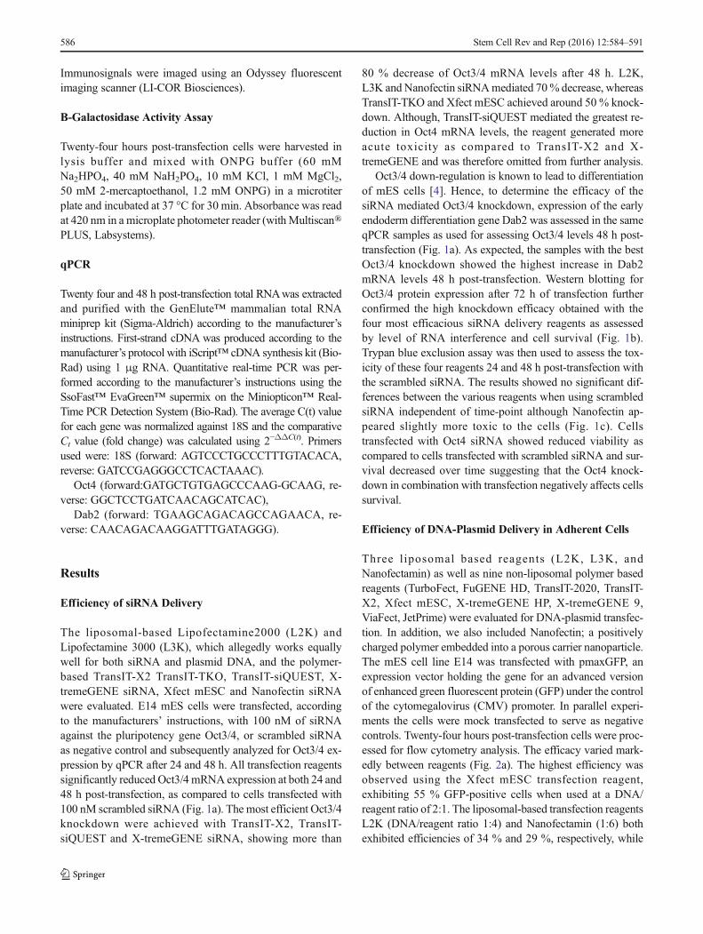
The liposomal-based Lipofectamine2000 (L2K) andLipofectamine 3000 (L3K), which allegedly works equallywell for both siRNA and plasmid DNA, and the polymer-based TransIT-X2 TransIT-TKO, TransIT-siQUEST, X-tremeGENE siRNA, Xfect mESC and Nanofectin siRNAwere evaluated. E14 mES cells were transfected, accordingto the manufacturers’ instructions, with 100 nM of siRNAagainst the pluripotency gene Oct3/4, or scrambled siRNAas negative control and subsequently analyzed for Oct3/4 ex-pression by qPCR after 24 and 48 h. All transfection reagentssignificantly reduced Oct3/4 mRNA expression at both 24 and48 h post-transfection, as compared to cells transfected with100 nM scrambled siRNA (Fig. 1a). The most efficient Oct3/4knockdown were achieved with TransIT-X2, TransIT-siQUEST and X-tremeGENE siRNA, showing more than
80 % decrease of Oct3/4 mRNA levels after 48 h. L2K,L3K and Nanofectin siRNAmediated 70% decrease, whereasTransIT-TKO and Xfect mESC achieved around 50 % knock-down. Although, TransIT-siQUEST mediated the greatest re-duction in Oct4 mRNA levels, the reagent generated moreacute toxicity as compared to TransIT-X2 and X-tremeGENE and was therefore omitted from further analysis.
Oct3/4 down-regulation is known to lead to differentiationof mES cells [4]. Hence, to determine the efficacy of thesiRNA mediated Oct3/4 knockdown, expression of the earlyendoderm differentiation gene Dab2 was assessed in the sameqPCR samples as used for assessing Oct3/4 levels 48 h post-transfection (Fig. 1a). As expected, the samples with the bestOct3/4 knockdown showed the highest increase in Dab2mRNA levels 48 h post-transfection. Western blotting forOct3/4 protein expression after 72 h of transfection furtherconfirmed the high knockdown efficacy obtained with thefour most efficacious siRNA delivery reagents as assessedby level of RNA interference and cell survival (Fig. 1b).Trypan blue exclusion assay was then used to assess the tox-icity of these four reagents 24 and 48 h post-transfection withthe scrambled siRNA. The results showed no significant dif-ferences between the various reagents when using scrambledsiRNA independent of time-point although Nanofectin ap-peared slightly more toxic to the cells (Fig. 1c). Cellstransfected with Oct4 siRNA showed reduced viability ascompared to cells transfected with scrambled siRNA and sur-vival decreased over time suggesting that the Oct4 knock-down in combination with transfection negatively affects cellssurvival.
Efficiency of DNA-Plasmid Delivery in Adherent Cells
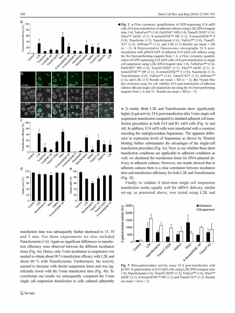
Three liposomal based reagents (L2K, L3K, andNanofectamin) as well as nine non-liposomal polymer basedreagents (TurboFect, FuGENE HD, TransIT-2020, TransIT-X2, Xfect mESC, X-tremeGENE HP, X-tremeGENE 9,ViaFect, JetPrime) were evaluated for DNA-plasmid transfec-tion. In addition, we also included Nanofectin; a positivelycharged polymer embedded into a porous carrier nanoparticle.The mES cell line E14 was transfected with pmaxGFP, anexpression vector holding the gene for an advanced versionof enhanced green fluorescent protein (GFP) under the controlof the cytomegalovirus (CMV) promoter. In parallel experi-ments the cells were mock transfected to serve as negativecontrols. Twenty-four hours post-transfection cells were proc-essed for flow cytometry analysis. The efficacy varied mark-edly between reagents (Fig. 2a). The highest efficiency wasobserved using the Xfect mESC transfection reagent,exhibiting 55 % GFP-positive cells when used at a DNA/reagent ratio of 2:1. The liposomal-based transfection reagentsL2K (DNA/reagent ratio 1:4) and Nanofectamin (1:6) bothexhibited efficiencies of 34 % and 29 %, respectively, while
586 Stem Cell Rev and Rep (2016) 12:584–591
the remaining reagents never achieved more than 25 % trans-fection efficiencies. The five best-performing reagents wereexamined under fluorescent microscopy (Fig. 2b) and a trypanblue exclusion assay was performed 24 h post-transfection toevaluate toxicity for these reagents. All tested reagents main-tained an 85 % survival rate or higher with no significantdifferences between groups (Fig. 2d).
Efficiency of DNA-Plasmid Delivery in Single CellSuspension
To assess whether 2i-cultured cells are more easily transfectedwhen dissociated into single cells, E14 mES cells weretrypsinized and then directly transfected using the sameDNA/reagent ratios as presented above. For all transfectionreagents 500.000 cells/sample were transfected with 1.5 μgpmaxGFP for two hours and subsequently seeded onto platesin 2i medium supplemented with 2% FBS. Twenty-four hourspost-transfection cells were processed for GFP expressionusing FACS analysis. Although Xfect generated the highesttransfection efficiency (Fig. 2c), this reagent was found to behighly toxic to cells in suspension and only a small fraction ofthe cells survived and could re-attach to plates after transfec-tion. This is also reflected by the lowest amount of β-galactivity compared to the other tested reagents (Fig. 3). In ef-fect, there were too few cells to even perform the trypan blueexclusion assay. In contrast, the liposomal-based transfectionreagents, L2K (1:4) and Nanofectamin (1:6) generated accept-able transfection rates of 63 % and 49 %, respectively with
good survival (Fig. 2c). Viability was assessed 24 h post-transfection for the five most efficient reagents using trypan blueexclusion assay. Lower viability was observedwhen transfectingtrypsinized cells as compared to adherent cultures and the toxic-ity level coincided with the transfection rate (Fig. 2e).
The five most efficient reagents from the initial screeningswere then used for transfecting cells with a β-galactosidase(β-gal) reporter construct. Theβ-gal activity results concurredwith the results presented above, both as to the efficiency ofthe different reagents but also that transfection rate of singlecells exceeds that of adherent cultures (Fig. 3).
Fast and Efficient DNA-Plasmid and siRNA Deliveryin Suspended mES Cells
In an attempt to decrease toxicity and further optimize trans-fection of trypsinized mES cells DNA/reagent ratios were al-tered and shorter transfection times were tested. Since L2Khad generated the most promising results we chose this re-agent for the initial experiments. A similar set-up, as presentedabove, was used using the GFP reporter construct at DNA/reagent ratios of 1:1, 1:2 and 1:4. The 1:2 and 1:4 DNA/reagent ratios was found to be significantly more efficient thanthe 1:1 ratio (supplemental Fig. 1a). However, since no differ-ence between the 1:2 and 1:4 ratios could be discerned, wecontinued with the 1:2 ratio to reduce toxicity. Transfectiontimes were then decreased from two hours to 60 and 30 min.Our results showed that the transfection rates were similarindependent of incubation time (supplemental Fig. 1b). The
Fig. 1 a Quantitative PCRanalysis for Oct3/4 (24 and 48 h)and Dab2 (48 h) after transfectionof E14 mES cells with either100 nM siOct3/4 or scrambledsiRNA using L2K (2 μl), L3K(1.5 μl), TransIT-X2® (3 μl),TransIT-TKO® (3 μl), TransIT-SiQuest® (5 μl), Xfect™ mESC(8 μl), X-tremeGENE™ siRNA(5 μl), and Nanofectin siRNA(8 μl). 18S expression was used fornormalization, and results arecomparative Ct value means ± SD(n = 3). bWestern blot analysis forβ-actin and Oct3/4 72 h post-transfection with siOct3/4 orscrambled siRNA using the fourbest-performing reagents in 1 a. cTrypan blue dye exclusion assay forcell viability 24 and 48 h post-transfection with siOct3/4 orscrambled siRNA using the samereagents as in 4b. Results aremean ± SD (n = 3)
Stem Cell Rev and Rep (2016) 12:584–591 587
transfection time was subsequently further shortened to 15, 10and 5 min. For these experiments we also includedNanofectamin (1:6). Again no significant differences in transfec-tion efficiency were observed between the different incubationtimes (Fig. 4a). Hence, only 5-min incubation in suspension wasneeded to obtain about 90 % transfection efficacy with L2K andabout 60 % with Nanofectamin. Furthermore, the toxicityseemed to decrease with shorter suspension times and was sig-nificantly lower with the 5-min transfection time (Fig. 4b). Tocorroborate our results we subsequently compared the 5-minsingle cell suspension transfection to cells cultured adherently
in 2i media. Both L2K and Nanofectamin show significantlyhigherβ-gal activity 24 h post-transfection after 5-min single cellsuspension transfection compared to standard adherent cell trans-fection procedures in both E14 and R1 mES cells (Fig. 4c and4d). In addition, E14mES cells were transfected with a constructencoding the endoglycosidase heparanase. The apparent differ-ence in expression levels of heparanase as shown by Westernblotting further substantiates the advantages of the single-celltransfection procedure (Fig. 4e). Next, to see whether these shorttransfection conditions are applicable to adherent conditions aswell, we shortened the transfection times for DNA-plasmid de-livery in adherent cultures. However, our results showed that inadherent cultures there is a clear correlation between incubationtime and transfection efficiency for both L2K and Nanofectamin(Fig. 4f).
Finally, to validate if short-time single cell suspensiontransfection works equally well for siRNA delivery, similarset-up, as presented above, was tested using L2K and
�Fig. 2 a Flow cytometry quantification of GFP-expressing E14 mEScells 24 h post-transfection of adherent cultures using L2K (DNA/reagentratio 1:4), TurboFect™ (1:4), FuGENE®HD (1:4), TransIT-2020® (1:4),Xfect™ mESC (2:1), X-tremeGENE™ HP (1:1), X-tremeGENE™ 9(1:6), Nanofectin (1:3), Nanofectamin (1:6), ViaFect™ (1:6), TransIT-X2® (1:2), JetPrime™ (1:2), and L3K (1:3) Results are mean ± SD(n = 2). b Representative fluorescence micrographs 24 h post-transfection with pMAX-GFP of adherent E14 mES cell cultures usingthe five best-performing reagents from 1 A. c Flow cytometry quantifi-cation of GFP-expressing E14 mES cells 24 h post-transfection in singlecell suspension using L2K (DNA/reagent ratio 1:4), TurboFect™ (1:4),FuGENE® HD (1:4), TransIT-2020® (1:2), Xfect™ mESC (2:1), X-tremeGENE™ HP (1:1), X-tremeGENE™ 9 (1:6), Nanofectin (1:3),Nanofectamin (1:6), ViaFect™ (1:6), TransIT-X2® (1:2), JetPrime™(1:2), and L3K (1:3) Results are mean ± SD (n = 2). d-e Trypan bluedye exclusion assay for cell viability 24 h post-transfection of adherentcultures (d) and single cell suspensions (e) using the five best-performingreagents from 1 A and 1C. Results are mean ± SD (n = 2)
Fig. 3 Beta-galactosidase activity assay 24 h post-transfection withpCMV β-galactosidase in E14 mES cells using L2K (DNA/reagent ratio1:4), Nanofectamin (1:6), TransIT-2020® (1:2), ViaFect™ (1:6), Xfect™mESC (2:1), X-tremeGENE™ HP (1:1), and TransIT-X2® (1:2). Resultsare mean ± sd (n = 2)
588 Stem Cell Rev and Rep (2016) 12:584–591

TransIT-X2 with 100 nM Oct3/4 siRNA. Oct3/4 expressionwas markedly reduced 24 h post-transfection with 5, 15and 30 min incubation (Fig. 4g). In fact, the level ofOct3/4 expression was clearly lower than after similartransfections in adherent 2i media cultures (Fig. 1a).Hence, as expected, these novel transfection conditions seemto work equally well for both plasmid-DNA as well as forsiRNA delivery.
Discussion
Over the years various biological, chemical, and physicalstrategies for introducing nucleic acids into cells have been
developed. Today there are numerous commercially availableproducts optimized for certain cell or nucleic acid types.Stem cells and primary cell cultures are often difficultto transfect. Consequently, working with these cells of-ten means accepting low transfection efficiencies or la-borious optimizations. Instead many studies are carriedout in easily transfectable cell lines, such as HEK293 orHeLa, despite being poor cell models. MultilayeredmES cell colonies give poor transfection efficiency dueto the lack of homogenous cellular access for transfec-tion reagents and nucleic acid complexes. As we havepreviously reported this quandary is even more trouble-some when growing the cells in 2i media [2]. Moreover,post-transfection intrusion on cellular proteostasis (e.g.
Fig. 4 a Flow cytometry quantification of GFP-expressing E14 mEScells 24 h after short-time (5, 10, 15 and 30 min) transfection of singlecell suspensions using L2K (DNA/reagent ratio 1:2) and Nanofectamin(1:6). Results are mean ± SD (n = 3) b Trypan blue dye exclusion assayfor cell viability 24 h 24 h after short-time (5, 10, 15 and 30 min) trans-fection of single cell suspensions using L2K (DNA/reagent ratio 1:2) andNanofectamin (1:6). Results are mean ± SD (n = 3). c-d Beta-galactosidase activity assay 24 h post-transfection with pCMV β-galactosidase using L2K (1:2) and Nanofectamin (1:6) of adherent cul-tures and single cell suspensions of E14 (c) and R1 (d) mES cells. e
Western blot analysis for β-actin and heparanase 24 h post-transfectionwith pCAGGs-heparanase of adherent cultures or single cell suspensionsof E14 mES cells. f Flow cytometry quantification of GFP-expressingE14 mES cells 24 h after short-time (5, 15 and 30 min) and standard-time(2 and 4 h) transfection of adherent cultures (DNA/reagent ratios 1:2).Results are mean ± SD (n = 3). g Quantitative PCR analysis for Oct3/424 h after short-time (5, 10, 15 and 30 min) transfection of single cellsuspensions with 100 nM siOct3/4 or scrambled siRNA using L2K and.18S expression was used for normalization, and results are comparativeCt value means ± SD (n = 2)
Stem Cell Rev and Rep (2016) 12:584–591 589

due to expression of plasmid DNA-encoded genes) andsubsequent decrease in proliferation rates result in cul-tures being promptly overtaken by the less affected non-transfected cells. Although the latter is a common con-cern it becomes more critical for mES cells due to theirexcessive proliferation rates and alternate day sub-culturing needs.
In the present study we evaluated a panel of com-monly used non-viral commercially available transfec-tion reagents with regard to their applicability to deliverplasmid DNA and siRNA into mES cells cultured in 2imedia. TransIT-X2 was found to be the most efficientreagent for siRNA transfection of adherent mouse EScells cultured in 2i medium. When transfecting adherentcultures with DNA plasmid, the Xfect mESC non-liposomal polymer was the only reagent that generatedmore than 50 % transfection rate. However, L2Kfollowed by Nanofectamin were the most efficient re-agents when transfecting dissociated single cell suspen-sions, suggesting that lipofection is the method ofchoice for this experimental approach. Although XfectmES reagents at first glance seemed equally efficient asL2K, the reagent was very toxic to cells in suspension.Suspension transfection is not mentioned in the manufac-turer’s instructions for the Xfect mESC reagent. However,the traditional Xfect transfection reagent has been shown towork for suspension cell cultures [10].
It is likely that the inefficient transfection of cells culturedin 2i medium is partly due to the densely packed cell culturesmaking the transfection agent less prone to access all thecells. Enzymatic dissociation induces shaving ofmembrane-associated factors, including glycoproteinswith highly sulfated glycosaminoglycan (GAGs) poly-saccharide chains [11]. The large number of sulfategroups renders the chains anionic, which allows interac-tion with a wide variety of positively charged substrates.Previous studies have shown that cell surface GAGsinteract with transfection reagent polymers and lipo-somes and inhibit cation-mediated gene transfer [12].Hence, the increase in transfection efficiency and trans-gene expression observed after single cell suspension trans-fections is likely a result of a combination of the homogenouscellular accessibility for the transfection complexes in additionto a temporary removal of GAGs from the plasma membranein response to trypsinization.
In an effort to fine-tune and reduce toxicity we optimizedthe use of L2K for plasmid-DNA delivery in single mES cellsuspension. When optimizing transfection – whether forstem cells or other cell types – various cell-related fac-tors such as culture health, passage numbers, and cultureconfluence need to be taken into consideration. However,those variables will be highly research group- and cell lab-dependent. Instead, we focused on parameters such as ratios
of nucleic acid to transfection reagent and incubation time.Weshow that a DNA/reagent ratio of 1:2 rendered the best effi-cacy without significantly increasing toxicity. Also, by reduc-ing incubation times to just 5 to 10 min we could maintain ahigh transfection efficacy but significantly increase cell via-bility. In fact, completely omitting the suspension incubation,and instead seeding the cells directly after adding the reagentand DNA-complexes, yielded only a slight decrease in trans-fection efficacy but a viability comparable to those obtained inadherent culture transfections (data not shown). Furthermore,similar results were obtained using R1 mES cells show-ing that the high efficiency and low toxicity is indepen-dent of cell line. Similar approaches have previouslybeen reported for other cell types [13–15]. Althoughthe focuses in those studies were on siRNA delivery,the efficiency for plasmid-DNA delivery was also test-ed. In concordance with our results these studies report-ed that a 10–15 min incubation of trypsinized cells withtransfection complexes using L2K increases transfectionefficiency 2–9 fold and that incubation times longerthan that decrease cell viability. Consequently, this rapidtransfection approach, which is both more time-efficientand gentle than the standard protocol, seems generallyapplicable to numerous cell types. In conclusion, thepreferred method for plasmid DNA delivery ofmESCs, cultured in 2i medium, is by transfecting thecells in suspension using L2K at a DNA:L2K ratio of1:2 for 5–10 min. This gives a transfection rate of>80 % with a survival of >75 %. Similarly, the mostefficient method for siRNA mediated knockdown is bytransfecting cells with TransIT-X2 or L2K for 5–30 min, which mediates a > 90 % knockdown ofOct3/4. In addition, transfecting trypsinized cells effec-tively shortens the experimental time by one day as aresult of not having to plate the cells 24 h prior totransfection as is required when transfecting adherentcells.
In the past, cationic lipid and polymer-based transfec-tion had yet to reach the levels observed with viraltransduction. With the presented method we have shownthat this is no longer the case and that high-efficienttransfections are achievable using non-viral methods.The optimized methods presented herein offers solutionsto the current need for efficient and fast transfection inorder to simplify and enhance studies of individualgenes in pluripotent stem cells. Moreover, the methodof transfecting trypsinized cells would be very advanta-geous in large scale and high-throughput transfectionapplications.
Acknowledgments The following individuals and provided reagents:Dr. Birgitta Tomkinson, Uppsala University (pCMV-beta-gal), Dr. Jin-Ping Li, Uppsala University (pCAGGS-heparanase and heparanase
590 Stem Cell Rev and Rep (2016) 12:584–591

antibody). This work was supported by the Faculty of Medicine, UppsalaUniversity.
Compliance with Ethical Standards
Conflict of Interest Dr. Annerén is an employee of GEHealthcare Bio-Sciences AB; the other authors do not have anything to disclose. Nocompeting financial interests exist.
Open Access This article is distributed under the terms of the CreativeCommons At t r ibut ion 4 .0 In te rna t ional License (h t tp : / /creativecommons.org/licenses/by/4.0/), which permits unrestricted use,distribution, and reproduction in any medium, provided you giveappropriate credit to the original author(s) and the source, provide a linkto the Creative Commons license, and indicate if changes were made.
References
1. Ying, Q. L., Wray, J., Nichols, J., et al. (2008). The ground state ofembryonic stem cell self-renewal. Nature, 453, 519–523.
2. Tamm, C., Pijuan Galitó, S., & Annerén, C. (2013). A comparativestudy of protocols for mouse embryonic stem cell culturing. PloSOne, 10, e81556.
3. Marks, H., Kalkan, T., Menafra, R., et al. (2012). The transcription-al and epigenomic foundations of ground state pluripotency. Cell,149, 590–604.
4. Guo, G., Pinello, L., Han, X., et al. (2016). Serum-based cultureconditions provoke Gene expression variability in mouse embryon-ic stem cells as revealed by single-cell analysis. Cell Reports, 14,956–965.
5. Stavridis, M. P., Lunn, J. S., Collins, B. J., & Storey, K. G. (2007).A discrete period of FGF-induced Erk1/2 signalling is required forvertebrate neural specification. Development, 134, 2889–2894.
6. Wray, J., Kalkan, T., Gomez-Lopez, S., et al. (2011). Inhibition ofglycogen synthase kinase-3 alleviates Tcf3 repression of thepluripotency network and increases embryonic stem cell resistanceto differentiation. Nature Cell Biology, 13, 838–845.
7. Kim, T. K., & Eberwine, J. H. (2010). Mammalian cell transfection:the present and the future. Analytical and Bioanalytical Chemistry,397, 3173–3178.
8. Schaffer, D. V., Koerber, J. T., & Lim, K. I. (2008). Molecularengineering of viral gene delivery vehicles. Annual Review ofBiomedical Engineering, 10, 169–194.
9. Tamm, C., Galito, S. P., & Anneren, C. (2011). Differential effectson cell motility, embryonic stem cell self-renewal and senescenceby diverse Src kinase family inhibitors. Experimental CellResearch, 318, 336–349.
10. Arukuusk, P., Parnaste, L., Oskolkov, N., et al. (2013). Newgeneration of efficient peptide-based vectors, NickFects, for thedelivery of nucleic acids. Biochimica et Biophysica Acta, 1828,1365–1373.
11. Vogel, K. G. (1978). Effects of hyaluronidase, trypsin, and EDTAon surface composition and topography during detachment of cellsin culture. Experimental Cell Research, 113, 345–357.
12. Ruponen,M., Honkakoski, P., Tammi, M., & Urtti, A. (2004). Cell-surface glycosaminoglycans inhibit cation-mediated gene transfer.The Journal of Gene Medicine, 6, 405–414.
13. Zhang, M., Guller, S., & Huang, Y. (2007). Method to enhancetransfection efficiency of cell lines and placental fibroblasts.Placenta, 28, 779–782.
14. Yang, H. Y., Vonk, L. A., Licht, R., et al. (2014). Cell type andtransfection reagent-dependent effects on viability, cell content,cell cycle and inflammation of RNAi in human primary mes-enchymal cells. European Journal of Pharmaceutical Sciences,53, 35–44.
15. Ma, Y., Jin, J., Dong, C., et al. (2010). High-efficiency siRNA-based gene knockdown in human embryonic stem cells. RNA, 16,2564–2569.
Stem Cell Rev and Rep (2016) 12:584–591 591


![Original Article MicroRNA-28-3p promotes fracture … promotes fracture healing through inhibition of Sox6 and ... Base () [9]. ... utilizing X- tremeGENE siRNA Transfection](https://img.pdfslide.us/doc/110x75/5b2827607f8b9a026e8b4b55/original-article-microrna-28-3p-promotes-fracture-promotes-fracture-healing-through.jpg)
![Design and Development of Fluorescent … and Development of Fluorescent Vemurafenib Analogs for In Vivo Imaging ... [9] Given its ... X-tremeGENE HP transfection reagent](https://img.pdfslide.us/doc/110x75/5b2827607f8b9a026e8b4b5e/design-and-development-of-fluorescent-and-development-of-fluorescent-vemurafenib.jpg)