Embed Size (px)
Citation preview

FESTSCHRIFT SECTION
' * „ ,
Familial polycythemia vera ROBIN L. MILLER, MD; JOSEPH D. PURVIS III, MD; JAMES K. WEICK, MD
• The occurrence of polycythemia vera in a father, mother, and two sons is reported. Thirteen kindreds with familial polycythemia vera in 31 members are reviewed. Comprehensive records were available for all four patients as well as other family members, since all were diagnosed and treated at the authors' in-stitution over a period of nearly 50 years. The mean age at diagnosis, sex predominance, symptoms, and incidence of chromosomal abnormalities, leukocytosis, thrombocytosis, and elevated leukocyte alkaline phosphatase levels were similar to those of nonfamilial cases. The mean RBC volume at diagnosis and the incidence of splenomegaly appear to be higher in familial than nonfamilial cases. The mode of inheritance is unclear, but genetic factors may be involved in the pathogenesis of this myeloproliferative disorder. • INDEX TERM: POLYCYTHEMIA VERA • CLEVE CLIN J MED 1989; 56:813-818
DOCUMENTED cases of familial polycy-themia vera are rare and insufficient to impli-cate a common genetic defect or basis of in-heritance.1-9 While numerous reports of
familial polycythemia have appeared since 1907, most cases lack modern diagnostic criteria for primary polycy-themia vera and, upon careful scrutiny, are better classified as secondary polycythemia due to either an al-tered hemoglobin molecule or abnormal erythropoietin production and regulation.10
True primary polycythemia or polycythemia rubra vera is a clonal, myeloproliferative disorder affecting erythrocytes, neutrophils, and platelets." Its cause is un-known; however, genetic and environmental factors have been implicated.12 The increased frequency of polycythemia vera among Jews,13 its decreased frequency in blacks,12 the occurrence of nonrandom chromosomal abnormalities,14-17 and the rare reports of familial cases1-9
From the Department of Hematology/Oncology, The Cleveland Clinic Foundation. Submitted March 1988; accepted May 1988.
Address reprint requests to J .K.W. , Department of Hema-tology/Oncology, T h e Cleveland Clinic Foundation, One Clinic Center, 9500 Euclid Avenue, Cleveland, Ohio 44195 .
suggest at least some genetic role in its etiology. Our cases represent, to our knowledge, the largest fa-
milial clustering of polycythemia vera reported to date. The affected patients span two generations and include a mother, father, and two sons. Figure 1 illustrates the af-fected family's pedigree. They were all Roman Catholics of German and Bavarian ancestry. Comprehensive re-cords were available for all four patients as well as other family members, since all were diagnosed and treated at our institution over a period of nearly 50 years. This unique family lends further support to the theory that there is some genetic predisposition in the etiology of the myeloproliferative disorders. Continued surveillance of the kindred may further illuminate this mode of in-heritance.
CASE REPORTS
Case 1 Patient 1 (father), who was employed as an organist,
presented in 1943 at age 58 with dizziness, fainting spells, and epigastric pain. Physical examination re-vealed a blood pressure of 200/130 mmHg, plethora, and splenomegaly (three fingerbreadths below the costal
NOVEMBER- DECEMBER 1989 CLEVELAND CLINIC JOURNAL OF MEDICINE 813
on December 17, 2021. For personal use only. All other uses require permission.www.ccjm.orgDownloaded from

POLYCYTHEMIA VERA • MILLER AND ASSOCIATES
D r O +78 +75 + +76
U U a a ¿¿A + + +78
MYELO-FIBROSIS
O - i +77 + 85 + 84 +
(AML)
Ô +69 (AML) 74
1 71
- o
ELDEST CHILD IS 43 19 20 22 28
• MALE
O FEMALE
I • POLYCYTHEMIA VERA
+ DEAD
F I G U R E 1. Pedigree of our cases of familial polycythemia vera. Current age or age at the time of death is recorded.
margin). R B C count was 10,120,000/|iL, hemoglobin 23 g/dL, and hematocrit 78%. T h e R B C volume was 124 mL/kg. T h e W B C count was 13,450/p.L (80 neutrophils, eight lymphocytes, two eosinophils, two monocytes, two basophils) and the platelet count was normal. Oxygen saturation, P50, leucocyte alkaline phosphatase (LAP) score, and B12 level were not obtained, and hemoglobin electrophoresis was not performed. He had no known cardiopulmonary disease. His father had died of un-known cause at age 78 and his mother had died of kid-ney disease at age 75. Two siblings were healthy. There was no family history of blood dyscrasias.
He was treated with phlebotomies initially and then a s ix-month regimen of nitrogen mustard. In 1950, P-32 treatment was started and he received 63 mCi (2 .331 G B q ) over the next 11 years. He experienced frequent headaches, substernal chest pain, and the onset of Parkinson's disease during this time.
Chlorambucil therapy was started in 1962. This was continued for 10 months, when anemia and thrombocy-topenia were discovered. Two months after stopping the chlorambucil, his hemoglobin level was 10.2 g/dL, W B C c o u n t 4,600/jaL, and p l a t e l e t c o u n t moderate ly decreased. T h e peripheral smear revealed oval, teardrop, and bizarre-shaped RBCs , compatible with either a my-elodysplastic syndrome or myelofibrosis. T h e sternal aspirate was hypocellular with no evidence of leukemia.
Testosterone was administered. He died of a massive stroke shortly thereafter.
Case 2 Patient 2 (mother) , a homemaker, presented in 1938
at age 52 with headaches, fainting episodes, and fatigue. Physical examinat ion revealed a blood pressure of 180/124 m m H g , fac ia l p le thora , and h e p a t o -splenomegaly (liver and spleen palpable 3 cm below the costal margins). R B C count was 7,940,000/jaL, hema-tocrit 56%, and hemoglobin 15 g/dL. T h e R B C volume was 59 mL/kg. T h e W B C count was 14,400/nL (84 neutrophils, eight lymphocytes, two eosinophils, three monocytes, and three basophils), and the platelet count was normal. Oxygen saturation, P50, L A P score, and B 1 2 level were not obtained, and hemoglobin electrophore-sis was not performed. T h e patient had no demonstrable cardiopulmonary disease. Her father had died of lung disease and her mother had died of a stroke at age 76. Four siblings were healthy. There was no family history of blood dyscrasias.
She was treated with phlebotomies initially. Therapy with P-32 was started in 1950. During the next 10 years, she received over 50 mCi (1.85 G B q ) of P-32. She ex-perienced episodes of gout, thrombophlebitis, epistaxis, and pruritus.
By 1962, she had progressive weakness and hepato-splenomegaly. Her hemoglobin level was 8.1 g/dL and the W B C count 17,000/|iL with 5 % blasts. Findings on bone marrow aspirate were consistent with acute myelo-genous leukemia. S h e was treated with 6 -mercap-topurine and prednisone but died in March 1963.
Case 3 Patient 3 (son), an attorney, presented in 1961 at age
52 with bath pruritus and a history of lightheadedness and fatigue. Physical examination revealed a blood pres-sure of 145/100 mmHg, plethora, a palpable spleen (4 cm below the costal margin) and a palpable liver. R B C count was 7,800,000/JJ.L, hemoglobin 22.8 g/dL, and he-matocrit 6 9 % . W B C count was 9,400/juL with a normal differential. Platelets were slightly increased. R B C mass, oxygen saturation, P50, LAP score, and B12 level were not obtained, and hemoglobin electrophoresis was not performed. There was no known cardiovascular disease.
He was treated with phlebotomies and chlorambucil ( 4 - 6 mg/day) from 1961 to 1972. In 1972, he was switched to melphalan for unknown reasons. This was discontinued after one year and chlorambucil resumed. Throughout the course of his disease, he had intermit-tent episodes of fatigue, bath pruritus, and thrombophle-
814 C :LEVELAND CLINIC ; JOURNAL OF MEDICINE VOLUME 56 NUMBER 8
on December 17, 2021. For personal use only. All other uses require permission.www.ccjm.orgDownloaded from

POLYCYTHEMIA VERA • MILLER AND ASSOCIATES
TABLE 1 SERIAL BLOOD COUNTS ON PATIENT #4 PRIOR TO TREATMENT
1961 1977
RBC (million/|jL) 4.8 5.27 Hemoglobin (g/dL) 14.6 15.2 WBC (/pL) 6,000 7,200 Platelets (/|JL) "normal" 468,000
bitis. An intravenous pyelogram obtained in 1972 be-cause of urinary urgency and frequency revealed mild right ureteropelvic junction obstruction.
In 1976, he complained of marked fatigue and was found to be anemic and thrombocytopenic. The WBC count was 10,200/p.L with 70% blasts. He was treated unsuccessfully with vincristine, prednisone, and 6-mer-captopurine and died in 1977.
Case 4 Patient 4 (son), an accountant, presented in 1983 at
age 70 with fatigue and weakness, occasional light-headedness, and tinnitus. He had a history of an inferior myocardial infarction and two episodes of amaurosis fugax of the right eye in 1977. Carotid angiograms were normal and coronary angiography revealed total occlu-sion of the proximal right coronary artery. Physical ex-amination was remarkable for a blood pressure of 180/100 mmHg and plethora. There was no splenomegaly. Because both parents and a brother had polycythemia vera, blood counts had been performed frequently during the preceding six years. These re-vealed gradually increasing hemoglobin levels and WBC and platelet counts (Table I). The RBC mass was 69.8 mL/kg, LAP score 82, and serum B12 level 410 pg/mL. The P50 was normal at 26.5 mmHg. Oxygen saturation was 91.3% with a POz of 68 mmHg. Full pulmonary function tests were normal except for elevated D L C O (37 mmHg, normal 21.9 mmHg). Chest radiographs re-vealed mild enlargement of the cardiac silhouette and a vague, diffuse interstitial infiltrate unchanged from five years previously. The patient had no cardiopulmonary symptoms and jogged 4 miles/day (6.4 km/day). He had a 40 pack-year smoking history, but had quit smoking 15 years previously. The serum erythropoietin level was 150 milli-immunochemical units (normal, 25-75 milli-im-munochemical units) before any phlebotomies. He had four healthy children.
He has been treated with phlebotomy to a hematocrit of less than 45%. He continues to complain of intermit-tent fatigue and pruritus and has a persistent thrombocy-tosis of greater than 1,000,000/ja.L.
1978 1980 1981 1983
5.37 5.80 6.54 7.70 17.2 17.4 19.4 20.7
8,000 11,100 10,700 11,200 784,000
DISCUSSION
Familial polycythemia may be either primary (polycy-themia vera) or secondary. Secondary causes include mutant hemoglobins with increased oxygen affinity, decreased erythrocyte diphosphoglycerate, abnormal erythropoietin production and/or regulation, congenital cardiopulmonary disease, methemoglobinemia, and other mechanisms.10,18 Most previously reported cases of familial polycythemia vera fall into the secondary cate-gory and are well reviewed by Adamson.10
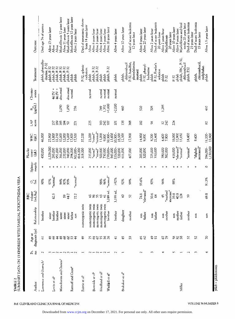
Familial cases of primary polycythemia (polycythemia vera) are distinctly unusual. Fourteen families have been reported (Table 2).1-919 In eight of these families, ade-quate laboratory data are available to meet the strict di-agnostic criteria of the Polycythemia Vera Study Group.18 In five families, one or more laboratory values are lacking so that the strict Polycythemia Vera Study Group criteria cannot be fulfilled. However, in all five cases, splenomegaly plus an elevated leukocyte or plate-let count are recorded, making a secondary form of poly-cythemia unlikely. In one family, that reported by Erf,19
insufficient information is available to diagnose familial polycythemia vera. Thus, 13 familial cases of polycy-themia vera are adequately documented. In only one family are more than two members affected.3 In four families, the affected members are siblings (non-twin),1"3,8 and in three families affected members are identical twins.5-7 In the remaining six families, the af-fected members are one parent-one child combinations.
The family we report is unique in that four members are affected (mother, father, and two sons) and a large pool of offspring in the third generation is available for continued follow-up. None of the 14 offspring in the third generation has demonstrated signs of polycythemia vera at this time; however, the oldest of them is cur-rently only in his 40s.
Our patient 4 meets the Polycythemia Vera Study Group diagnostic criteria18 except for an oxygen satura-tion of 91.3% instead of 92%. He has no respiratory symptoms and full pulmonary function tests are normal except for elevated D L C O . Vague interstitial infiltrates
NOVEMBER- DECEMBER 1989 CLEVELAND CLINIC JOURNAL OF MEDICINE 815
on December 17, 2021. For personal use only. All other uses require permission.www.ccjm.orgDownloaded from
3 O
è*
Cu <
O n -ca -d
rt O g i r
ò c a. a; ao Q. e cn e
o è ^ « 3 5
a 2
So o bi)
fi e
re £ re
O -a
a> v <u v
t- i- »_ CO re re re re <l> w a; i> >• o n ' t fO (N
<U <U W U U > > > > > >
— d)
re e CU t->
H 3 re
re o
<v _re 52 £ 12 re 3 D (J > * re
"o
re iri C ^ re (L> <u T3 «
g re J J
re « .52 u re 3 '
re e 3J 3
i J i !
5 < < < 2 < 2 2 <
j H !
a ,g&,£ucuo«eucu JO 'S J3 J3 _Q _Q _D _Q 3 - " D,
< < < < < < < < -a ' V '
Q
« CU re r\ « .. ^ ^ D r e o "^Xfiu « re re ^ OON rs)^- ^r-a ce ^rH « i»
O > >"3 ¿¡ -DLn-om CsJ £ -C. (U (S <UH
< < < 5 < 2 < < S S a
> TJ o >
<y C m f i co "ci •§ cù a< a,
S S 2 2 2 2 2 2 cu o. o. a. d. cu o.
= c <J ,rt 3 Ss -2 3 e £ 3
- 2 - q - S - S n -3 m
£
+ + "co "re
J3 i E E IH
Cu Cu O 0 KO SO G
re c
- E a
o o \d -4-
0 0 0 0 0 0 0 0 0 0 1 0 0 0 0 0 0 O i ( » N O O o 00 10 N ^(N r<ì 1—1 1 1 1—<
0 0 0 0 0 0 0 A 0 0 0 0 0 0 0 0 0 R 0 0 X" ° o o_ O S o_ o ^ o\m'O t^ o\ pi w ^ ì ~ — — o ^ -N OS f^ N m N
JS 2 -Q
h OJ U u « J3 j ; f) .2 p p 1/3 t! fc
1n <n yi VI C/ì Crt t
I (N -H N f ì I
0 O
O
e O
J5 0 C
6 2 a cu ci.
- 3 M J li N O 3 ^ « Ju »
rv) ro
1 I 0 * , ^J -O 0 -Q 1 0) -p <u 1 2 2 -5 2 h i i D. CL
3 <= -C aj £ "ra « J2
-Q O 0 1 3 u (J
2 o £ 3 o. o.
r 5 e e
O VO
o o o o CO
0 0 0 o O O vo O
- t/j vj S ' s
r 3 3
0 0 O 1 O 0 0 O O O O O O 0 0 O O 0 0 O O O O O O 0 0 O o_ 0 O o_ o_ o_ O o_ O 0 . o_ 0 o~ 00 ro ro ro
O" sd : - vo 0" 10 O" O rs O r i 00 "J- ro ro ro
r l O 00 r l ON 00 m 00
10 ro ro ro r-- SO E — rO (H-Ì
0 "b o ^ rt q S £ 00 > fe 0i) CJ
I O o o 0 . 0 00 1
+ + + + +
^ ^ E 00 vo c
E 00 vom m ^ O,
c c c c c I ' I ' 1 1 1 y o o y o 5-o o o o o -fi OC OC 00 OC OC Q
>- c >. >. c 0 0 0 0 0 - ° c a c c e 0 0 0 0 0 E E S E e
co o Co - s S o ; g 2
c (j ^ - c e
J3 2 xi
15 c T3
JÌ e.
816 CLEVELAND CLINIC JOURNAL OF MEDICINE VOLUME 56 NUMBER 8
on December 17, 2021. For personal use only. All other uses require permission.www.ccjm.orgDownloaded from

POLYCYTHEMIA VERA • MILLER AND ASSOCIATES
of unknown etiology are present on chest radiographs, but have been unchanged for five years. His symptoms and clinical course are classic, however, and he had a normal P50, ruling out a mutant hemoglobin with in-creased oxygen affinity as a cause for his polycythemia. He did, however, have an unexplained mildly elevated serum erythropoietin assay prior to treatment. Erythro-poietin levels were not reported in any other cases of fa-milial polycythemia vera. There has been no evidence of renal disease or occult neoplasm during the 18 months since his diagnosis.
It is highly unlikely that abnormal production and/or regulation of erythropoietin is responsible for this family's polycythemia because splenomegaly and ele-vated neutrophil and/or platelet count were present in the other members, as was transformation to either acute nonlymphocytic leukemia or myelofibrosis. None of these findings would be expected with secondary causes of familial polycythemia.
The other three members of our family did not meet the strict Polycythemia Vera Study Group diagnostic criteria because they were diagnosed at a time when oxy-gen saturation, LAP score, and B12 levels were not routinely measured. Their classic clinical symptoms, splenomegaly, leukocytosis and/or thrombocytosis, and their progression to myelofibrosis or acute nonlympho-cytic leukemia strongly support the diagnosis of polycy-themia vera.
The age at diagnosis (mean 57 years, median 58 years, range 26-82 years) for the previously reported familial cases plus our four cases is similar to that for nonfamilial polycythemia vera (mean 60 years, range 20-85 years).18
Likewise, a male predominance is found in familial cases as well as nonfamilial ones; the male/female ratio for fa-milial cases was 1.8/1, while that of nonfamilial ones was 1 .2/1 . 1 8
In the familial cases, the mean RBC volume was 60 mL/kg, slightly higher than the 49 mL/kg seen in nonfa-milial cases studied by the Polycythemia Vera Study Group.18 Oxygen saturation was reported in 14 of 31 fa-milial cases and found to be > 92% in 12 cases. Splenomegaly was present in 30/31 familial cases (97%) whereas it is found in only 70% of nonfamilial cases.18 A platelet count greater than 400,000/(J.L or "elevated" was reported in 17/31 (55%) familial cases and 43% of nonfamilial cases.18 WBC count greater than 12,000/|O.L or "raised" was reported in 22/31 (71%) familial cases and 63% of nonfamilial ones.18 LAP score was reported in 19 of the familial cases and was elevated (greater than 100) in 15 (79%). Seventy percent of patients studied by the Polycythemia Vera Study Group had an elevated
NOVEMBER- DECEMBER 1989
LAP score.18 Thus familial cases have a similar age of onset, male predominance, and incidence of elevated WBC count, platelets, and LAP scores. They have a slightly higher mean RBC volume at diagnosis and an increased incidence of splenomegaly.
Symptoms reported in patients with familial polycy-themia vera are similar to those in nonfamilial cases and consist mainly of pruritus, headaches, weakness, and diz-ziness.
It has been reported that polycythemia vera is more frequent among Jews of European extraction13 and less frequent in blacks than whites.12 For most familial cases, race and religion have not been specified; however, the family reported by Ratnoff and Gress4 was Jewish and that reported by Waddell et al8 was black. The family we report was white, of Bavarian-German ancestry, and Roman Catholic.
Consanguinity was not mentioned in any of the pre-viously reported familial cases. In the family reported here, there was no known blood relationship between the affected mother and father.
Of the 31 reported patients with familial polycy-themia vera, five (16%) have disease that has progressed to myelofibrosis (5, 10, 12, 20, and 20 years after diagno-sis) and four (13%) have developed acute leukemia (12, 15, 20, and 25 years after diagnosis). The actual inci-dence of acute leukemia or myelofibrosis in familial polycythemia vera cannot be determined as yet since many of the patients are still alive and have had limited follow-up time since diagnosis.
Cytogenic studies in patients with polycythemia vera reveal a nonrandom pattern of abnormalities that most frequently involves chromosomes 1, 8, 9, and 20.14 The incidence of abnormal karyotypes in untreated patients is 13%—26%, and in treated patients it jumps to 38%-44%.14-16 Abnormal karyotypes present early in the dis-ease do not predict eventual leukemic transformation, but a change in karyotype during the course of disease may herald leukemic transformation.14
Of 13 reported kindreds with familial polycythemia vera, chromosome studies were done in six.2-5,7,8 Four of these families had normal karyotypes4,5,7,8 and two revealed abnormalities.2,3 The two brothers reported by Levin et al2
were Philadelphia-chromosome positive. One brother was studied prior to treatment with P-32 and the other after. Two of the sisters with familial polycythemia vera reported by Manoharan and Garson3 were studied cytogenetically after treatment with P-32. On one occasion, they each had normal karyotypes and, at other times, one sister demon-strated 46,XX,-E,+Er and 47,XX,+C while the other sister demonstrated 46,XX,-A,+mar. In this limited group of fa-
CLEVELAND CLINIC JOURNAL OF MEDICINE 817
on December 17, 2021. For personal use only. All other uses require permission.www.ccjm.orgDownloaded from

POLYCYTHEMIA VERA • MILLER AND ASSOCIATES
milial polycythemia vera patients, the incidence of cyto-genetic abnormalities was not higher than that in polycy-themia vera patients in general, and no consistent abnor-malities were found.
Exploring the etiology of familial aggregations of polycythemia vera, one must consider environmental as well as genetic factors. Scattered case reports of polycy-themia in patients exposed to various toxic agents are found in the literature.12 In most instances, the patients demonstrated a polycythemia, but not primary polycy-themia vera. A few cases of true polycythemia vera as-sociated with benzene exposure have been reported, and benzene has also been implicated in the etiology of other myeloproliferative disorders.12 Ratnoff and Gress4 re-
ported a familial occurrence of polycythemia vera in a father and son exposed to organic solvents. Friedland et al7 reported identical twins with polycythemia vera who also had organic solvent exposure. Other reports of fa-milial polycythemia vera do not mention any associated environmental agents. No unusual environmental expo-sure could be documented in the family we studied. They hailed from a small town in Ohio, and the patients worked as a musician, housewife, attorney, and account-ant, with no known industrial exposure. The incidence of familial erythrocytosis is not of a frequency to warrant routinely testing the family members of affected in-dividuals unless they exhibit compatible symptoms or signs of absolute erythrocytosis.
REFERENCES
1. Lawrence JH, Goetsch AT. Familial occurrence of polycythemia and leukemia. Calif Med 1950; 73:361-364.
2. Levin WC, Houston EW, Ritzmann SE. Polycythemia vera with Ph1
chromosomes in two brothers. Blood 1967; 30 :503-512. 3. Manoharan A, Garson OM. Familial polycythemia vera: a study of 3
sisters. Scand ] Haematol 1976; 17 :10-16. 4- Ratnoff WD, Gress RE. The familial occurrence of polycythemia vera:
report of a father and son, with consideration of the possible etiologic role of exposure to organic solvents, including tetrachloroethylene. Blood 1980; 56 :233-236.
5. Fairrie G, Black AJ, McKenzie AW. Polycythemia rubra vera and con-genital deafness in monozygotic twins. Br Med J 1981; 283 :192-193.
6. Burnside P, Salmon DC, Humphrey CA, Robertson JH, Morris TCM. Polycythemia rubra vera in monozygotic twins (letter). Br Med J 1981; 283 :560-561.
7. Friedland ML, Wittels EG, Robinson RJ: Polycythemia vera in identical twins. Am J Hematol 1981; 10 :101-103.
8. Waddell CC, Brown JA, Riggs SA, White MR. Polycythemia vera oc-curring in two brothers. South Med J 1982; 75:1010-1011.
9. Brubaker LH, Wasserman LR, Goldberg JD, et al. Increased prevalence of polycythemia vera in parents of patients on Polycythemia
Vera Study Group protocols. Am J Hematol 1984; 16 :367-383. 10. AdamsonJW. Familial polycythemia. Semin Hematol 1975; 12:383-
396. 11. Hoffman R, Wasserman LR. Natural history and management of
polycythemia vera. Adv Int Med 1979; -285.24:255, 1979 12. Modan B. Polycythemia: a review of epidemiological and clinical
aspects. J Chronic Dis 1965; 18:605-645. 13. Modan B, Kallner H, Zemer D, Yoran C. A note on the increased risk
of polycythemia vera in Jews. Blood 1971; 37 :172-176. 14. Testa JR, Kanofsky JR, Rowley JD, Baron JM, Vardiman JW.
Karyotypic patterns and their clinical significance in polycythemia vera. Am J Hematol 1981; 11:29-45.
15. Lawler SD. Cytogenetic studies in Philadelphia chromosome-negative myeloproliferative disorders, particularly polycythaemia rubra vera. Clin Hematol 1980; -174.9:159, 1980
16. Wurster-Hill D, Whang-Peng J, Mclntyre OR, et al. Cytogenetic studies in polycythemia vera. Semin Hematol 1976; 13 :13-32.
17. Carbonell F, Ganser A, Heimpel H. Cytogenetic studies in chronic myeloproliferative disorders. Acta Haematol 1983; 69:145-151,
18. Berlin Nl. Diagnosis and classification of the polycythemias. Semin Hematol 1975; 12 :339-351.
19. Erf LA. Radioactive phosphorus in the treatment of primary polycythemia (vera). Prog Hematol 1956; 1:153-165.
818 C :LEVELAND CLINIC ; JOURNAL OF MEDICINE VOLUME 56 NUMBER 8
on December 17, 2021. For personal use only. All other uses require permission.www.ccjm.orgDownloaded from


![Thromboembolic events in polycythemia vera · 2019. 4. 15. · cardiovascular disease are more prevalent in polycythemia vera (PV) than in other myeloproliferative disorders [2–4]](https://img.pdfslide.us/doc/110x75/60e1db808b7c7d25000871e0/thromboembolic-events-in-polycythemia-vera-2019-4-15-cardiovascular-disease.jpg)