Embed Size (px)
Citation preview
Failure of foot salvage in patients end-stage renal disease after surgical revascularization
with
Brad L. Johnson, MD, Marc H. Glickman, MD, Dennis F. Bandyk, MD, and Glenn E. Esses, MD, Norfolk, Va., and Tampa, Fla.
Purpose: This report ascertained factors responsible for failure of foot salvage in patients with end-stage renal disease (ESRD) after undergoing infrainguinal bypass for critical ischemia. Methods: A retrospective review of 69 distal arterial reconstructions performed in 53 patients with ESRD (hemodialysis [n = 37], kidney transplantation [n = 10], peritoneal dialysis [n = 6]) for foot gangrene (n = 28), nonhealing ulcer (n = 25), or ischemic rest pain (n = 16) was conducted. Endpoints of surgical morbirty, limb loss, and graft patency were correlated with extent of preoperative tissue loss and presence of diabetes mellims. Results: The 30-day operative mortality rate was 10%, and the patient survival rate at 2 years was 38%. The primary graft patency rate was 96% at 30 days, 72% at 1 year, and 68% at 2 years. Eleven of 22 foot amputations performed during the mean follow-up period of 14 months (range 3 to 96 months) occurred within 2 months of revasculariza- tion. Mechanisms responsible for limb loss included graft failure (n -- 9), foot ischemia despite a patent bypass (n = 8), and uncontrolled infection (n = 5). Overall, 59% of amputations were performed in limbs with a patent bypass to popliteal or tibial arteries. Healing of forefoot amputations was prolonged, but all limb loss beyond 9 months of revascularization was due to graft failure. The limb salvage rate at 1 year decreased (p = 0.13) from 74% to 51% in patients admitted with gangrene. Only two of seven patients admitted with forefoot gangrene experienced foot salvage. Conclusion: Failure of foot salvage in patients with ESRD and critical ischemia was due to wound healing problems rather than graf* thrombosis. Earlier referral for revasculariza- tion, before development of extensive tissue ischemia and infection, is recommended. Primary amputation should be considered in patients admitted with forefoot gangrene, particttlarly if it is complicated by infection. (J VASC SURG 1995;22:280-6.)
Lower limb revascularization has been advocated to patients with end-stage renal disease (ESRD) when critical ischemia develops to improve quality of life. Surgical revascularization can be arduous be- cause of comorbid heart and pulmonary disease, as weil as anticipated difficulties with wound healing and infection resulting from uremia, malnutrition,
From Virginia Vascular Associates, Eastern Virginia School of Medicine, Norfolk, and the Division of Vascular Surgery, University of South Florida College of Medicine, Tampa.
Presented at the Nineteenth Annual Meeting of the Southern Association for Vascular Surgery, Cancün, Mexico, Jan. 25-28, 1995.
Reprint requests: Dennis F. Bandyk, MD, Harbourside Medical Tower No. 730, 4 Columbia Dr., Tampa, FL 33606
Copyright © 1998 by The Society for Vascular Surgery and International Society for Cardiovascular Surgery, North Ameri- can Chapter.
0741-5214/95/$5.00 + 0 24/6/66439
280
diabetes mellims, and long-term immunosuppres- sion. A role for primary amputation has been suggested, but the patient cohort in whom the expense and manpower of revascularization can be avoided has not been clearly defined. With modern day techniques of lower limb revascularization and free muscle flap tissue transfer, even patients with forefoot or hindfoot gangrene are often deemed suitable candidates for limb salvage surgery if a suitable nmoff artery and venous conduit are avail- able. Unfortunately, in the patient population with ESRD and foot ischemia, these advancements in distal artery bypass grafting and tissue healing have not uniformly resulted in foot salvage, particularly in patients with long-standing insulin-dependent diabe- tes mellitus. Several authors have reported clinical failure with limb amputation despite a functioning bypass graft. I-7
IOURNAL OF VASCULAR SURGERY Volume 22, Number 3 Johnson et al. 281
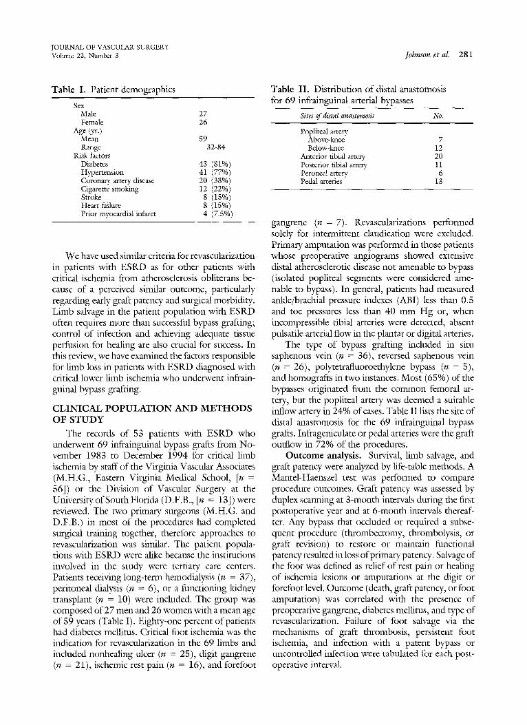
Table I. Patient demographics
Sex Male 27 Female 26
Age (yr.) Mean 59 Range 32-84
Risk factors Diabetes 43 (81%) Hypertension 41 (77%) Coronary artery disease 20 (38%) Cigarette smoking 12 (22%) Stroke 8 (15%) Heart failure 8 (15%) Prior myocardial infarct 4 (7.5%)
We have used similar criteria for revascularization in patients with ESRD as for other patients with critical ischemia from atherosclerosis obliterans be- cause of a perceived similar outcome, particularly regarding early graft patency and surgical morbidity. Limb salvage in the patient population with ESRD offen requires more than successful bypass grafting; control of infection and achieving adequate tissue perfusion for healing are also crucial for success. In this review, we have cxamined the factors responsible for limb loss in patients with ESRD diagnosed with critical lower limb ischemia who undelxvent infrain- guinal bypass grafting.
CLINICAL POPULATION AND METHODS OF STUDY
The records of 53 patients with ESRD who underwent 69 infrainguinal bypass grafts from No- vember 1983 to December 1994 for critical limb ischemia by staff of the Virginia Vascular Associates (M.H.G., Eastern Virginia Medical School, [n = 56]) or the Division of Vascular Surgery at the Universiry ofSouth Florida (D.F.B., [n = 13]) were reviewed. The two primary surgeons (M.H.G. and D.F.B.) in most of the procedures had completed surgical training together, therefore approaches to revascularization was similar. The patient popula- tions with ESRD were alike becanse the institutions involved in the study were tertiary care centers. Patients receiving long-term hemodialysis (n = 37), peritoneal dialysis (n = 6), or a functioning kidney transplant (n = 10) were included. The group was composed of 27 men and 26 women with a mean age of 59 years (Table I). Eighty-one percent of patients had diabetes meHitus. Critical foot ischemia was the indication for revascularization in the 69 limbs and included nonhealing ulcer (n = 25), digit gangrene (n = 21), ischemic rest pain (n = 16), and forefoot
Table II. Distribution of distal anastomosis for 69 infrainguinal arterial bypasses
Sites of distal anastomosis No.
Popliteal artery Above-knee 7 Below-knee 12
Anterior tibial artery 20 Posterior tibial artery 11 Peroneal artery 6 Pedal arteries 13
gangrene (n = 7). Revascularizations pcrformed solely for intermittent claudication were excluded. Primary amputation was performed in those patients whose preoperative angiograms showed extensive distal atherosclerotic disease not amenable to bypass (isolated popliteal segments were considered ame- nable to bypass). In general, patients had measured ankle/brachial pressure indexes (A_BI) less than 0.5 and toe pressures less than 40 mm Hg or, when incompressible tibial arteries were detected, abseht pulsatile arterial flow in the plantar or digital arteries.
The type of bypass grafting included in situ saphenous vein (n = 36), reversed saphenous vein (n = 26), polytetrafluoroethylene bypass (n = 5), and homografts in two instances. Most (65%) of the bypasses originated from the common femoral ar- tery, but the popliteal artery was deemed a suitable inflow artery in 24% of cases. Table II 1ists the site of distal anastomosis for the 69 infrainguinal bypass grafts. Infrageniculate or pedal arteries were the graft outflow in 72% of the procedures.
Outcome analysis. Survival, limb salvage, and graft patency were analyzed by life-table methods. A Mantel-Haenszel test was performed to compare procedure outcomes. Graft patency was assessed by duplex scanning at 3-month intervals during the first postoperative year and at 6-month intervals thereaf- ter. Any bypass that ocduded or required a subse- quent procedure (thrombectomy, thrombolysis, or graft revision) to restore or maintain functional patency resulted in loss ofprimary patency. Salvage of the foot was defined as relief of rest pain or healing of ischemia lesions or amputations at the digit or forefoot level. Outcome (death, graft patency, or foot amputation) was correlated with the presence of preoperative gangrene, diabetes mellitus, and type of revascularization. Failure of foot salvage via the mechanisms of graft thrombosis, persistent foot ischemia, and infection with a patent bypass or uncontrolled infection were tabulated for each post- operative interval.
JOURNAL OF VASCULAR SURGERY 282 Johnson et al. September 1995
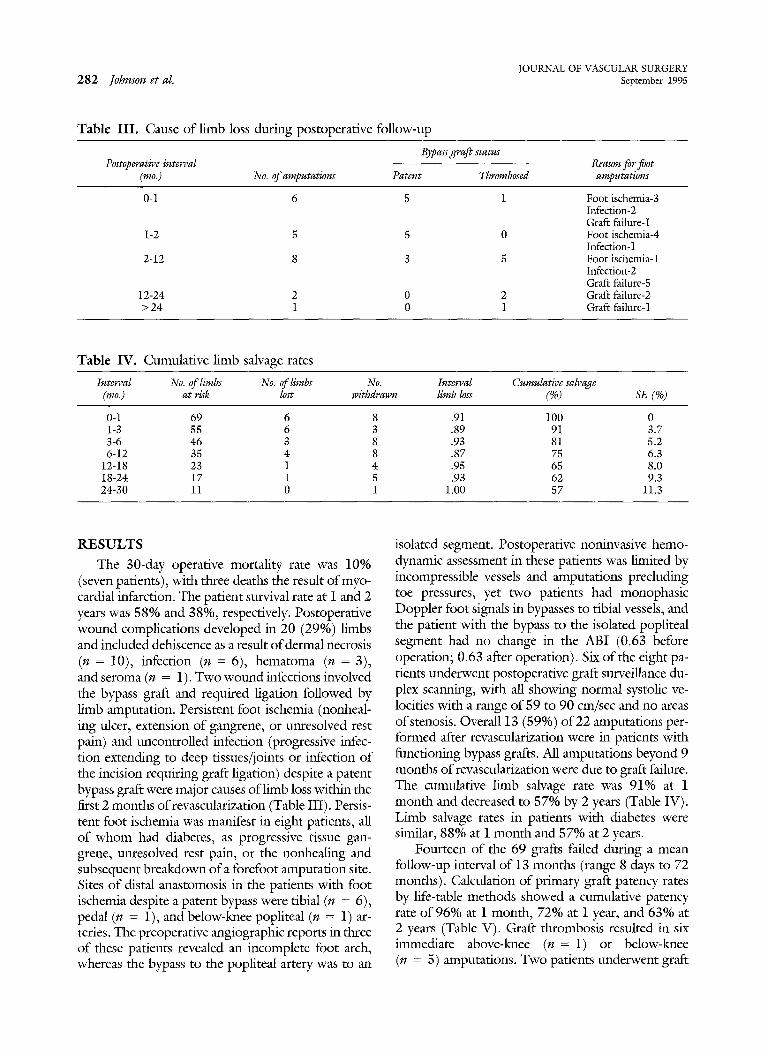
Table III. Cause of limb loss during postoperative follow-up
Bypass graft status Postoperative interval Reason for foot
(mo.) No. of amputations Patent Thrombosed amputations
0-1 6 5 1 Foot ischemia-3 Infection-2 Graft failure- 1
1-2 5 5 0 Foot ischemia-4 Infection-1
2-12 8 3 5 Foot ischemia- 1 Infection-2 Graf* failure-5
12-24 2 0 2 Graft failure-2 > 24 1 0 1 Graft failure-1
Table IV. Cumulative limb salvage rates
Interval No. of limbs No. of limbs No. Interval Cumulative salvage (mo.) at risk lost withdrawn limb loss (%) SE (%)
0-1 69 6 8 .91 100 0 1-3 55 6 3 .89 91 3.7 3-6 46 3 8 .93 81 5.2 6-12 35 4 8 .87 75 6.3
12-18 23 1 4 .95 65 8.0 18-24 17 1 5 .93 62 9.3 24-30 11 0 1 1.00 57 11.3
RESULTS
The 30-day operative mortality rate was 10% (seven pafients), with three deaths the result ofmyo- cardial infarcfion. The patient survival rate at I and 2 years was 58% and 38%, respectively. Postoperative wound complications developed in 20 (29%) limbs and included dehiscence as a result ofdermal necrosis (n = 10), infection (n = 6), hematoma (n = 3), and seroma (n = 1). Two wound infections involved the bypass graft and required ligation followed by limb amputation. Persistent foot ischemia (nonheal- ing ulcer, extension of gangrene, or unresolved rest pain) and uncontrolled infection (progressive infec- tion extending to deep tissues/joints or infection of the incision requiring graft ligation) despite a patent bypass graft were major causes oflimb loss within the first 2 months of revascularization (Table III). Persis- tent foot ischemia was manifest in eight patients, all of whom had diabetes, as progressive tissue gan- grene, unresolved rest pain, or the nonhealing and subscquent breakdown of a forefoot amputation site. Sites of distal anastomosis in the patients with foot ischemia despite a patent bypass were tibial (n = 6), pedal (n = 1), and below-knee popliteal (n = 1) ar- teries. The preoperative angiographic reports in three of these patients revealed an incomplete foot arch, whereas the bypass to the popliteal artery was to an
isolated segment. Postoperative noninvasive hemo- dynamic assessment in these patients was limited by incompressible vessels and amputations precluding toe pressures, yet two patients had monophasic Doppler foot signals in bypasses to tibial vessels, and the patient with the bypass to the isolated popliteal segment had no change in the ABI (0.63 before operation; 0.63 after operation). Six of the eight pa- tients tmderwent postoperative graft surveillance du- plex scanning, with all showing normal systolic ve- locities with a range of 59 to 90 cm/sec and no areas ofstenosis. Overal113 (59%) o f 22 arnputations per- formed after revascularization were in patients with functioning bypass grafts. All amputations beyond 9 months of revascularization were due to graft failure. The cumulative limb salvage rate was 91% at 1 month and decreased to 57% by 2 years (Table IV). Limb salvage rates in patients with diabetes were similar, 88% at i month and 57% at 2 years.
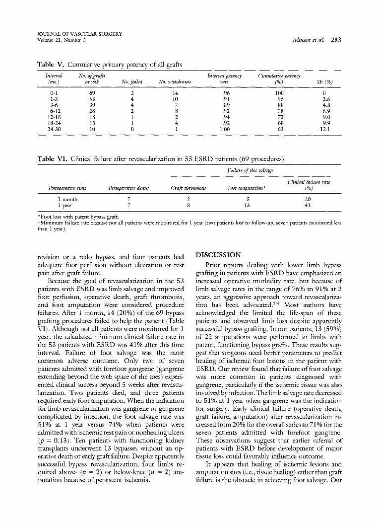
Fourteen of the 69 grafts falled during a mean follow-up interval of 13 months (range 8 days to 72 months). Calculation of primary graft patency rates by life-table methods showed a cumulative patency rate of 96% at 1 month, 72% at 1 year, and 63% at 2 years (Table V). Graft thrombosis resulted in six immediate above-knee (n = 1) or below-lmee (n = 5) amputätions. Two patients underwent graft
JOUKNAL OF VASCULAR SURGERY Volurne 22, Number 3 Johnson et al. 2 8 3
Table V. Cumulative primary patency of all grafts
Interval No. of grafls Interval patency Cumulative patency (mo.) at risk No. failed No. withdrawn rate (%) SE (%)
0-1 69 2 14 .96 100 0 1-3 53 4 10 .91 96 2.6 3-6 39 4 7 .89 88 4.8 6-12 28 2 8 .92 78 6.9
12-18 18 1 2 .94 72 9.0 18-24 15 1 4 .92 68 9.9 2 4 4 0 10 0 1 1.00 63 12.1
Table VI. Clinical failure after revascularization in 53 ESRD patients (69 procedures)
Failure of foot salvage
Clinical failure rate Postoperative time Perioperative death Graft thrombosis Foot amputation « (%)
1 month 7 2 5 20 1 year 7 8 13 41-~
«Foot lost with patent bypass graft. tMinimum failure rate because not all patients were monitored for 1 year (two patients lost to follow-up, seven patients monitored less than 1 year).
revision or a redo bypass, and four patients had adequate foot perfusion without ulceration or rest pain after graft failure.
Because the goal of revascularization in the 53 patients with ESRD was limb salvage and improved foot perfusion, operative death, graft thrombosis, and foot amputation were considered proeedure failures. After 1 month, 14 (20%) of the 69 bypass grafting procedures failed to help the patient (Table VI). Although not all patients were monitored for 1 year, the calculated minimum clinical failure rate in the 53 patients with ESRD was 41% after this time interval. Failure of foot salvage was the most common adverse outcome. Only two of seven patients admitted with forefoot gangrene (gangrene extending beyond the web space of the toes) experi- enced clinical success beyond 5 weeks after revascu- larization. Two patients died, and three patients required early foot amputation. When the indication for limb revascularization was gangrene or gangrene complicated by infection, the foot salvage rate was 51% at i year versus 74% when patients were admitted with ischemic rest pain or nonhealing ulcers (p = 0.13). Ten patients with functioning kidney transplants underwent 13 bypasses without an op- erative death or early graft failure. Despite apparently successful bypass revascularization, four limbs re- quired above- (n = 2) or below-knee (n = 2) am- putation because of persistent ischemia.
D I S C U S S I O N
Prior reports dealing wi~ lower limb bypass grafting in patients with ESRD have emphasized an increased operative morbidity rate, hut because of limb salvage rates in the range of 76% to 91% at 2 years, an aggressive approach toward revasculariza- tion has been advocated. 2-4 Most authors have acknowledged the limited the life-span of these patients and observed limb loss despite apparently succëssful bypass grafting. In our patients, 13 (59%) of 22 amputations were performed in limbs with patent, functioning bypass grafts. These results sug- gest that surgeons need bet-ter parameters to predict healing of ischemic foot lesions in the patient with ESRD. Our review found that failure of foot salvage was more common in patients diagnosed with gangrene, particularly if the ischemic tissue was also involved by infection. The limb salvage rate deereased to 51% at 1 year when gangrene was the indication for surgery. Early clinical failure (operative death, graft failure, amputation) after revascularization in- creased from 20% for the overall series to 71% for the seven patients admitted with forefoot gangrene. These observations suggest that earlier referral of patients with ESRD before development of major tissue loss could favorably influence outcome.
It appears that healing of ischemic lesions and amputation sites (i.e., tissue healing) rather than graft failure is the obstacle in achieving foot salvage. Our
JOURNAL OF VASCULAR SURGERY 284 Johnson et al. September 1995
retrospective review cannot reliably identify patients in whom primary amputation would be the "best" treatment, but this alternative should be considered when extensive forefoot gangrene or infection is present. We found that early limb loss ( < 2 months) was not due to graft thrombosis but to progressive gangrene, absence of wound healing, and infection. Of the 11 amputations in the first 2 months, 10 (91%) were in patients with patent bypasses with persistent foot ischemia or uncontrolled infection. Beyond 2 months from revascularization, eight (73%) of 11 amputations were the result of bypass failure. If the problem of limb loss despite a patent bypass could have been avoided, the fimb salvage rate in our patients would have improved from 65% to 85% at 1 year. Therefore achieving higher early limb savage rates depends on bet-ter selection of patients for revascularization or improvements in wound healing and control ofinfection after surgery. Criteria for primary amputation in patients with ESRD and critical ischemia were suggested by Edwards et al.,1 who found that the size (> 2 cm) of foot ulcers in patients with diabetes could predict failure of limb salvage. Subsequent studies have failed to confirm this, and in out study only three of 13 patients with a patent bypass when undergoing amputation had a nonhealing ulcer as the initial indication for surgery. Wassermann et al.,s in a study of 42 bypasses in paticnts with ESRD, believed that a preoperative ABI <0.3 was predictive of poor limb salvage. Larger studies of patients undergoing infrainguinal bypass have failed to find preoperative ABIs statisti- cally significant for predicting limb salvage. 8
Out review highlights the problem of clinical failure despite successful bypass grafting that oc- curred in 13 patients in out series. Multiple studies have reported this problem, especially in patients with diabetes, large ischemic ulcers, and bypasses to isolated popliteal segments) ,<6,7,9 Orte reason for the failure of bypass grafting to prevent foot loss in patients with ESRD could be due to the incorrect placement of the distal anastomosis. In our study, seven of eight limbs with a patent bypass requiring amputation because of tissue ischemia had the distal anastomosis performed to a tibial or pedal artery. This should have provided adequate perfusion to prcvent progression of gangrene or to achieve ulcer healing. The angiographic reports showed that three of these patients had an incomplete foot arch. One patient had a bypass to an isolated popliteal artery. Postoperative noninvasive hemodynamic assessment in these patients was limited by incompressible vessels, amputations precluding toe pressures and
nonavailability of laser Doppler velocimeter and transcutaneous oximetry. When these data were available it showed monophasic Doppler foot signals in bypasses to tibial vessels in two patients and no improvement in the ABI of the patient with the bypass to the isolated popliteal segment. The in- creased percentage of limbs amputated with a patent bypass in patients with ESRD suggests that, al- though successful revascularization and foot salvage require operative technical precision, they also de- pend greatly on careful preoperative selection of operative candidates, with detailed angiographic assessment of foot vessels and consideration for primary amputation in those patients with only an isolated popliteal artery.
Even with adequate perfusion, failure of tissue healing in patients with ESRD could be explained by associated conditions such as anemia, malnutrition, and depressed immune function. Delayed wound healing occurs with uremia, which reduces formation ofgranulation tissue in wounds when compared with controls. 1° In addition, diabetes, a disease present in most patients with ESRD, is also associated with a deficiency of coUagen content in granulation tissue. Although patients undergoing transplantation are not affected by uremia, they receive immunosuppres- sive medications that impair wound healing. It has been suggested that use of nonnephrotoxic antibiot- ics such as cefoperazone in patients with lddney transplants can reduce wound infection rates and thereby improve wound healing, n The use ofrecom- binant human tissue growth factor may be needed in patients with ESRD to promote tissue regeneration and healing. This hormone has been shown to enhance protein balance and increase wound healing and immune function in the malnourished patient undergoing hemodialysis. 12
Problems of impaired healing and resistance to infection orten result in wound complications in patients with ESRD after they undergo lower limb bypass grafting. In this series, 20 (29%) procedures were associated with wound complications, ofwhich eight required a return to the operating room for treatment. We used an in situ bypass technique in 52% of the procedures, which may have contributed to the high number of wound problems. Of note, both grafts that required ligation because of a wound complication were in situ saphenous vein bypasses. Wassermann et al.s reported a 36% incidence of wound complications with only reversed saphenous vein grafting but did not have any wound problems that resulted in graft ligation or rupture. These authors believed that tunneling the vein graft in an
JOURNAL OF VASCULAR SURGERY Volume 22, Number 3 Johnson et al. 285
anatomic or subfascial plane was an impor t an t technical maneuver w h e n opera t ing on patients wi th E S R D .
I t is impor t an t w h e n considering any major opera t ion to be cognizant o f its overall o u t c o m e and benefit to the patient. Perioperat ive death, graft th rombos is , and amputa t ions wi th pa ten t grafts obviously do not benefit the pat ient wi th E S R D and critical ischemia. We found the clinical failure rate to be 22% at 1 m o n t h and 4 1 % at 1 year. These sober ing results occurred despite a high technical success rate wi th bypäss graft ing (30-day pr imary graft patency rate o f 96%) . I f t h e mos t f requent cause o f failure, ampu ta t ion wi th a pa tent graft, could be el iminated, the ou t come o f surgical revascularization could be improved significantly. W e cont inue to r e c o m m e n d infrainguinal bypass graf t ing to patients wi th E S R D when a mult idisciplinary evaluation indicates that l imb salvage is likely. In patients wi th gangrene and infection, consul tat ion wi th plastic surgery and infectious disease are f requent ly ob- tained. Al though we were not able to identify conclusively which factors will predict l imb loss, we believe that referral at the first signs o f foo t ischemia and considerat ion for p r imary ampu ta t ion in patients wi th forefoot gangrene or poo r runof f (documented by angiographic assessment wi th comple te foo t films) should decrease the failures o f foo t salvage seen wi th in the first 2 mon ths after infrainguinal bypass g ra f ring.
REFERENCES
1. Edwards JM, Taylor LM, Porter JM. Limb salvage in end-stage renal disease. Arch Surg 1988;123:1164-8.
2. Chang BB, Paty PSK, Shah DM, Kaufman JL, Leather RP. Results of infrainguinal bypass for limb salvage in patients with end-stage renal disease. Surgery I990;108:742-7.
3. Harrington EB, Harrington ME, Schanzer H, Haimov M. End-stage renal disease-is infrainguinal limb salvage justi- fied? J VASC SURG 1990;12:691-6.
4. Lumsden AbB Besman A, Jaffe M, MacDonald MJ, Allen RC. Infrainguinal revascularization in end-stage renal disease. Ann Vasc Surg 1994:8:107-I2.
5. Wassermann RJ, Saroyan RM, Rice JC, Kerstein MD. Infrainguinal revascularization for limb salvage in patients with end-stage renat disease. S Med J i991;84:I90-2.
6. Dietzek AM, Gupta SK, Kram HB, Wengerter KR, Veith FJ. Limb loss with patent infrainguinal bypasses. Eur J Vasc Surg 1990;4:413-7.
7. Sanchez LA, Goldsmith J, Rivers SP, et al. Limb salvage in end stage renal disease: is it worthwhile. J Cardiovasc Surg 1992',33:344-8.
8. Samson RH, Gupta SK, Veith FJ, Ascer E, Scher L. Perioperative noninvasive hemodynamic ankle indices as predictors of infrainguinal graft patency. J VASC SURG 1985:2:307-11.
9. Taylor LM, Hamre D, Dalman RL, Porter JM. Limb salvage vs amputation for critical isehemia: the role of vascular surgery. Arch Surg 1991;I26:I251-8.
I0. Yue DK, McLennan MM, Mai YW, Spaliviero J, Delbridge L, Reeve T, Turtle JR. Effects of experimental diabetes, uremia, and malnutrition on wound healing. Diabetes 1987;36: 295-9.
I l . Koyle MA, Glassock RJ, Ward HJ, Rajfer J, Twomey PA. Declining ineidence of wound infection in cadaveric renal transplant recipient. Urology Feb 1988;31:103-6.
12. Kopple JD. The rationale for the use of growth hormone or insulin-like growth factor I in adult patients with renal failure. Min Electrolyte Metab I992;18: 269-75.
Submitted Feb. 14, 1995; accepted May 20, 1995.
D I S C U S S I O N
Dr. Frank J. Veith (Bronx, N.Y.) I generally agree with the findings and condusions of this study. My agreement is based on a recent 10-year analysis that Dr. Luis Sanchez 7 carried out on 141 threatened limbs in 112 patients with ESRD. When gangrene and infection were extensive and involved the mid foot, as was the case in 21 limbs, we performed a primary amputation. We attempted limb salvage by balloon angioplasty in 40 limbs and by bypass in the remaining 80 limbs. Our overall 30-day and late mortality rates were almost identical to Dr. Johnson's. Our results with bypasses were much better than those with balloon angioplasty, eren though the bypasses were perforrned in the rnore difficult situations with worse arterial disease. The 6-month limb salvage rates were
83% +- 5% with bypasses and only 50% _+ 11% with percutaneous transluminal angioplasty (PTA). Moreover, PTA of-ten (seven limbs) delayed the rescue bypass so that, eren when the bypass was successful, the limb was lost. We therefore believe PTA to be a poor option for treating limb-threatening ischemia in patients with ESRD. Like Dr. Johnson, we had a high incidence in these patients of limb loss despite a patent bypass and a pälpable foot pulse. This always occurred in the face of extensive infection or gangrene as he found, and it is in this group of patients with ESRD that we believe primary amputation to be appropriate; however, if necrosis is limited, bypasses are worthwhile eren in patients with diabetes and ESRD. Some good results can be obtained in this setting. Surgeons
JOURNAL OF VASCULAR SURGERY 286 Johnson et aL September 1995
undertaking these procedures must, however, be aware that these patients with ESRD and diabetes have calcified, difficult arteries to deal with and high operative mortality and morbidity rates.
What are your views on the role of PTA in patients with ESRD? How precisely do you determine when forefoot infection and gangrene are extensive enough to justify primary amputation?
Dr. Brad L. Johnson. We have not used PTA in our patients very often and, given the severity of disease in this patient population, I do not believe that it would meet with rauch success, especially given the degree of gangrene seen in these patients.
We also have difficulty trying to choose those patients who should proceed to undergo primary amputation with forefoot gangrene, hut given that only two ofseven patients experienced clinical success, we believe that probably this group of patients should proceed directly to primary amputation.
Dr. James Seeger (Gainesville, Fla.). You stated that you performed some amputations eren with patent bypass grafts presumably because you couldn't control the infec- tion in the foot. What information do you have that documents good perfusion to the foot, even out into the toes in these patients! Did you have toe pressures that confirm the hemodynamics of the foot? Alternatively, do you have any information about the arterial anatomy in the foot, not whether there were patent pedal arches but rather whether digital arteries were present so that there was a potential of getting perfusion out to the area that was infected?
Dr. Johnson. Yes, we were concerned about whether
we had adequate distal perfusion. No, we have not reviewed angiograms to assess patent arches or digital arteries.
Dr. William Suggs (Bronx, N.Y.). I have believed that the flow should be adequate for wound healing because of pulsatile flow coming from small digital calcified vessels, and yet in 2 däys after what appears to be a clean wound, the muscle turns gray and the foot is unsalvageable. Has that been your experience, that eren when the wound looks clean and the amputation looks adequate, 2 or 3 days later there is a local wound healing problem that then leads to a below-knee amputation?
Dr. Johnson. We had patients in whom we believed we had successful limb salvage with good perfusion hut who continued to progress with gangrene. We had one patient who had an ulcer that eventually eroded into bis ankle joint, which led to limb loss, so, yes, it still occurs when you believe you have adequate perfusion to the distal foot.
Dr. T imothy R. S. Ha rward (Gainesville, Fla.). I want to answer the question pertaining to small-vessel disease in the patient with diabetes. Dr. Strandness, ämong others, has shown quite nicely that small-vessel disease does not occur in the feet of patients with diabetes. In patients with diabetes and ESRD, tibial arteries are noncompress- ible because of medial calcinosis hut orte can still compress the digital artery. With a photoplethysmograph and a small cuff, orte can very accurately measure toe pressures in the second, third, or fourth toes, whereas ifyou move up to the an!de or transmetatarsal areas, you are measuring pressure in the dorsalis pedis arteries, which are calcified; therefore you can't compress the artery.