Embed Size (px)
Citation preview
FAD-I, a Fusobacterium nucleatum Cell Wall-Associated DiacylatedLipoprotein That Mediates Human Beta Defensin 2 Induction throughToll-Like Receptor-1/2 (TLR-1/2) and TLR-2/6
Sanghamitra Bhattacharyya,a* Santosh K. Ghosh,a Bhumika Shokeen,b Betty Eapan,a Renate Lux,b Janna Kiselar,c
Stanley Nithianantham,a* Andrew Young,d Pushpa Pandiyan,a Thomas S. McCormick,a,d Aaron Weinberga
Department of Biological Science, Case School of Dental Medicine, Cleveland, Ohio, USAa; School of Dentistry, University of California—Los Angeles, Los Angeles,California, USAb; Center for Proteomics, Case School of Medicine, Cleveland, Ohio, USAc; Department of Dermatology, Case School of Medicine, Cleveland, Ohio, USAd
We previously identified a cell wall-associated protein from Fusobacterium nucleatum, a Gram-negative bacterium of the oralcavity, that induces human beta defensin 2 (hBD-2) in primary human oral epithelial cells (HOECs) and designated it FAD-I(Fusobacterium-associated defensin inducer). Here, we report differential induction of hBD-2 by different strains of F. nuclea-tum; ATCC 25586 and ATCC 23726 induce significantly more hBD-2 mRNA than ATCC 10953. Heterologous expression of plas-mid-borne fadI from the highly hBD-2-inducing strains in a �fadI mutant of ATCC 10953 resulted in hBD-2 induction to levelscomparable to those of the highly inducing strains, indicating that FAD-I is the principal F. nucleatum agent for hBD-2 induc-tion in HOECs. Moreover, anti-FAD-I antibodies blocked F. nucleatum induction of hBD-2 by more than 80%. RecombinantFAD-I (rFAD-I) expressed in Escherichia coli triggered levels of hBD-2 transcription and peptide release in HOECs similar tothose of native FAD-I (nFAD-I) isolated from F. nucleatum ATCC 25586. Tandem mass spectrometry revealed a diacylglycerolmodification at the cysteine residue in position 16 for both nFAD-I and rFAD-I. Cysteine-to-alanine substitution abrogatedFAD-I’s ability to induce hBD-2. Finally, FAD-I activation of hBD-2 expression was mediated via both Toll-like receptor-1/2(TLR-1/2) and TLR-2/6 heterodimerization. Microbial molecules like FAD-I may be utilized in novel therapeutic ways to bolsterthe host innate immune response at mucosal surfaces.
The epithelial surfaces of the oral cavity are sites of active bac-terial colonization. While colonizing, certain bacteria pro-
mote activation of human beta defensin (hBD) expression in theoral mucosa (1–3). By virtue of their antimicrobial and immuno-regulatory properties, these epithelial-cell-derived innate re-sponse peptides contribute to the homeostasis between the bacte-rium and the host (4). Human beta defensin 2 (hBD-2) and hBD-3are the two inducible members of the hBD peptide family that weand others have described in the oral cavity (1, 5–10). Interest-ingly, while hBD-3 is associated with the highly proliferating, non-differentiated stratum basale of the oral mucosa, hBD-2 is com-partmentalized in the more superficial stratum spinosum andstratum granulosum; i.e., nonproliferating yet differentiating re-gions of the oral mucosa (11, 12). This, along with other resultsshowing that hBD-2 is induced as a result of inflammation viaMAPK or NF�� (5) while hBD-3 is activated through epidermalgrowth factor receptor (13, 14), strongly suggests that the latter ismore involved in wound healing while the former plays a moreactive role in inhibiting microbial invasion during mucosal-bar-rier disruption (15–17). Moreover, in addition to their antimicro-bial properties (18, 19), both peptides have been shown to act aschemokines in recruiting lymphoid and myeloid cells from thebloodstream (20).
Fusobacterium nucleatum, a ubiquitous Gram-negative bacte-rium of the human oral cavity, has been extensively studied for itsproperties of adhesion to other bacteria, an important feature inoral biofilm formation (21, 22). In addition to its role in oralcommunity architecture, we and others have shown that the cellwall of F. nucleatum induces hBD-2 expression in normal primaryhuman oral epithelial cells (HOECs) (1, 5, 10, 23). The presence ofF. nucleatum in oral biofilms colonizing oral surfaces may be a
reason why the generally inducible hBD-2 is constitutively ex-pressed in the upper strata of the oral mucosa, a trait apparentlyunique to this site compared to other mucosal body sites (5, 11,17). We have also shown that, as well as in HOECs, F. nucleatum isalso capable of inducing hBD-2 in skin keratinocytes (14).
Recently, we reported the identification, isolation, and func-tional evaluation of a cell wall-associated protein from F. nuclea-tum that induces hBD-2 in HOECs (1). We named this proteinFusobacterium-associated defensin inducer (FAD-I). Expressionof FAD-I in a bacterium (Porphyromonas gingivalis ATCC 33277)that did not promote hBD-2 expression resulted in its ability to doso (1). Here, we present new evidence showing that (i) FAD-I isthe principal F. nucleatum molecule responsible for hBD-2 induc-
Received 19 October 2015 Returned for modification 20 November 2015Accepted 13 February 2016
Accepted manuscript posted online 29 February 2016
Citation Bhattacharyya S, Ghosh SK, Shokeen B, Eapan B, Lux R, Kiselar J,Nithianantham S, Young A, Pandiyan P, McCormick TS, Weinberg A. 2016. FAD-I, aFusobacterium nucleatum cell wall-associated diacylated lipoprotein that mediateshuman beta defensin 2 induction through Toll-like receptor-1/2 (TLR-1/2) andTLR-2/6. Infect Immun 84:1446 –1456. doi:10.1128/IAI.01311-15.
Editor: S. M. Payne
Address correspondence to Aaron Weinberg, [email protected].
* Present address: Sanghamitra Bhattacharyya, Department of Pathology, CaseSchool of Medicine, Cleveland, Ohio, USA; Stanley Nithianantham, Department ofMolecular and Cellular Biology, University of California, Davis, California, USA.
Supplemental material for this article may be found at http://dx.doi.org/10.1128/IAI.01311-15.
Copyright © 2016, American Society for Microbiology. All Rights Reserved.
crossmark
1446 iai.asm.org May 2016 Volume 84 Number 5Infection and Immunity
on February 17, 2018 by guest
http://iai.asm.org/
Dow
nloaded from
tion; (ii) FAD-I is posttranslationally modified at its cysteine inposition 16 (C16) by a diacylglycerol, which is essential for FAD-I-dependent hBD-2 activation in HOECs; and (iii) FAD-I induceshBD-2 in HOECs through both Toll-like receptor-1/2 (TLR-1/2)and TLR-2/6. Since most mucosal body sites do not express con-stitutive levels of hBD-2, FAD-I, or its derivatives, offers the pos-sibility of inducing the body’s own innate antimicrobial agents invulnerable mucosa.
MATERIALS AND METHODSHOEC culture and treatment. Tissue acquisition for primary cell isola-tion was conducted in accordance with our Institutional Review Board(IRB)-approved protocol (NHR-15-19) for the use of discarded tissue.Primary HOECs were expanded from tissues overlying impacted thirdmolars, as previously described (24), and grown as monolayers to �70%confluence. The cells were cultured in EpiLife medium (Gibco, Life Tech-nologies) supplemented with 1% penicillin-streptomycin, 0.2% Fungi-zone, and 1% human keratinocyte growth supplement (HKGS) (Gibco)at 37°C and 5% CO2 prior to challenge with various agents.
HOECs that reached �70% confluence were trypsinized, split, andseeded in 12- or 24-well plates at concentrations of 1.7 � 105 and 3.5 �105 cells/well, respectively. The plates were incubated for 1 or 2 days untilthe cells reached �80% confluence. These cells were then used for thevarious experiments described below. Incubations with the various agentswere for �18 h.
Construction of F. nucleatum mutant and complementationstrains. For generation of an F. nucleatum ATCC 10953 mutant derivativelacking FAD-I, allelic-exchange mutagenesis was used to replace theATCC 10953 fadI gene with the catP gene resistance cassette. Briefly, theconstruct was generated by fusing 1 kb of DNA upstream and downstreamof fadI with the catP gene, along with its promoter. The upstream regionwas amplified from F. nucleatum ATCC 10953 using primer pair BS933/BS934, while the downstream region was amplified using the primer pairBS937/BS938. The catP gene was PCR amplified from pHS30 (25) usingprimers BS935 and BS936. The primers contained an overlap of 25 to 30bp to allow fusion in PCRs, and the fusion PCR was carried out as de-scribed by Shevchuk et al. (26). The fusion product was cloned into pCR-BluntII-TOPO (Invitrogen) and confirmed by sequencing. The plasmidDNA was electroporated into F. nucleatum ATCC 10953 as described byHaake et al. (25) and plated on selective medium containing thiampheni-col to obtain the corresponding �fadI derivative. The genomic DNA ofthe obtained colonies was analyzed by PCR for the presence of the catPgene and the absence of fadI. One of the colonies was designated the �fadIinactivated mutant for complementation with heterologous expressedfadI from highly hBD-2-inducing F. nucleatum strains ATCC 23726 andATCC 25586 under the control of the fomA promoter. The fadI genes of F.nucleatum ATCC 23726 and ATCC 25586 were amplified from the respec-tive genomes using the primer pair BS926/BS927. The fomA promoter wasamplified from F. nucleatum 10953 using the primer pair BS924/BS925.The fused products of the fomA promoter and the respective fadI geneswere cloned into the shuttle vector pHS58 (27). All the resulting plasmidswere confirmed by sequencing and transformed into the �fadI derivativeof F. nucleatum ATCC 10953 for complementation and heterologous ex-pression of the different fadI genes. The presence of the fusion constructbetween the fomA promoter and fadI from ATCC 25586 and ATCC 23726in the transformants was confirmed by PCR. The primer pairs used aredescribed in Table S1 in the supplemental material.
Isolation of FnCW. F. nucleatum cell wall (FnCW) extracts from dif-ferent F. nucleatum parent and mutant strains were prepared as previouslydescribed (28).
Isolation of nFAD-I. Native FAD-I (nFAD-I) was isolated fromFnCW extracts derived from F. nucleatum ATCC 25586 by immunopre-cipitation using a polyclonal anti-FAD-I antibody (1). To eliminate non-specific interactions, 100 �g of the bacterial cell wall fraction was initiallyincubated with 50 �l of preimmune serum at 4°C overnight in the pres-
ence of antibody binding buffer (Thermo Scientific, Waltham, MA) andprotease inhibitors (Thermo Scientific). Precleared lysates were obtainedby adding 25 �l of protein A magnetic beads (Novex, Life Technologies)to the mixture. Thirty micrograms of the anti-FAD-I antibody was addedto the supernatants and continuously mixed at 4°C overnight. The mag-netic beads were washed with antigen-antibody binding buffer (ThermoScientific). nFAD-I was eluted out of the complex at room temperaturewith Gentle antigen elution buffer (Thermo Scientific) by 40 min of incu-bation at room temperature with constant rotation. The isolated proteinwas further dialyzed against Tris buffer (pH 7.5) at 4°C overnight, and theprotein content was measured with a Dc protein assay kit (Bio-Rad, Her-cules, CA). The protein was identified by mass spectrometric (MS) anal-ysis (Proteomics Core, Case Western Reserve University [CWRU]).
Cloning and bacterial overexpression of rFAD-I. The full-lengthFAD-I gene (FN1527) (1) was amplified by PCR from genomic DNA of F.nucleatum strain ATCC 25586 using primers (see Table S2 in the supple-mental material) with Nde1 and Xho1 restriction sites at their ends (NewEngland BioLabs). The PCR product was gel purified using a PCR purifi-cation kit (Qiagen, Germantown, MD) and cloned in the pET-20b vector(Novagen). Recombinant FAD-I (rFAD-I) protein was overexpressedfrom one positive clone in the host Escherichia coli BL21(DE3) (Invitro-gen, Life technologies). The E. coli culture, maintained at 37°C with 1 mMIPTG (isopropyl-�-D-thiogalactopyranoside), generated the recombi-nant protein within 4 h. Bacterial cells were harvested by centrifugation at4,000 rpm for 10 min, and the cell pellet was washed with 1� phosphate-buffered saline (PBS) and stored at 80°C until further processed. Dif-ferent mutant forms of FAD-I (�15, C16A, and 15-amino-acid signalsequence) (see Fig. S1 in the supplemental material) were generated usinga Quick Site-Directed Mutagenesis kit (Stratagene, La Jolla, CA) with thefull-length FAD-I gene of F. nucleatum strain ATCC 25586 as the tem-plate. The primers used for creating the mutants are listed in Table S2 inthe supplemental material. Cloning and overexpression were conductedin pET20b following the same protocol used to generate the full-lengthFAD-I protein.
Purification of overexpressed proteins from E. coli. E. coli cell pelletsoverexpressing complete FAD-I, the �15 and C16A derivatives, or thesignal peptide (SP) were suspended in 50 mM Na2HPO4, 100 mM NaCl(pH 7.5), 0.1 mM phenylmethylsulfonyl fluoride (PMSF), and 1� Haltprotease inhibitor (Thermo Scientific) and sonicated on ice using a FisherSonic Dismembrator 300 (Thermo Fisher, Bridgewater, NJ) on ice. Thecells were subjected to 20 cycles of 5-s pulses repeated 5 times with a waittime of 2 to 3 min after each cycle. The lysed cells were centrifuged at18,000 rpm and 4°C for 30 min. The supernatant underwent further pro-cessing to isolate the desired protein using a Qiaexpress Ni-nitrilotriaceticacid (NTA) column (Qiagen), following the manufacturer’s protocol. TheHis-tagged column eluent was dialyzed against 10 mM Tris-HCl, pH 7.5,and 100 mM NaCl. The dialyzed protein was concentrated using an Ami-con tube concentrator with a 5-kDa cutoff (Millipore, MA). The recom-binant signal sequence was expressed in inclusion bodies. Recovery of theoverexpressed protein from the inclusion bodies and its purification weredone using inclusion body solubilization reagent (Thermo Scientific) ac-cording to the manufacturer’s protocol.
Mass spectrometric analysis. A C18 reverse-phase column was uti-lized for identification of possible posttranslational modifications (PTM),including phosphorylation, myristoylation, and sulfonation. Approxi-mately 300 ng of both the rFAD-I and the nFAD-I peptide mixtures wasloaded onto a 100-�m by 2-cm Acclaim PepMap100 C18 reverse-phasetrapping column to preconcentrate and wash away excess salts using anano-UltiMate-3000 Rapid Separation LC system (Dionex, Sunnyvale,CA). The reverse-phase separation was performed on a 75-�m by 25-cm(particle size, 2 �m, and pore size, 100 A) Acclaim PepMap100 C18 re-verse-phase column (Dionex) using a linear gradient of 5 to 60% buffer B(100% acetonitrile-0.1% formic acid [FA]) over 55 min.
A C4 reverse-phase column was utilized for identification of lipidmodification only. The approximately 300 ng of digest mixture derived
TLR-1/2- and TLR-2/6-Mediated Induction of hBD-2 by FAD-I
May 2016 Volume 84 Number 5 iai.asm.org 1447Infection and Immunity
on February 17, 2018 by guest
http://iai.asm.org/
Dow
nloaded from
from both the rFAD-I and the nFAD-I proteins was loaded onto a 100-�mby 2-cm Acclaim PepMap100 C18 reverse-phase trapping column to pre-concentrate and wash away excess salts using a nano-UltiMate-3000Rapid Separation LC system (Dionex, Sunnyvale, CA). The reverse-phaseseparation was performed on a 75-�m by 15-cm (particle size, 5 �m, andpore size, 300 A) Acclaim PepMap300 C4 reverse-phase column (Dionex)using a linear gradient of 20 to 100% buffer B over 64 min.
Proteolytic peptides eluting from either C18 or C4 columns were di-rected to an LTQ-FT mass spectrometer (Thermo Fisher Scientific, Wil-mington, DE) equipped with a nanospray ion source with a needle voltageof 2.4 kV. All mass spectra were obtained from data-dependent experi-ments in the positive ion mode. MS and tandem-MS (MS-MS) spectrawere acquired with full-scan MS recorded in the FT analyzer at a resolu-tion (R) of 100,000, followed by MS-MS of the eight most intense peptideions in the LTQ analyzer. The data were analyzed by searching tan-dem-MS spectra using Mascot software against a database containingFN1527 protein, considering variable modification of S, T, and Y residuesby 79.966 Da; S, T, and Y residues by 79.957 Da; C residues by 210.198 Da;and C residues by 576.511 Da, which correspond to phosphorylation,sulfation, myristoylation, and S-diacylglycerol cysteine, respectively.
RNA isolation, quantitative PCR (qPCR), and enzyme-linked im-munosorbent assay (ELISA). RNA was extracted from HOECs with theRNeasy minikit (Qiagen) following the manufacturer’s instructions. TheRNA concentration was measured by UV absorbance at 260/280 nm usinga Nanodrop 1000 (Thermo Fisher Scientific). Quantitative real-time PCRwas performed with a Bio-Rad CFX96 system with a high-capacity cDNAreverse-transcription kit (Applied Biosystems, Carlsbad, CA) and SyBrGreen Supermix (Bio-Rad) according to the manufacturer’s protocol.The primer sequences for hBD-2 and the glyceraldehyde-3-phosphatedehydrogenase (GAPDH) housekeeping gene are shown in Table S2 in thesupplemental material. Primers were designed using Primer3 primer de-sign software (29) and were analyzed using Oligo Analysis (IntegratedDNA Technologies [IDT]) in order to avoid secondary structures. Themedium supernatants from HOECs were analyzed for hBD-2 protein lev-els following our previously described protocol (9, 10).
Flow cytometry. Biotin-conjugated anti-TLR-1, allophycocyanin(APC)-conjugated anti-TLR-2, and phycoerythrin (PE)-conjugated anti-TLR-6 antibodies were used for flow cytometry (Ebiosciences, San Diego,CA). For surface staining, the cells were first harvested in cell dissociationbuffer and then washed and stained in PBS- bovine serum albumin (BSA)(0.5%)-EDTA (0.5 mM) buffer for 1 h and immediately analyzed (LSR IIFortessa; BD Biosciences, CA). Cells were harvested in trypsin-EDTA andwashed in PBS-BSA (0.5%)-EDTA (0.5 mM) buffer to remove surfaceproteins. Unconjugated antibodies were used to further block the remain-
ing surface proteins. The cells were then fixed using 1� fixation buffer(Ebiosciences) overnight, followed by permeabilization and staining, us-ing the conjugated antibodies mentioned above, in 1� permeabilizationbuffer (Ebiosciences) for 2 h, and analyzed by flow cytometry. Surfacedetection of the same receptor proteins was done with and without cy-tochalasin D (CytD) (Life Technologies) treatment. HOECs were pre-treated with 40 �M cytochalasin D at 37°C 1 h prior to FAD-I stimulation.We also used fluorescein isothiocyanate (FITC)-labeled FAD-I (Bio BasicCanada Inc., Markham, Ontario, Canada) to measure the receptor-ligandassociation at the surface, as detected by TLR-2 and FAD-I–FITC-double-positive cells by flow cytometry.
Statistical analysis. All statistical analyses (Student t test or analysis ofvariance [ANOVA]) were performed using Graph Pad (La Jolla, CA)Prism v.6 software. P values of 0.05 were considered statistically signif-icant.
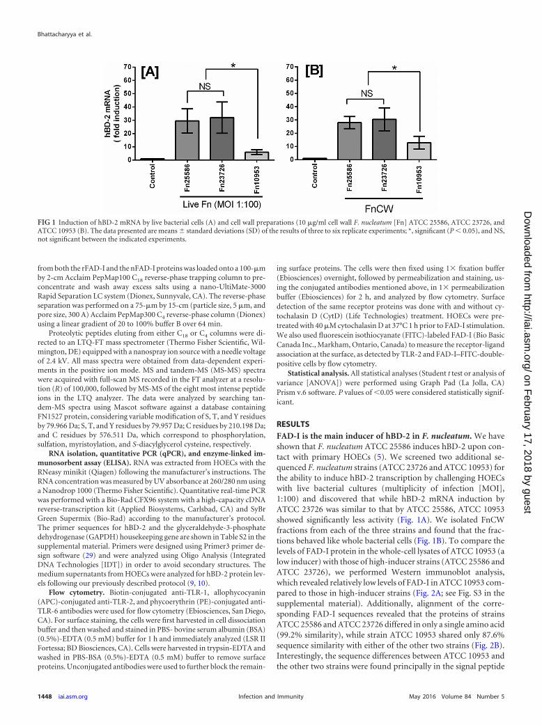
RESULTSFAD-I is the main inducer of hBD-2 in F. nucleatum. We haveshown that F. nucleatum ATCC 25586 induces hBD-2 upon con-tact with primary HOECs (5). We screened two additional se-quenced F. nucleatum strains (ATCC 23726 and ATCC 10953) forthe ability to induce hBD-2 transcription by challenging HOECswith live bacterial cultures (multiplicity of infection [MOI],1:100) and discovered that while hBD-2 mRNA induction byATCC 23726 was similar to that by ATCC 25586, ATCC 10953showed significantly less activity (Fig. 1A). We isolated FnCWfractions from each of the three strains and found that the frac-tions behaved like whole bacterial cells (Fig. 1B). To compare thelevels of FAD-I protein in the whole-cell lysates of ATCC 10953 (alow inducer) with those of high-inducer strains (ATCC 25586 andATCC 23726), we performed Western immunoblot analysis,which revealed relatively low levels of FAD-I in ATCC 10953 com-pared to those in high-inducer strains (Fig. 2A; see Fig. S3 in thesupplemental material). Additionally, alignment of the corre-sponding FAD-I sequences revealed that the proteins of strainsATCC 25586 and ATCC 23726 differed in only a single amino acid(99.2% similarity), while strain ATCC 10953 shared only 87.6%sequence similarity with either of the other two strains (Fig. 2B).Interestingly, the sequence differences between ATCC 10953 andthe other two strains were found principally in the signal peptide
FIG 1 Induction of hBD-2 mRNA by live bacterial cells (A) and cell wall preparations (10 �g/ml cell wall F. nucleatum [Fn] ATCC 25586, ATCC 23726, andATCC 10953 (B). The data presented are means � standard deviations (SD) of the results of three to six replicate experiments; *, significant (P 0.05), and NS,not significant between the indicated experiments.
Bhattacharyya et al.
1448 iai.asm.org May 2016 Volume 84 Number 5Infection and Immunity
on February 17, 2018 by guest
http://iai.asm.org/
Dow
nloaded from
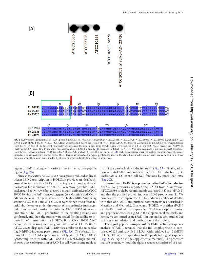
region of FAD-I, along with various sites in the mature-peptideregion (Fig. 2B).
Since F. nucleatum ATCC 10953 has a greatly reduced ability totrigger hBD-2 transcription in HOECs, it provides an ideal back-ground to test whether FAD-I is the key agent produced by F.nucleatum for induction of hBD-2. To remove possible FAD-Ibackground activity, we first created a mutant derivative of ATCC10953 lacking the FAD-I-encoding gene (see Materials and Meth-ods for details). The fadI genes of the highly hBD-2-inducingstrains ATCC 25586 and ATCC 23726 were cloned into a fusobac-terial shuttle vector under the control of a constitutive fusobacte-rial promoter and transformed into the ATCC 10953 �fadI mu-tant strain. The FAD-I production of the resulting strains wasconfirmed, and then the strains were tested for the ability to in-duce hBD-2 transcription in HOECs. Both ATCC 10953 �fadIderivatives expressing heterologous FAD-I of ATCC 25586 orATCC 23726 displayed FAD-I activities similar to the respectivehighly hBD-2-inducing parent strains (Fig. 3A). The Western im-munoblot for FAD-I expression in cell lysates of ATCC 10953�fadI complemented with FAD-I of ATCC 23726 (a high inducer)showed a level of expression of FAD-I in cell lysates comparable to
that of the parent highly inducing strain (Fig. 2A). Finally, addi-tion of anti-FAD-I antibodies reduced hBD-2 induction by F.nucleatum ATCC 25586 cell wall fractions by more than 80%(Fig. 3C).
Recombinant FAD-I is as potent as native FAD-I in inducinghBD-2. We previously reported that FAD-I from F. nucleatumATCC 25586 could be recombinantly expressed in E. coli (rFAD-I)and that the purified protein induces hBD-2 production (1). Wenow wanted to compare the hBD-2-inducing ability of rFAD-Iwith that of nFAD-I and purified both proteins (as described inMaterials and Methods). Challenge of HOECs with either rFAD-Ior nFAD-I resulted in comparable hBD-2 transcript expressionand peptide release (see Fig. S1 in the supplemental material), andhence, we continued using rFAD-I in our subsequent studies dueto easier manipulation and purification of the protein.
The signal peptide is important for FAD-I activity. Sequenceanalysis of FAD-I revealed that the full-length protein is com-prised of 129 amino acids (14 kDa), with residues 1 to 15 (MKKILLLLSSLFLFA) corresponding to the predicted signal sequence(Fig. 2; see Fig. S2 in the supplemental material). The processedmature protein, without the signal sequence, consists of 114 resi-
FIG 2 (A) Western immunoblot of FAD-I protein in whole-cell lysates of F. nucleatum ATCC 25586, ATCC 23726, ATCC 10953, ATCC 10953 �fadI, and ATCC10953 �fadI/pFAD-I 23726 (ATCC 10953 �fadI with plasmid-based expression of FAD-I from ATCC 23726). For Western blotting, whole-cell lysates derivedfrom 1.5 � 107 cells of the different Fusobacterium strains at the mid-logarithmic growth phase were resolved in a 4 to 12% SDS-PAGE precast gel (NuPAGE,Invitrogen, USA) according to standard protocols, and anti-FAD-I antibody (1) was used to detect FAD-I. (B) Multiple-sequence alignment of FAD-I peptidesfrom three F. nucleatum strains (ATCC 25586, ATCC 23726, and ATCC 10953). The Clustal W (54) Web-based service was used to align the sequences. The arrowindicates a conserved cysteine; the box at the N terminus indicates the signal peptide sequences; the dark-blue-shaded amino acids are common to all threeproteins, while the amino acids shaded light blue or white indicate differences in sequences.
TLR-1/2- and TLR-2/6-Mediated Induction of hBD-2 by FAD-I
May 2016 Volume 84 Number 5 iai.asm.org 1449Infection and Immunity
on February 17, 2018 by guest
http://iai.asm.org/
Dow
nloaded from
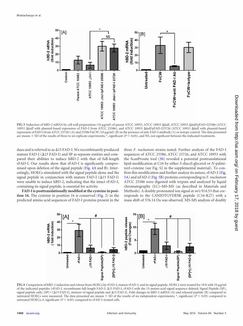
dues and is referred to as �15 FAD-I. We recombinantly producedmature FAD-I (�15 FAD-I) and SP as separate entities and com-pared their abilities to induce hBD-2 with that of full-lengthrFAD-I. Our results show that rFAD-I is significantly compro-mised upon deletion of the signal peptide (Fig. 4A and B). Inter-estingly, HOECs stimulated with the signal peptide alone and thesignal peptide in conjunction with mature FAD-I (�15 FAD-I)were unable to induce hBD-2, indicating that the intact rFAD-I,containing its signal peptide, is essential for activity.
FAD-I is posttranslationally modified at the cysteine in posi-tion 16. The cysteine in position 16 is conserved (Fig. 2) in thepredicted amino acid sequences of FAD-I proteins present in the
three F. nucleatum strains tested. Further analysis of the FAD-Isequences of ATCC 25586, ATCC 23726, and ATCC 10953 withthe ScanProsite tool (30) revealed a potential posttranslationallipid modification at C16 by either S-diacyl-glycerol or N-palmi-toyl-cysteine (see Fig. S2 in the supplemental material). To con-firm this modification and further analyze its nature, rFAD-I (Fig.5A) and nFAD-I (Fig. 5B) proteins corresponding to F. nucleatumATCC 25586 were digested with trypsin and analyzed by liquidchromatography (LC)–MS-MS (as described in Materials andMethods). A doubly protonated ion signal at m/z 914.53 that cor-responds to the CANIDTGVDESK peptide (C16-K27) with amass shift of 576.14 Da was observed. MS-MS analysis of doubly
FIG 3 Induction of hBD-2 mRNA by cell wall preparations (10 �g/ml) of parent ATCC 10953, ATCC 10953 �fadI, ATCC 10953 �fadI/pFAD-I25586 (ATCC10953 �fadI with plasmid-based expression of FAD-I from ATCC 25586), and ATCC 10953 �fadI/pFAD-I23726 (ATCC 10953 �fadI with plasmid-basedexpression of FAD-I from ATCC 23726) (A) and 25586 FnCW (10 �g/ml) (B) in the presence of anti-FAD-I antibody (1) or isotype control. The data presentedare means � SD of the results of three to six replicate experiments; *, significant (P 0.05), and NS, not significant between the indicated treatments.
FIG 4 Comparison of hBD-2 induction and release from HOECs by rFAD-I, mature rFAD-I, and its signal peptide. HOECs were treated for 18 h with 10 �g/mlof the indicated peptides {rFAD-I, recombinant full-length FAD-I; �15 FAD-I, rFAD-I with the 15-amino-acid signal sequence deleted; Signal Peptide (SP),signal peptide only; [SP]�[�15 FAD-I], mixture of signal peptide and �15 FAD-I}. Fold changes in hBD-2 mRNA (A) and released peptide (B) compared tountreated HOECs were measured. The data presented are means � SD of the results of six independent experiments; *, significant (P 0.05) compared tountreated HOECs; #, significant (P 0.05) compared to rFAD-I-treated cells.
Bhattacharyya et al.
1450 iai.asm.org May 2016 Volume 84 Number 5Infection and Immunity
on February 17, 2018 by guest
http://iai.asm.org/
Dow
nloaded from
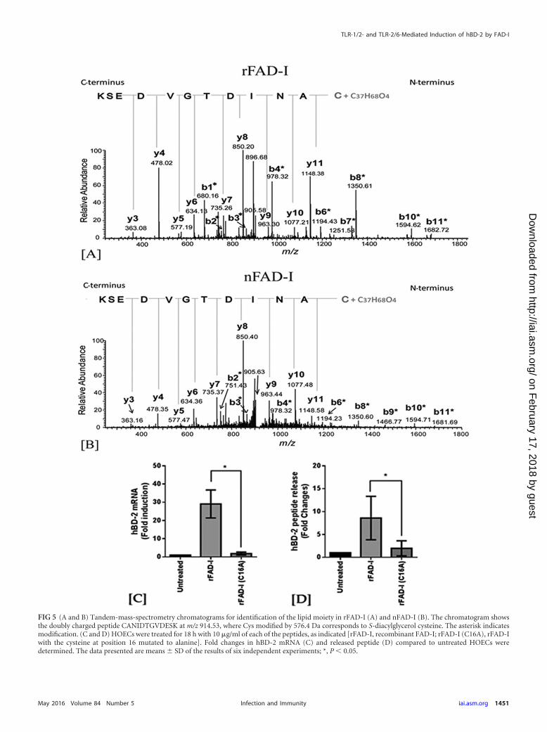
FIG 5 (A and B) Tandem-mass-spectrometry chromatograms for identification of the lipid moiety in rFAD-I (A) and nFAD-I (B). The chromatogram showsthe doubly charged peptide CANIDTGVDESK at m/z 914.53, where Cys modified by 576.4 Da corresponds to S-diacylglycerol cysteine. The asterisk indicatesmodification. (C and D) HOECs were treated for 18 h with 10 �g/ml of each of the peptides, as indicated [rFAD-I, recombinant FAD-I; rFAD-I (C16A), rFAD-Iwith the cysteine at position 16 mutated to alanine]. Fold changes in hBD-2 mRNA (C) and released peptide (D) compared to untreated HOECs weredetermined. The data presented are means � SD of the results of six independent experiments; *, P 0.05.
TLR-1/2- and TLR-2/6-Mediated Induction of hBD-2 by FAD-I
May 2016 Volume 84 Number 5 iai.asm.org 1451Infection and Immunity
on February 17, 2018 by guest
http://iai.asm.org/
Dow
nloaded from
protonated ion signals at m/z 914.53 derived from the FAD-I tryp-tic digest produced a spectrum in which all the observed b-frag-ment ions, including b1 to b11 ions, were shifted by �576.14 Da.In contrast, the observed y-fragment ions, including y1 to y11ions, were unchanged. The shifting of the b1 to b11 ions but notthe y1 to y11 ions by 576.4 Da is indicative of S-diacyl-glycerolmodification at the cysteine residue of the CANIDTGVDESK pep-tide (i.e., the cysteine at position 16 of the full-length FAD-I pep-tide).
To investigate the importance of the lipidated C16 in hBD-2induction, the cysteine was replaced by alanine (A) in rFAD-I.This significantly reduced the ability of FAD-I to induce hBD-2,indicating the importance of the cysteine-diacylglycerol modifica-tion in hBD-2 induction (Fig. 5C and D).
FAD-I-mediated hBD-2 induction is TLR-1/2 and TLR-2/6dependent. Previously, we reported that the induction of hBD-2by FAD-I is TLR-2 dependent (1). Here, we investigated if thisTLR dependence included heterodimerization of TLR-2 with ei-ther TLR-1 or TLR-6. When we treated HOECs with FAD-I in thepresence or absence of anti-TLR-1 or anti-TLR-6 antibody, weobserved significantly diminished hBD-2 induction, indicatingthat TLR-2 interaction with both TLR-1 and TLR-6 is involved ininduction of hBD-2 by FAD-I in HOECs (Fig. 6A).
To further investigate this observation, we conducted flow cy-tometric analysis to reveal the presence or absence of TLR recep-tors on HOEC surfaces after FAD-I challenge. Representative flowcytometric analysis demonstrated that both TLR-1/2 and TLR-2/6are reduced on the surfaces of HOECs following FAD-I challenge,with more surface expression reduction of TLR-1 than of TLR-6(Fig. 6B and D). Additionally, while both TLR-1 and TLR-6 wereinternalized in response to FAD-I, there was a greater percentageof TLR-1 internalization than of TLR-6 compared to untreatedcells (Fig. 6B and C).
The FAD-I–TLR-2 interaction was additionally investigatedusing CytD, an inhibitor of actin polymerization, to see if blockingof receptor internalization can inhibit the reduction in TLR-2 sur-face expression in the presence of FAD-I. HOECs in the presenceof CytD showed little change in TLR2 surface expression afterchallenge with FAD-I compared to FAD-I-treated HOECs alone(Fig. 6D). This was confirmed by using FITC-labeled rFAD-I inthe presence and absence of CytD (Fig. 6E and F).
DISCUSSION
Previously, the cell wall of F. nucleatum, a ubiquitous bacterium ofthe oral cavity, was shown to induce hBD-2 in HOECs (5). Morerecently, using systematic biochemical fractionation of FnCW, weidentified FAD-I as the hBD-2-inducing factor. This was furtherconfirmed via functional heterologous expression of FAD-I in P.gingivalis (1). In our present study, we report differential induc-tion of hBD-2 by different strains of F. nucleatum; ATCC 25586and ATCC 23726 trigger significantly more hBD-2 mRNA pro-duction than ATCC 10953. ATCC 25586 and ATCC 23726 aretherefore designated high inducers, while ATCC 10953 is desig-nated a low inducer of hBD-2. Via heterologous expression of fadIfrom highly hBD-2-inducing strains in a �fadI derivative of thelow-hBD-2-inducing strain ATCC 10953, we were able to restoreFAD-I induction in the low inducer to levels comparable to thoseobserved for the highly inducing strains.
In addition, by using antibodies specifically targeting FAD-I,we inhibited the high-inducer cell wall fraction from inducing
hBD-2 by more than 80%. These two independent approachessupport our contention that FAD-I is the principal cell wall-asso-ciated fusobacterial agent responsible for hBD-2 induction inHOECs.
Our mass spectroscopy analysis and replacement of cysteine(C16) with alanine revealed the importance of the posttransla-tional C16 diacylglycerol moiety in FAD-I in promoting hBD-2expression in HOECs. Additionally, our in silico analysis revealedthat all three F. nucleatum strains expressed a diacylglycerol moi-ety bound to cysteine in position 16. Therefore, that alone couldnot explain the strain-related differences in hBD-2 induction.
The Western immunoblot data using whole-cell lysates of F.nucleatum hBD2 low- and high-inducer strains showed reducedFAD-I expression in the whole-cell lysates of the low inducer(ATCC 10953) compared to the high inducers (ATCC 25586 andATCC 23726), which was even more apparent when we used cellwall fractions for these strains (see Fig. S3 in the supplementalmaterial). The mechanism of protein secretion in eukaryotes andprokaryotes requires the signal sequence to facilitate transport ofthe translated protein to the membrane (31), and it also helps theprotein attain the right conformation required to maintain itsfunctionality (32). The fact that the major sequence differencebetween the FAD-I of ATCC 10953 and those of the other twostrains is in the signal peptide domain of FAD-I (Fig. 2; see Fig. S2in the supplemental material) suggests that the variability in levelsof FAD-I production and cell wall integration in ATCC 10953compared to ATCC 25586 and ATCC 23726 could be due to vari-ability in the efficiency of FAD-I signal peptide anchoring to theouter membranes of the respective strains. However, we cannotrule out the possibility that a conformational difference, as dem-onstrated by amino acid sequence differences in the mature-pep-tide region of FAD-I proteins from the hBD2 low- and high-in-ducer strains could also contribute to variability in hBD-2induction in HOECs. The theoretically predicted structures ofFAD-I proteins from ATCC 10953 and ATCC 25586 based onI-TASSER (iterative threading assembly refinement) (33, 34)showed differences in the residues predicted to interact with theTLRs (data not shown). Therefore, differences in FAD-I presen-tation from its cognate receptors, along with differences in levelsof expression of FAD-I on the outer membranes of F. nucleatumstrains, may collectively be responsible for variability in hBD-2induction in HOECs. Deciphering this intriguing hypothesis willrequire additional work, which is under way in our laboratory.
Purified FAD-I proteins, resolved under denaturing condi-tions, resulted in two prominent bands, i.e., at 14 kDa and 12 kDa,as detected by Coomassie staining and Western blotting (see Fig.S4 in the supplemental material), indicating the presence of full-length and mature peptide in rFAD-I. Another F. nucleatum out-er-membrane-associated protein, FadA, which has been shown toplay an important role in binding and invasion of host cells by theorganism, also exhibits a premature and a mature peptide whenresolved by SDS-PAGE. The amino acid sequence analysis ofFAD-I indicated the presence of a 15-amino-acid signal peptide.Challenging HOECs with recombinantly produced matureFAD-I, i.e., without the signal peptide (�15 FAD-I), significantlyreduced its activity, and adding the signal peptide to the assaymixture in the presence of the mature peptide did not restoreFAD-I activity to an appreciable extent. This suggests that thesignal sequence and the mature peptide must be bound as a singleunit to maintain conformational integrity for FAD-I activity when
Bhattacharyya et al.
1452 iai.asm.org May 2016 Volume 84 Number 5Infection and Immunity
on February 17, 2018 by guest
http://iai.asm.org/
Dow
nloaded from
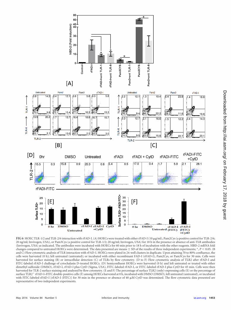
FIG 6 HOEC TLR-1/2 and TLR-2/6 interaction with rFAD-I. (A) HOECs were treated with either rFAD-I (10 �g/ml), Pam2Cys (a positive control for TLR-2/6;20 ng/ml; Invivogen, USA), or Pam3Cys (a positive control for TLR-1/2; 20 ng/ml; Invivogen, USA) for 18 h in the presence or absence of anti-TLR antibodies(Invivogen, USA) as indicated. The antibodies were incubated with HOECs for 60 min prior to 18 h of incubation with the other reagents. HBD-2 mRNA foldchanges compared to untreated HOECs were determined. The data presented are means � SD of the results of three independent experiments; *, P 0.05. (Band C) Flow cytometric analysis of TLR interaction with rFAD-I. HOECs were plated in 24-well clusters in duplicate. Upon attaining 70 to 80% confluence, thecells were harvested (0 h); left untreated (untreated); or incubated with either recombinant FAD-I (rFAD-I), Pam2Cys, or Pam3Cys for 30 min. Cells wereharvested for surface staining (B) or intracellular detection (C) of TLRs by flow cytometry. (D to F) Flow cytometric analysis of TLR2 after rFAD-I andFITC-labeled rFAD-I challenge of cytochalasin D-treated HOECs. (D) Semiconfluent HOECs were harvested (0 h) and left untreated or treated with eitherdimethyl sulfoxide (DMSO), rFAD-I, rFAD-I plus CytD (Sigma, USA), FITC-labeled rFAD-I, or FITC-labeled rFAD-I plus CytD for 45 min. Cells were thenharvested for TLR-2 surface staining and analyzed by flow cytometry. (E and F) The percentage of surface TLR2 (only)-expressing cells (E) or the percentage ofsurface TLR2� rFAD-I–FITC double-positive cells (F) among HOECs harvested at 0 h, incubated with DMSO (DMSO), left untreated (untreated), or incubatedwith FITC-labeled rFAD-I [rFAD-I (FITC)] for 30 min in the presence or absence of 40 �M CytD was determined. The flow cytometric data presented arerepresentative of two independent experiments.
May 2016 Volume 84 Number 5 iai.asm.org 1453Infection and Immunity
on February 17, 2018 by guest
http://iai.asm.org/
Dow
nloaded from
presented to HOECs as a standalone molecule. However, this maynot be relevant in vivo when F. nucleatum comes in contact withHOECs, since the outer membrane, in which FAD-I would beembedded, would provide conformational stability to the mole-cule. According to Mascioni et al. (35), the amino terminus ofbacterial lipoproteins may not only function to direct outer mem-brane localization, but also serve as an extended linker sequence,making the protein available at the extracellular surface.
Our current results show that nFAD-I and rFAD-I behave sim-ilarly in their ability to induce hBD-2. Mass spectrometric analysisdemonstrated the presence of a PTM associated with the onlycysteine in the molecule in both proteins. Interestingly, rFAD-Igenerated in E. coli retained the same posttranslational modifica-tion as nFAD-I, which we isolated from F. nucleatum, i.e., diacyl-glycerol and not triacylglycerol. The classical lipoprotein modifi-cation pathway in bacteria is composed of three sequentialenzymatic reactions catalyzed by two acetyltransferases and onesignal peptidase (36). The enzymes lipoprotein diacylglyceryltransferase (Lgt) and signal peptidase II (Lsp) are involved in pro-ducing diacylated lipoproteins (36), while triacylated lipoproteinsare the result of additional catalysis steps by apolipoprotein N-acyltransferase (Lnt) (36, 37). We searched for these enzymes inthe F. nucleatum ATTC 25586 genome database (38) and foundLgt (GeneID 991758; gene symbol, FN0489) and Lsp (GeneID991403; gene symbol, FN0068) homologues, but not lnt, consis-tent with our finding that FAD-I is diacylated and not triacylated.Gram-negative bacteria are known to have lnt, which promotestriacylated-lipoprotein generation (39). E. coli, like other Gram-negative bacteria, expresses all three enzymes; however, in thegeneration of lipoproteins, lnt causes diacylated moieties to be-come triacylated, as it is the default enzyme that adds an acyl groupto the N terminus of the diacylated molecule (36, 39). Thus, thelipid modification of FAD-I appears to be unique, as it is a diacylpeptide produced by a Gram-negative bacterium (F. nucleatum).It is also surprising that even when produced in E. coli it is diacy-lated, in spite of the presence of machinery for triacylation. Wecould speculate that there could be a specific recognition motifthat determines the number of acyl groups added to the lipopro-tein; however, while this opens up a new avenue for further inves-tigation, it is beyond the scope of the present study.
The lipid moiety of a bacterial lipoprotein plays a crucial role inbacterium-host cell interactions. For example, lipidated peptidesare discriminated by host TLRs on the basis of the degree of fattyacid acylation. Among the TLRs, TLR-2 plays a major role in therecognition of Gram-positive bacteria (40). Biochemical studiesusing cell wall component-deficient mutant bacteria demon-strated that bacterial lipoproteins, but not lipoteichoic acid orpeptidoglycan, act as real native TLR-2 ligand molecules (41–43).Only the lipoproteins are real TLR-2 ligands, and the others maycontain lipoproteins as contaminants during their preparation(44). In the present report, we show that FAD-I is a diacylatedlipopeptide, and previously, we demonstrated that it induceshBD-2 in HOECs through TLR-2 (1). TLR-2 is unique in its abilityto form heterodimer complexes with TLR-1 or TLR-6. Triacyl anddiacyl synthetic lipopeptides, such as N-palmitoyl-S-dipalmitoyl-glyceryl CSK4 and MALP-2, have been used as TLR-1/2 and TLR-2/6 agonists, respectively, leading to a model in which triacylatedlipopeptides/lipoproteins activate through the TLR-1/2 het-erodimer, whereas diacylated lipopeptides/lipoproteins activatethrough the TLR-2/6 heterodimer (45–47). Here, we report that
induction of hBD-2 by diacylated FAD-I induces hBD-2 throughboth TLR-1/2 and TLR-2/6 in HOECs. Triacylated SitC lipopro-tein purified from Staphylococcus aureus cells has been shown tostimulate immune cells via both TLR-1/2 and TLR-2/6 het-erodimers (43), and some diacylated bacterial lipopeptides canalso activate cells in a TLR-6-independent manner (48, 49).
In the context of induction of innate response elements in hu-man oral mucosa, we envision that variability in hBD-2 inductioncould be the result of both the FAD-I expression level and confor-mation differences, as demonstrated by low- and high-inducer F.nucleatum strains, along with interpersonal variability in TLR lev-els of expression in oral mucosal cells (50).
Teleologically, bacterial molecules such as FAD-I, from variousoral bacterial species, could be contributing to the regulation ofhomeostasis in the oral mucosa and thus explain why hBD-2,while inducible, is constitutively expressed in the oral mucosawith little concomitant inflammation. Interestingly, hBD-2 ap-pears only in the presence of infection or inflammation in mosttissues, including the skin (51), trachea (52), and gut epithelium(53, 54), sites where F. nucleatum is not a normal inhabitant. Fur-thermore, we have noticed that FAD-I does not induce interleukin8 (IL-8) in HOECs (data not shown). Therefore, the interaction ofFAD-I with the host mucosal epithelium, which results in expres-sion of antimicrobials, such as hBD-2 (1) and CCL20 (10), with-out simultaneously triggering inflammatory mediator(s), may re-flect an inherent strategy of symbiosis between certain bacteriaand the host mucosa. The growing problem of resistance to con-ventional antibiotics and the need for new antimicrobials hasstimulated interest in the development of antimicrobial peptides(AMPs), such as hBDs, as human therapeutics. Unlike conven-tional antibiotics, resistance by an organism to AMPs is surpris-ingly rare and difficult to generate (55–57). This is probably due tothe nonspecific nature of the electrostatic/hydrophobic interac-tion of the AMP with various anionic components of the bacterialmembrane (55, 56). We hypothesize that a new class of thera-peutics could be developed that would facilitate the productionof endogenous AMPs when needed and locally where applied.Identification and characterization of bacterial molecules, suchas FAD-I, including understanding their interactions with thehost, may one day offer a new paradigm in immunoregulatorytherapeutics to promote mucosal protection at vulnerablebody sites.
ACKNOWLEDGMENTS
This work was supported by NIH/NIDCR grants R01 DE018276 (A.W.)and RO1 DE021108 (R.L.).
We thank J. R. Blakemore, E. K. Schneider, W. S. Blood, S. Alperin,and F. Faddoul for providing normal human oral tissue.
A.W., T.S.M., R.L., and S.K.G. conceived the study. A.W., S.K.G,T.S.M., R.L., and P.P. analyzed and interpreted data. S.K.G. and A.W.wrote and prepared the manuscript. T.S.M. and R.L. edited the manu-script. S.B. purified nFAD-I and different mutants of rFAD-I and per-formed cell cultures, qPCR, ELISA, and Western immunoblotting. B.S.generated different mutants of F. nucleatum. S.N. and A.Y. developed theprotocol and generated rFAD-I. J.K. performed mass spectrophotometricanalysis of FAD-I. B.E. isolated primary human oral epithelial cells. P.P.performed flow cytometry for TLR internalization.
We declare that we have no conflicts of interest with the contents ofthis article.
Bhattacharyya et al.
1454 iai.asm.org May 2016 Volume 84 Number 5Infection and Immunity
on February 17, 2018 by guest
http://iai.asm.org/
Dow
nloaded from
FUNDING INFORMATIONThis work, including the efforts of Aaron Weinberg, was funded by HHS| NIH | National Institute of Dental and Craniofacial Research (NIDCR)(R01 DE018276). This work, including the efforts of Renate Lux, wasfunded by HHS | NIH | National Institute of Dental and CraniofacialResearch (NIDCR) (RO1 DE021108).
REFERENCES1. Gupta S, Ghosh SK, Scott ME, Bainbridge B, Jiang B, Lamont RJ,
McCormick TS, Weinberg A. 2010. Fusobacterium nucleatum-associated beta-defensin inducer (FAD-I): identification, isolation, andfunctional evaluation. J Biol Chem 285:36523–36531. http://dx.doi.org/10.1074/jbc.M110.133140.
2. Gomes PDS, Fernandes MH. 2010. Defensins in the oral cavity: distribu-tion and biological role. J Oral Pathol Med 39:1–9. http://dx.doi.org/10.1111/j.1600-0714.2009.00832.x.
3. Gursoy UK, Könönen E. 2012. Understanding the roles of gingival beta-defensins. J Oral Microbiol 2012:4. http://dx.doi.org/10.3402/jom.v4i0.15127.
4. Hancock RE, Sahl HG. 2006. Antimicrobial and host-defense peptides asnew anti-infective therapeutic strategies. Nat Biotechnol 24:1551–1557.http://dx.doi.org/10.1038/nbt1267.
5. Krisanaprakornkit S, Kimball JR, Weinberg A, Darveau RP, BainbridgeBW, Dale BA. 2000. Inducible expression of human beta-defensin 2 byFusobacterium nucleatum in oral epithelial cells: multiple signaling path-ways and role of commensal bacteria in innate immunity and the epithelialbarrier. Infect Immun 68:2907–2915. http://dx.doi.org/10.1128/IAI.68.5.2907-2915.2000.
6. Dale BA, Krisanaprakornkit S. 2001. Defensin antimicrobial peptides inthe oral cavity. J Oral Pathol Med 30:321–327. http://dx.doi.org/10.1034/j.1600-0714.2001.300601.x.
7. Ji S, Shin JE, Kim YS, Oh JE, Min BM, Choi Y. 2009. Toll-like receptor2 and NALP2 mediate induction of human beta-defensins by fusobacte-rium nucleatum in gingival epithelial cells. Infect Immun 77:1044 –1052.http://dx.doi.org/10.1128/IAI.00449-08.
8. Dommisch H, Reinartz M, Backhaus T, Deschner J, Chung W, JepsenS. 2012. Antimicrobial responses of primary gingival cells to Porphy-romonas gingivalis. J Clin Periodontol 39:913–922. http://dx.doi.org/10.1111/j.1600-051X.2012.01933.x.
9. Ghosh SK, Gerken TA, Schneider KM, Feng Z, McCormick TS, Wein-berg A. 2007. Quantification of human beta-defensin-2 and -3 in bodyfluids: application for studies of innate immunity. Clin Chem 53:757–765.http://dx.doi.org/10.1373/clinchem.2006.081430.
10. Ghosh SK, Gupta S, Jiang B, Weinberg A. 2011. Fusobacterium nuclea-tum and human beta-defensins modulate the release of antimicrobialchemokine CCL20/macrophage inflammatory protein 3 . Infect Immun79:4578 – 4587. http://dx.doi.org/10.1128/IAI.05586-11.
11. Jin G, Kawsar HI, Hirsch SA, Zeng C, Jia X, Feng Z, Ghosh SK, ZhengQY, Zhou A, McIntyre TM, Weinberg A. 2010. An antimicrobial peptideregulates tumor-associated macrophage trafficking via the chemokine re-ceptor CCR2, a model for tumorigenesis. PLoS One 5:e10993. http://dx.doi.org/10.1371/journal.pone.0010993.
12. Kawsar HI, Weinberg A, Hirsch SA, Venizelos A, Howell S, Jiang B, JinG. 2009. Overexpression of human beta-defensin-3 in oral dysplasia: po-tential role in macrophage trafficking. Oral Oncol 45:696 –702. http://dx.doi.org/10.1016/j.oraloncology.2008.10.016.
13. Doss M, White MR, Tecle T, Hartshorn KL. 2010. Human defensins andLL-37 in mucosal immunity. J Leukoc Biol 87:79 –92. http://dx.doi.org/10.1189/jlb.0609382.
14. Feng Z, Jia X, Adams MD, Ghosh SK, Bonomo RA, Weinberg A.2014. Epithelial innate immune response to Acinetobacter baumanniichallenge. Infect Immun 82:4458 – 4465. http://dx.doi.org/10.1128/IAI.01897-14.
15. Ganz T. 2003. Defensins: antimicrobial peptides of innate immunity. NatRev Immunol 3:710 –720. http://dx.doi.org/10.1038/nri1180.
16. Kimball JR, Nittayananta W, Klausner M, Chung WO, Dale BA. 2006.Antimicrobial barrier of an in vitro oral epithelial model. Arch Oral Biol51:775–783. http://dx.doi.org/10.1016/j.archoralbio.2006.05.007.
17. Yin L, Dale BA. 2007. Activation of protective responses in oral epithelialcells by Fusobacterium nucleatum and human beta-defensin-2. J MedMicrobiol 56:976 –987. http://dx.doi.org/10.1099/jmm.0.47198-0.
18. Pazgier M, Prahl A, Hoover DM, Lubkowski J. 2007. Studies of the
biological properties of human beta-defensin 1. J Biol Chem 282:1819 –1829. http://dx.doi.org/10.1074/jbc.M607210200.
19. Harder J, Gläser R, Schröder JM. 2007. Human antimicrobial proteins:effectors of innate immunity. J Endotoxin Res 13:317–338. http://dx.doi.org/10.1177/0968051907088275.
20. Weinberg A, Jin G, Sieg S, McCormick TS. 2012. The yin and yang ofhuman beta-defensins in health and disease. Front Immunol 3:294. http://dx.doi.org/10.3389/fimmu.2012.00294.
21. Kolenbrander PE, Palmer RJ, Jr, Periasamy S, Jakubovics NS. 2010.Oral multispecies biofilm development and the key role of cell-cell dis-tance. Nat Rev Microbiol 8:471– 480. http://dx.doi.org/10.1038/nrmicro2381.
22. Kolenbrander PE. 2011. Multispecies communities: interspecies interac-tions influence growth on saliva as sole nutritional source. Int J Oral Sci3:49 –54. http://dx.doi.org/10.4248/IJOS11025.
23. Krisanaprakornkit S, Kimball JR, Dale BA. 2002. Regulation of humanbeta-defensin-2 in gingival epithelial cells: the involvement of mitogen-activated protein kinase pathways, but not the NF-kappaB transcriptionfactor family. J Immunol 168:316 –324. http://dx.doi.org/10.4049/jimmunol.168.1.316.
24. Oda D, Watson E. 1990. Human oral epithelial cell culture. I. Improvedconditions for reproducible culture in serum-free medium. In Vitro CellDev Biol 26:589 –595.
25. Haake SK, Yoder SC, Attarian G, Podkaminer K. 2000. Native plasmidsof Fusobacterium nucleatum: characterization and use in development ofgenetic systems. J Bacteriol 182:1176 –1180. http://dx.doi.org/10.1128/JB.182.4.1176-1180.2000.
26. Shevchuk NA, Bryksin AV, Nusinovich YA, Cabello FC, Sutherland M,Ladisch S. 2004. Construction of long DNA molecules using long PCR-based fusion of several fragments simultaneously. Nucleic Acids Res 32:e19. http://dx.doi.org/10.1093/nar/gnh014.
27. Kaplan A, Kaplan CW, He X, McHardy IH, Shi W, Lux R. 2014.Characterization of aid1, a novel gene involved in Fusobacterium nuclea-tum interspecies interactions. Microb Ecol 68:379 –387. http://dx.doi.org/10.1007/s00248-014-0400-y.
28. Kennell W, Holt SC. 1990. Comparative studies of the outer membranesof Bacteroides gingivalis, strains ATCC 33277, W50, W83, 381. Oral Mi-crobiol Immunol 5:121–130. http://dx.doi.org/10.1111/j.1399-302X.1990.tb00409.x.
29. Rozen S, Skaletsky H. 2000. Primer3 on the WWW for general users andfor biologist programmers. Methods Mol Biol 132:365–386.
30. de Castro E, Sigrist CJ, Gattiker A, Bulliard V, Langendijk-GenevauxPS, Gasteiger E, Bairoch A, Hulo N. 2006. ScanProsite: detection ofPROSITE signature matches and ProRule-associated functional andstructural residues in proteins. Nucleic Acids Res 34:W362–W365. http://dx.doi.org/10.1093/nar/gkl124.
31. Emr SD, Silhavy TJ. 1983. Importance of secondary structure in the signalsequence for protein secretion. Proc Natl Acad Sci U S A 80:4599 – 4603.http://dx.doi.org/10.1073/pnas.80.15.4599.
32. Singh P, Sharma L, Kulothungan SR, Adkar BV, Prajapati RS, Ali PS,Krishnan B, Varadarajan R. 2013. Effect of signal peptide on stability andfolding of Escherichia coli thioredoxin. PLoS One 8:e63442. http://dx.doi.org/10.1371/journal.pone.0063442.
33. Roy A, Kucukural A, Zhang Y. 2010. I-TASSER: a unified platform forautomated protein structure and function prediction. Nat Protoc 5:725–738. http://dx.doi.org/10.1038/nprot.2010.5.
34. Zhang Y. 2008. I-TASSER server for protein 3D structure prediction.BMC Bioinformatics 9:40. http://dx.doi.org/10.1186/1471-2105-9-40.
35. Mascioni A, Bentley BE, Camarda R, Dilts DA, Fink P, Gusarova V,Hoiseth SK, Jacob J, Lin SL, Malakian K, McNeil LK, Mininni T, MoyF, Murphy E, Novikova E, Sigethy S, Wen Y, Zlotnick GW, Tsao DH.2009. Structural basis for the immunogenic properties of the meningococ-cal vaccine candidate LP2086. J Biol Chem 284:8738 – 8746. http://dx.doi.org/10.1074/jbc.M808831200.
36. Kovacs-Simon A, Titball RW, Michell SL. 2011. Lipoproteins of bacterialpathogens. Infect Immun 79:548 –561. http://dx.doi.org/10.1128/IAI.00682-10.
37. Tokuda H. 2009. Biogenesis of outer membranes in Gram-negative bac-teria. Biosci Biotechnol Biochem 73:465– 473. http://dx.doi.org/10.1271/bbb.80778.
38. Kapatral V, Anderson I, Ivanova N, Reznik G, Los T, Lykidis A,Bhattacharyya A, Bartman A, Gardner W, Grechkin G, Zhu L, VasievaO, Chu L, Kogan Y, Chaga O, Goltsman E, Bernal A, Larsen N, D’Souza
TLR-1/2- and TLR-2/6-Mediated Induction of hBD-2 by FAD-I
May 2016 Volume 84 Number 5 iai.asm.org 1455Infection and Immunity
on February 17, 2018 by guest
http://iai.asm.org/
Dow
nloaded from
M, Walunas T, Pusch G, Haselkorn R, Fonstein M, Kyrpides N, Over-beek R. 2002. Genome sequence and analysis of the oral bacterium Fuso-bacterium nucleatum strain ATCC 25586. J Bacteriol 184:2005–2018.http://dx.doi.org/10.1128/JB.184.7.2005-2018.2002.
39. Tschumi A, Nai C, Auchli Y, Hunziker P, Gehrig P, Keller P, Grau T,Sander P. 2009. Identification of apolipoprotein N-acyltransferase (Lnt)in mycobacteria. J Biol Chem 284:27146 –27156. http://dx.doi.org/10.1074/jbc.M109.022715.
40. Takeuchi O, Hoshino K, Kawai T, Sanjo H, Takada H, Ogawa T,Takeda K, Akira S. 1999. Differential roles of TLR2 and TLR4 inrecognition of gram-negative and gram-positive bacterial cell wallcomponents. Immunity 11:443– 451. http://dx.doi.org/10.1016/S1074-7613(00)80119-3.
41. Hashimoto M, Tawaratsumida K, Kariya H, Aoyama K, Tamura T,Suda Y. 2006. Lipoprotein is a predominant Toll-like receptor 2 ligand inStaphylococcus aureus cell wall components. Int Immunol 18:355–362.
42. Hashimoto M, Tawaratsumida K, Kariya H, Kiyohara A, Suda Y,Krikae F, Kirikae T, Götz F. 2006. Not lipoteichoic acid but lipoproteinsappear to be the dominant immunobiologically active compounds inStaphylococcus aureus. J Immunol 177:3162–3169. http://dx.doi.org/10.4049/jimmunol.177.5.3162.
43. Kurokawa K, Lee H, Roh KB, Asanuma M, Kim YS, Nakayama H,Shiratsuchi A, Choi Y, Takeuchi O, Kang HJ, Dohmae N, Nakanishi Y,Akira S, Sekimizu K, Lee BL. 2009. The triacylated ATP binding clustertransporter substrate-binding lipoprotein of Staphylococcus aureus func-tions as a native ligand for Toll-like receptor 2. J Biol Chem 284:8406 –8411. http://dx.doi.org/10.1074/jbc.M809618200.
44. Nakayama H, Kurokawa K, Lee BL. 2012. Lipoproteins in bacteria:structures and biosynthetic pathways. FEBS J 279:4247– 4268. http://dx.doi.org/10.1111/febs.12041.
45. Takeda K, Takeuchi O, Akira S. 2002. Recognition of lipopeptides byToll-like receptors. J Endotoxin Res 8:459 – 463. http://dx.doi.org/10.1177/09680519020080060101.
46. Takeuchi O, Akira S. 2010. Pattern recognition receptors and inflamma-tion. Cell 140:805– 820. http://dx.doi.org/10.1016/j.cell.2010.01.022.
47. Takeuchi O, Kawai T, Mühlradt PF, Morr M, Radolf JD, Zychlinsky A,Takeda K, Akira S. 2001. Discrimination of bacterial lipoproteins byToll-like receptor 6. Int Immunol 13:933–940. http://dx.doi.org/10.1093/intimm/13.7.933.
48. Buwitt-Beckmann U, Heine H, Wiesmüller KH, Jung G, Brock R, AkiraS, Ulmer AJ. 2005. Toll-like receptor 6-independent signaling by diacy-
lated lipopeptides. Eur J Immunol 35:282–289. http://dx.doi.org/10.1002/eji.200424955.
49. Buwitt-Beckmann U, Heine H, Wiesmüller KH, Jung G, Brock R, AkiraS, Ulmer AJ. 2006. TLR1- and TLR6-independent recognition of bacteriallipopeptides. J Biol Chem 281:9049 –9057. http://dx.doi.org/10.1074/jbc.M512525200.
50. Kinane DF, Shiba H, Stathopoulou PG, Zhao H, Lappin DF, Singh A,Eskan MA, Beckers S, Waigel S, Alpert B, Knudsen TB. 2006. Gingivalepithelial cells heterozygous for Toll-like receptor. Genes Immun 7:190 –200. http://dx.doi.org/10.1038/sj.gene.6364282.
51. Ong PY, Ohtake T, Brandt C, Strickland I, Boguniewicz M, Ganz T,Gallo RL, Leung DY. 2002. Endogenous antimicrobial peptides and skininfections in atopic dermatitis. N Engl J Med 347:1151–1160. http://dx.doi.org/10.1056/NEJMoa021481.
52. Starner TD, Agerberth B, Gudmundsson GH, McCray PB, Jr. 2005.Expression and activity of beta-defensins and LL-37 in the developinghuman lung. J Immunol 174:1608 –1615. http://dx.doi.org/10.4049/jimmunol.174.3.1608.
53. O’Neil DA, Porter EM, Elewaut D, Anderson GM, Eckmann L, Ganz T,Kagnoff MF. 1999. Expression and regulation of the human beta-defensins hBD-1 and hBD-2 in intestinal epithelium. J Immunol 163:6718 – 6724.
54. Wehkamp J, Fellermann K, Herrlinger KR, Baxmann S, Schmidt K,Schwind B, Duchrow M, Wohlschläger C, Feller AC, Stange EF. 2002.Human beta-defensin 2 but not beta-defensin 1 is expressed preferentiallyin colonic mucosa of inflammatory bowel disease. Eur J GastroenterolHepatol 14:745–752. http://dx.doi.org/10.1097/00042737-200207000-00006.
55. Diamond G, Beckloff N, Weinberg A, Kisich KO. 2009. The roles ofantimicrobial peptides in innate host defense. Curr Pharm Des 15:2377–2392. http://dx.doi.org/10.2174/138161209788682325.
56. Zasloff M. 2002. Antimicrobial peptides of multicellular organisms. Na-ture 415:389 –395. http://dx.doi.org/10.1038/415389a.
57. Ge Y, MacDonald DL, Holroyd KJ, Thornsberry C, Wexler H, ZasloffM. 1999. In vitro antibacterial properties of pexiganan, an analog ofmagainin. Antimicrob Agents Chemother 43:782–788.
58. Larkin MA, Blackshields G, Brown NP, Chenna R, McGettigan PA,McWilliam H, Valentin F, Wallace IM, Wilm A, Lopez R, ThompsonJD, Gibson TJ, Higgins DG. 2007. Clustal W and Clustal X version 2.0.Bioinformatics 23:2947–2948. http://dx.doi.org/10.1093/bioinformatics/btm404.
Bhattacharyya et al.
1456 iai.asm.org May 2016 Volume 84 Number 5Infection and Immunity
on February 17, 2018 by guest
http://iai.asm.org/
Dow
nloaded from