Embed Size (px)
Citation preview
Faculty of Resource Science and Technology
Antibiotic Screening of Soil Microorganisms Isolated from Remote Village in Nanga Merit,
Sarawak
Nurul Saiyidah Binti Abdul Rahim (19633)
Bachelor of Science with Honours
(Resource Biotechnology)
2010
DECLARATION
I hereby declare that no portion of this dissertation has been submitted in support of an
application for another degree of qualification of this or any other university or institution of
higher learning.
............................................................
(NURUL SAIYIDAH BINTI ABDUL RAHIM)
Resource Biotechnology Programme
Department of Molecular Biology
Faculty of Resource Science and Technology
Universiti Malaysia Sarawak.
ii
ACKNOWLEDGEMENT
First of all, praise to Allah s.w.t, the Mighty One, for His bless in giving me the strength
and good health to complete this study.
Most appreciation to Prof. Dr. Ismail bin Ahmad for being such a dedicated and
responsible supervisor. Thank you for the knowledge and guidance that has been given
throughout conducting the study. This appreciation is also dedicated to Lab Assistant, Mr.
Zaidi and Mr. Iskandar for providing the equipments and laboratory needs, and thank you
to all MASTER students of Virology Lab for the guidance and advice.
To my dear colleagues and friends, especially Fatin Fatanah Ramle, Munirah Abdul Talip,
Aisha Izam, Nurhamizah Merali, Joan Alicia Joseph Blandoi, Nurulain Mustafa Udin, and
Azyyati Zatil Iman Muhammad, thank you for the support and sharing of information. To
my dear table partner, Chai Sin Lin, lots of thanks for always helping and sharing
knowledge with me. Not to forget, to my beloved family, thank you for the pray and for
always being there when time needed.
iii
Table of Contents
Acknowledgement …………………………………………………………….. ii
Table of Contents ………………………………………………………………iii
List of Abbreviations ………………………………………………………….. v
List of Tables and Figures …………………………………………………….. vi
Abstract ………………………………………………………………………... 1
1.0 Introduction ………………………………………………………………... 3
2.0 Literature Review ………………………………………………………….. 5
2.1 Antibiotics …………………………………………………………. 5
2.2 Antibiotics-Resistant Bacteria ……………………………………... 7
2.2.1 Staphylococcus aureus……………………………………. 8
2.2.2 Methicillin-Resistant Staphylococcus aureus (MRSA) ….. 8
2.3 Antibiotic-Producing Microorganisms …………………………….. 9
2.4 Isolation of Soil Microorganisms ………………………………….. 10
3.0 Material and Methods ………………………………………………………11
3.1 Media Preparation …………………………………………………. 11
3.2 Sample preparation …………………………………………………11
3.3 Isolation of Microorganisms from Soil ...………………………….. 11
3.3.1 Isolation and Subculturing ……………...…………………11
3.3.2 Subculturing of Bacteria and Fungi Colonies ..…………...12
3.4 Preliminary Test of Antibiotic-Producing Bacteria and Fungi …..….12
3.4.1 Preliminary Screening …..………………………………...12
3.4.1 Secondary Screening ……………………………………...13
iv
3.5 Antibiotic Extraction ………………………………………………..14
3.6 Antibiotic Screening for Methanol Extraction ………….………… 14
3.7 Antifungal Test ……………………………………………………..15
3.8 Morphological Characterization ……………………………………16
3.8.1 Macroscopic Observation ……………………………….. 16
3.8.2 Microscopic Observation ………………………………... 16
4.0 Result ……….………………………………………………………………17
4.1 Isolation and Subculturing of Microorganisms from soil ..………... 17
4.2 Preliminary Test of Antibiotic-Producing Bacteria and Fungi ……..18
4.2.1 Preliminary Screening .…...…………………………........ 18
4.2.2 Secondary Screening ……………………………………...19
4.3 Antibiotic Screening for Methanol Extraction ………………………24
4.4 Antifungal Test ……………………………………………………... 27
4.5 Morphological Characterization ……………………………………. 27
5.0 Discussion …………………………………………………………………... 29
5.1 Location of Soil Sampling ………………………………………….. 30
5.2 Isolation and Subculturing of Microorganisms from Soil ………….. 30
5.3 Peliminary and Secondary Screening ………………………………. 32
5.4 Antibiotic Screening of Methanol Extraction ………………………. 33
5.5 Morphological Characterization ……………………………………. 33
6.0 Conclusion and Recommendation ………………………………………….. 35
References …………………………………………………………………….… 36
v
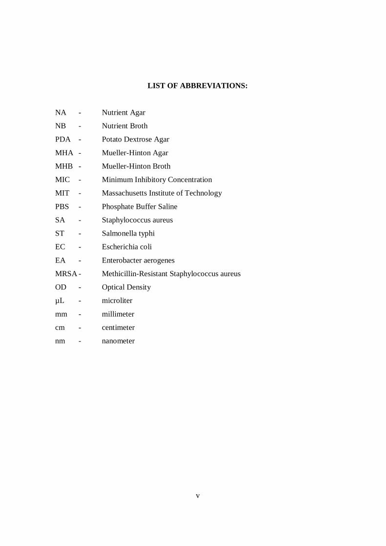
LIST OF ABBREVIATIONS:
NA - Nutrient Agar
NB - Nutrient Broth
PDA - Potato Dextrose Agar
MHA - Mueller-Hinton Agar
MHB - Mueller-Hinton Broth
MIC - Minimum Inhibitory Concentration
MIT - Massachusetts Institute of Technology
PBS - Phosphate Buffer Saline
SA - Staphylococcus aureus
ST - Salmonella typhi
EC - Escherichia coli
EA - Enterobacter aerogenes
MRSA - Methicillin-Resistant Staphylococcus aureus
OD - Optical Density
µL - microliter
mm - millimeter
cm - centimeter
nm - nanometer
vi
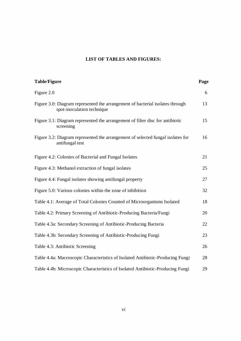
LIST OF TABLES AND FIGURES:
Table/Figure Page
Figure 2.0 6
Figure 3.0: Diagram represented the arrangement of bacterial isolates through 13
spot-inoculation technique
Figure 3.1: Diagram represented the arrangement of filter disc for antibiotic 15
screening
Figure 3.2: Diagram represented the arrangement of selected fungal isolates for 16
antifungal test
Figure 4.2: Colonies of Bacterial and Fungal Isolates 21
Figure 4.3: Methanol extraction of fungal isolates 25
Figure 4.4: Fungal isolates showing antifungal property 27
Figure 5.0: Various colonies within the zone of inhibition 32
Table 4.1: Average of Total Colonies Counted of Microorganisms Isolated 18
Table 4.2: Primary Screening of Antibiotic-Producing Bacteria/Fungi 20
Table 4.3a: Secondary Screening of Antibiotic-Producing Bacteria 22
Table 4.3b: Secondary Screening of Antibiotic-Producing Fungi 23
Table 4.3: Antibiotic Screening 26
Table 4.4a: Macroscopic Characteristics of Isolated Antibiotic-Producing Fungi 28
Table 4.4b: Microscopic Characteristics of Isolated Antibiotic-Producing Fungi 29
ANTIBIOTIC SCREENING OF SOIL MICROORGANISMS ISOLATED FROM
REMOTE VILLAGE AREA IN NANGA MERIT, KAPIT, SARAWAK
NURUL SAIYIDAH BINTI ABDUL RAHIM
This project is submitted in partial fulfillment of the requirement for the degree of
Bachelor of Science with Honours
(Resource Biotechnology)
Faculty of Resource Science and Technology
UNIVERSITI MALAYSIA SARAWAK
2010
1
Antibiotic Screening of Soil Microorganisms Isolated from Remote Village Area in Nanga
Merit, Kapit, Sarawak
Nurul Saiyidah Binti Abdul Rahim
Resource Biotechnology Programme Faculty of Resource Science and Technology
University Malaysia Sarawak
ABSTRACT
A study was conducted to isolate antibiotic-producing microorganisms from soil samples
collected from a remote village in Nanga Merit Kapit, Sarawak. The soil was suspended
and homogenized with phosphate buffer saline (PBS), and then was inoculated on potato
dextrose agar (PDA) or nutrient agar (NA) by spread-plate method. Screening for the
antibiotic-producing activity was accomplished through preliminary and secondary
screening, by using agar overlay technique. These procedures were followed with
antibiotic screening using disc diffusion test. Through preliminary screening, 16 bacterial
and 8 fungi isolates that showed zone of inhibition were selected for secondary screening.
After secondary screening, only six of the fungal isolates were selected for further
experiment which involves extraction of antibiotic by methanol. This procedure involved
drying of agar containing the selected isolates, followed by immersion of the dried agar in
10ml of absolute methanol. Antibiotic screening was then performed to screen antibiotic
activity for the methanol extractions. Result showed that two types of fungal isolates,
respectively coded as L10.1.F3 and L12.1.F1 carried the potential of producing antibiotic
with strong zone of inhibition against test bacteria. Another four fungal isolates, coded as
L11.2.F1, L11.2.F3, L11.2.F4, and L12.1.F1, only showed zone of inhibition on primary
and secondary screening. Probabilities of having insufficient amount of antibiotic extracts
may be the reason for these fungi to showed negative activity throughout the antibiotic
screening. Even so, these finding suggest that soil from a remote village in Nanga Merit
Kapit, Sarawak does contain with antibiotic-producing microorganisms. Isolation of novel
strains of antibiotic-producing microorganisms from this location seems to be possible.
Key words: Antibiotic, antibiotic-producing microorganisms, soil sample
2
ABSTRAK
Kajian untuk memencilkan mikroorganisma penghasil antibiotik daripada sampel tanah
yang diambil dari kawasan pedalaman sebuah perkampungan di Nanga Merit, Kapit,
Sarawak, telah dijalankan. Sampel tanah yang diambil dilarut bersama larutan ‘phosphate
buffer saline’ (PBS) dan diikuti dengan homogenisasi. Kemudian, sedikit daripada larutan
tanah yang dihomogen diinokulasi pada permukaan agar ‘potato dextrose’ (PDA) dan
agar nutrien (NA) melalui kaedah piring sebaran (spread-plate). Penyaringan untuk
mengenal pasti tindakan penghasilan antibiotik dijalankan menerusi saringan awal dan
saringan sekunder dengan menggunakan teknik pelapisan agar. Kemudian, diikuti dengan
saringan antibiotik menerusi ujian penyerapan disk. Sebanyak 16 jenis bakteria dan 8
jenis kulat yang menghasilkan zon perencatan telah dipilih daripada saringan awal untuk
melalui saringan sekunder. Hanya 6 daripada kulat yg dipencil telah dipilih daripada
saring sekunder untuk melalui eksperimen seterusnya yang mana melibatkan proses
pengekstrakan antibiotik menggunakan metanol. Prosedur ini memerlukan agar yang
mengandungi kulat yang dipilih, dikeringkan terlebih dahulu sebelum direndam bersama
10ml 100% metanol. Procedur penyaringan antibiotik dijalankan untuk menyaring
tindakan antibiotik daripada ekstrak metanol. Hasil kajian menunjukkan dua jenis kulat,
masing-masing dilabel sebagai L10.1.F3 dan L12.1.F1, berupaya menghasilkan antibiotik
yang kuat dan mampu merencat tindakan bakteria penguji. Hasil ujian daripada empat
kulat pencilan yang lain masing-masing dilabel dengan L11.2.F1, L11.2.F3, L11.2.F4, dan
L12.1.F2 hanya menunjukkan zon perecatan pada saringan awal dan sekunder. Hal ini
berkemungkinan disebabkan oleh kandungan antibiotik yang diekstrak keluar adalah
kurang. Meski pun begitu, secara keseluruhannya kajian ini menunjukkan bahawa tanah
dari kawasan pedalaman sebuah perkampungan di Nanga Merit, Kapit, Sarawak
mengadungi mikroorganisma penghasil antibiotik, dan tidak mustahil mikroorganisma
jenis baru dan berupaya menghasilkan antibiotik dapat dipencil daripada lokasi ini.
Kata kunci: Antibiotik, mikrooganisma penghasil antibiotik, sampel tanah
3
1.0 INTRODUCTION
The term antibiotic refers to chemical substances that provide the capacity to inhibit the
growth or even destroy bacteria and other microorganisms that cause infectious diseases in
human and animal (Hairston et al., 2000). The discovery of sulfonamide drug and later the
penicillins have led to other discoveries of different antibiotics. Since then, more than 100
varieties of these drugs have been developed by the pharmaceutical industry and are
broadly used until now (Bertrand et al., 2004). Bacteria begin to evolve and gained the
ability to resist the antibiotics‟ effect. Accordingly, a steady decline in the number of
effective antibiotics each year has been reported (Bertrand et al., 2004). Therefore, new
antibiotics are required to be developed. The antibiotics should be not only susceptible to
the resistant strain, but should also be able to replace some effective antibiotics such as
streptomycin which has been abandoned due to its negative side effects (Hairston et al.,
2000).
Staphylococcus aureus are pathogenic bacteria that can infect various tissues in the
body and leading to development of varieties of diseases. The difficulty in controlling S.
aureus infection is related to its ability to acquire resistant to antibiotic as soon as it is
exposed towards those antibiotics (García-Lara, 2005). It is therefore not surprising that the
number of methicillin-resistant S. aureus (MRSA) in hospital acquired infections, has
increased in recent years by 10-15% in countries such as Germany, United Kingdom, and
the United States (García-Lara, 2005). Its impact on health care problem is so serious, that
the antibiotic resistance and virulence in S. aureus is claimed to be one of depressing
evolutionary progression (Deresinski, 2005). This is due to the ability of this strain to
become resistant not only towards methicillin and β-lactams, but also to drugs of last resort
such as vancomycin, linezolid, and daptomycin (Memmi et al., 2008).
4
Earlier studies had found that three unrelated groups of microbes were mostly
involved in the production of natural antibiotics used in medicine. Those are the
Penicillium and Cephalosporium molds, the actinomycetes and the Bacillus species (Todar,
2009). Actinomycetes for example, have become the mainstay of the antibiotics industry.
This group of bacteria species has produces most of the clinically-useful antibiotics that are
not beta-lactams such as tetracyclines, streptomycin, and erythromycin (Todar, 2009). As a
matter of fact, these groups of microbes naturally inhabiting the soil. Therefore the soil
microorganisms from remote village in Nanga Merit, Kapit, Sarawak might provide with
possibilities of isolating novel antibiotics to which the resistant bacteria are susceptible
(Todar, 2009).
OBJECTIVES:-
1. To isolate microorganisms from the soil sample collected from a remote Village in
Nanga Merit, Kapit, Sarawak.
2. To extract antibiotics from the selected microorganism isolates.
3. To determine the susceptibility of the selected test bacteria towards the antibiotic
extracts.
5
2.0 LITERATURE REVIEW
2.1 Antibiotics
Antibiotic is derived from the word anti, which means „against‟ and bios which is referring
to „life‟- since a bacterium is a life forms (Nordqvist, 2009). Therefore, antibiotic was
originally defined as a substance produced by a microorganism, which has the ability to
inhibit the growth of other microorganisms (Russell, 1992). However, due to the
introduction of synthetic methods, the definition of antibiotic has undergone some
modifications. Currently, the definition of antibiotic is referred to a substance produced by
a microorganism, or a chemically similar substance that will either inhibit the growth or
destroy the other pathogenic microorganisms when applied at low concentration (Russel,
1992; Romanowski, 2007).
According to Todar (2009), most of microbiologists have distinguished two groups
of antimicrobial agents that are used for treated infectious diseases. Those that involve
natural substances produced by certain group of microorganisms are known as antibiotics,
while those that are chemically synthesized are referred to chemotherapeutic agents. In
addition to these groups, there is another group which is known as semisynthetic antibiotic.
This group of antibiotic contains molecular part produced by fermentation process using
the appropriate microorganisms and the product is then further modified through a
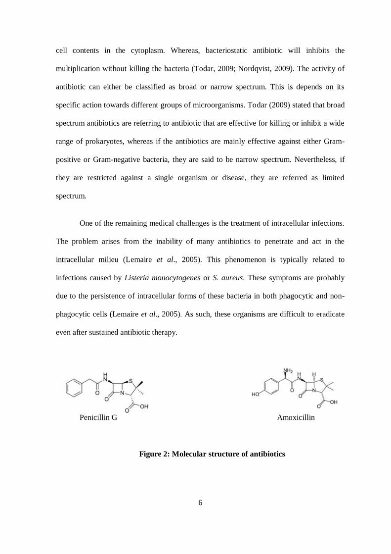
chemical process (Russell, 1992). Amoxicillin is one of the semisynthetic antibiotics in
which the main part of the molecule, 6-aminopenicillanic acid produced by a fungus, is
chemically modified by the addition of side chains (Figure 2) (Todar, 2009).
The mechanism of action of an antibiotic is described via the characteristic of the
antibiotics, either bactericidal or bacteriostatic (Nordqvist, 2009). Bactericidal antibiotic
kills the pathogenic bacteria by interfere with the formation of the bacteria cell wall or the
6
cell contents in the cytoplasm. Whereas, bacteriostatic antibiotic will inhibits the
multiplication without killing the bacteria (Todar, 2009; Nordqvist, 2009). The activity of
antibiotic can either be classified as broad or narrow spectrum. This is depends on its
specific action towards different groups of microorganisms. Todar (2009) stated that broad
spectrum antibiotics are referring to antibiotic that are effective for killing or inhibit a wide
range of prokaryotes, whereas if the antibiotics are mainly effective against either Gram-
positive or Gram-negative bacteria, they are said to be narrow spectrum. Nevertheless, if
they are restricted against a single organism or disease, they are referred as limited
spectrum.
One of the remaining medical challenges is the treatment of intracellular infections.
The problem arises from the inability of many antibiotics to penetrate and act in the
intracellular milieu (Lemaire et al., 2005). This phenomenon is typically related to
infections caused by Listeria monocytogenes or S. aureus. These symptoms are probably
due to the persistence of intracellular forms of these bacteria in both phagocytic and non-
phagocytic cells (Lemaire et al., 2005). As such, these organisms are difficult to eradicate
even after sustained antibiotic therapy.
Penicillin G Amoxicillin
Figure 2: Molecular structure of antibiotics
7
2.2 Antibiotic-Resistant Bacteria
The possibility on the development of antibiotic resistant bacteria had already been
expected by Dr. Alexander Fleming since the time he published his discovery of penicillin
(Roberts, 1998). The increase in usage of antibiotics has brought about the emergence of
bacteria that are resistant to currently used antibiotics. As a result, the numbers of
antibiotics that are effective have begun to decline (Bertrand et al., 2004). The
development of penicillin resistance was indicated through an increase in the MIC value of
the antibiotic. Such observation is related to the alterations in the enzymatic targets of β-
lactam antibiotics which is the penicillin-binding proteins (PBPs) (Tomasz, 1988).
Hughes and Datta, cited by Roberts (1998), had examined a collection of
Escherichia coli isolated at the beginning of the 20th century at which time, antibiotic
therapy was not yet available. Through the examination, they had found that these early
bacteria were not antibiotic resistant; hence they postulated a hypothesis claiming that the
antibiotic resistant E. coli was developed in response to antibiotic use in the last 50 years.
Therefore, it is believed that if disease-causing bacteria are continuously exposed to
antibiotics, there is a high probability that the bacteria will accelerate their adaptation to
these chemicals through evolutionary mutation (Hairston, 2000). This also being supported
by Tomasz (1988), as he stated that “ The prompt emergence of bacterial strains resistant
to these drugs (antibiotics) show that the ingenuity of clinical chemists is more than
matched by the resourcefulness of bacteria in finding counter measures that allow them to
evade the inhibitory effects of these antibiotics”.
8
2.2.1 Staphylococcus aureus
Staphylococcus aureus is a commonly occurring bacterium that resides on the skin and in
the nose of healthy persons (The Federal Bureau of Prison, 2005). This bacterium has the
ability to grow comparatively well under conditions of high osmotic pressure and low
moisture (Tortoro, 2004). Amongst all the Staphylococcus groups, S. aureus is the one that
causes most of infections (Stöppler, 2009). The bacteria gained their pathogenicity via
production of many toxins that increases the ability to invade the body or damage the
tissue (Tortoro, 2004; Stöppler, 2009).
In hospital, the surgical wound infection by S. aureus is a common problem.
However, its ability to quickly develop resistance toward antibiotics such as penicillin has
become a threat to patients in hospital environments (Tortoro, n.d.; García-Lara et al.,
2005). Moreover, the formation of intercellular aggregates and biofilms in which increases
evasion of host defenses and antibiotics tolerance have provide an explanation regarding
the ability of S. aureus to adhere to inert surfaces like medical devices and colonize living
tissue such as cartilage and heart valves (García-Lara et al., 2005).
2.2.2 Methicillin-Resistant Staphylococcus aureus (MRSA)
Methicillin-Resistant Staphylococcus aureus (MRSA) is a strain of S. aureus that is
resistant to methicillin and other β-lactam antibiotics including penicillin, ampicillin,
amoxicillin, and oxacillin (Federal Bureau of Prison, 2005; Stöppler, 2009). As stated by
Stöppler (2009), MRSA infections are usually mild superficial infections of the skin that
can be treated successfully with proper skin care and antibiotics. Nevertheless, due to
insufficient effective antibiotics available, MRSA can be difficult to treat and can progress
to life-threatening blood or bone infections.
9
Currently, bacteria known as vancomycin-intermediate resistant S. aureus (VISA)
and vancomycin-resistant S. aureus (VRSA) have been identified. These bacteria have
developed the strains that are resistant towards antibiotics vancomycin, eventhough this
type of antibiotics is normally effective against Staph infections (Stöppler, 2009).
2.3 Antibiotic-Producing Microorganisms
Acknowledge with the ability of certain fungi and bacteria to produce chemical substances
which inhibits or destroy pathogenic organisms (Waksman, 1952) has supported the fact
that, these unrelated groups of microbes have involved in most of the natural antibiotics
production (Todar, 2009). These substances are hypothesized to confer a selective
advantage to the producer when competition is significant to microbial fitness (Kinkel,
2006). A fungus such as Penicillium chrysogenum is an important industrial organism due
to its ability to produce several types of β-lactam antibiotics. Different antibacterial
properties could be obtained through the substitution of R-group substituent of the
penicillin nucleus (Onyegeme-Okerenta, 2009).
In September 1943, American microbiologist, Selman Waksman and associates
had managed to isolate the first streptomycin-producing microorganism from the soil
(Waksman, 1952). These were then followed with the isolation of other potential different
antibiotics, produced by the same species of Streptomyces such as actinomycin,
streptothrin, and neomycin (Romanowski, 2007). The specialized feature of Streptomyces
which are able to produce more than one antibiotic within a single strain (Hopwood, 1988)
might be the reason for actinomycetes to be the organisms that are recently significant to
antibiotics industry.
10
2.4 Isolation of Soil Microorganisms
As different microbes require different needs as well as different environmental conditions,
therefore it is impossible to devise only a single isolation technique that can be applied on
every type of microbes (Parkinson et al., 1971). One technique involves the isolation from
the soil suspension, by which the soil sample is suspended in medium or diluting fluid
(Parkinson et al., 1971). Besides, there is also method known as soil plate method.
Through this method, it allows the isolation of fungi from a large number of separate soil
samples, without the need of using plenty amount of dilution blanks.
Recently, study involves the isolation of soil-dwelling type bacteria named as
Rhodococcus is being conducted. These bacteria use to be known as non-antibiotics
producer. However, it has currently been discovered to gain the ability of producing
antibiotic that can be utilized for treatment of Helicobacter pylori, which causes stomach
ulcers in humans (Trafton, 2008). Due to this new discovery, it is believes to enhance the
development of strategies for finding other new antibiotics mainly from the soil.
11
3.0 MATERIALS AND METHODS
3.1 Media Preparation
In this study, PDA and NA media were used for growing and culturing the soil
microorganisms. The media were prepared according to the directions of the manufacturer,
DifcoTM
(USA) and OXIOD, CM0003 (England), respectively.
3.2 Sample Preparation
Soil samples were provided by UNIMAS Soil Laboratory. The soil samples were
originally collected from a remote village in Nanga Merit, Kapit, Sarawak. The samples
were obtained from seven different sites with two different depths, which are 0-20 cm and
20-40 cm. The samples were then brought to the laboratory and were air dried at room
temperature. From the original sample, about 10g was randomly sampled and put into
smaller plastic bag. These samples were then stored in the refrigerator at 4°C.
3.3 Isolation of Microorganisms from Soil
3.3.1 Isolation and Subculturing
Firstly, 1g of soil sample was transferred into a Falcon tube. Then the sample was
suspended with 10ml of PBS. After that, the suspension was homogenized by a vortex
machine and was left to stand for 1 hour in order to allow the large particles to settle.
Culturing was done by spread-plate method, in which 100µL of the aliquot was pipetted
onto the petri plates and was then spread by using sterilized cotton swab. For each sample,
there were four replicates of NA and four replicates of PDA. Then, the plates were
incubated for 5 days at room temperature.
12
3.3.2 Subculturing of Bacteria and Fungi Colonies
After 5 days of incubation, bacterial or fungal colonies that showed inhibition towards the
growth of other microorganisms were isolated and subcultured on new agar media to
obtain the pure culture. The plates were then incubated for another 3 days at room
temperature while the rest of the petri plates were transferred into a refrigerator at 4°C
before being subjected to preliminary screening of antibiotic activity.
3.4 Preliminary Test of Antibiotic-Producing Bacteria and Fungi
The preliminary test was conducted in two phases, which involves preliminary screening
and then followed by secondary screening. The agar overlay technique (Nkanga &
Hagedorn, 1978) was applied in both phases. This technique involves the preparation of
homogeneous bacterial lawn within a thin layer of agar across the surface of a plate
(Fankhauser, 2005). The bacterial lawn was prepared by adding appropriate concentration
of bacteria into soft NA (0.75%), followed by homogenizing it. In this study, three Gram
negative bacteria which were E. coli, Salmonella typhi, and Enterobacter aerogenes were
used as the test bacteria, whilst S. aureus was used as Gram positive test bacterium.
3.4.1 Preliminary Screening
Sample plates with the growth of bacterial and fungal isolates were took out from the
refrigerator and allowed to adapt to room temperature. Four replicates of each sample were
labelled as EC, EA, SA, and ST, respectively. Aliquots of 3 ml of both NB and soft NA
were prepared in the bijou bottles. The desired test bacteria were inoculated into NB and
were left at 37°C for an overnight incubation. After 18 hours of incubation, optical density
(OD) of the incubated test bacteria culture was measured and then adjusted to 0.6 OD at
13
520nm. Bijou bottles containing hot melted soft agar (NA), were placed inside water bath
to allow them to cool to 45°C, and the temperature was maintained.
Aliquots of 100µl of test bacteria were pipetted into the melted agar followed by
homogenizing using the vortex machine. Then, the agar mixture was immediately poured
onto the agar plate of sample. The plate was tilted back and forth, and shook gently to
ensure an even distribution. After the agar mixture had solidified, the plate was inverted,
and then incubated overnight at room temperature (28 °C). After the incubation, colonies
producing inhibition zone were observed and subcultured. The selected bacterial and
fungal isolates with same physical morphology were eliminated.
3.4.2 Secondary Screening
Fresh agar plates (NA for isolated bacteria, and PDA for isolate fungi) were prepared and
labelled. Pure selected fungal isolates were inoculated, and the plates were then incubated
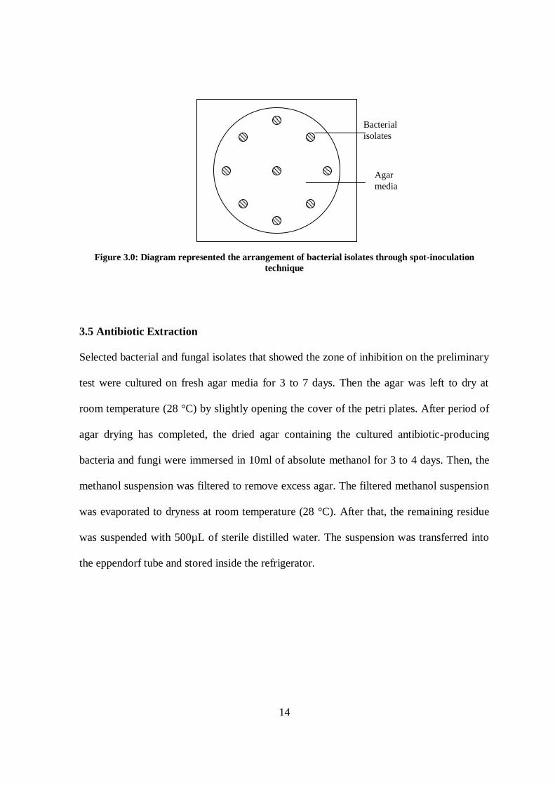
for 4 days at room temperature (28°C). For bacterial isolates, spot-inoculation technique
(Huck et al., 1991) was applied to inoculate pure culture of the selected bacterial isolates
(Figure 3.0). The plates also were incubated at room temperature for 2 days. After the days
of incubation, same agar overlay technique as applied in the preliminary screening was
conducted. Isolates that produced zone of inhibition were once again observed. The pure
cultures of positive activity of bacterial and fungal isolates were cultured on slant agar
media for storage.
14
Figure 3.0: Diagram represented the arrangement of bacterial isolates through spot-inoculation
technique
3.5 Antibiotic Extraction
Selected bacterial and fungal isolates that showed the zone of inhibition on the preliminary
test were cultured on fresh agar media for 3 to 7 days. Then the agar was left to dry at
room temperature (28 °C) by slightly opening the cover of the petri plates. After period of
agar drying has completed, the dried agar containing the cultured antibiotic-producing
bacteria and fungi were immersed in 10ml of absolute methanol for 3 to 4 days. Then, the
methanol suspension was filtered to remove excess agar. The filtered methanol suspension
was evaporated to dryness at room temperature (28 °C). After that, the remaining residue
was suspended with 500µL of sterile distilled water. The suspension was transferred into
the eppendorf tube and stored inside the refrigerator.
Bacterial
isolates
Agar
media
15
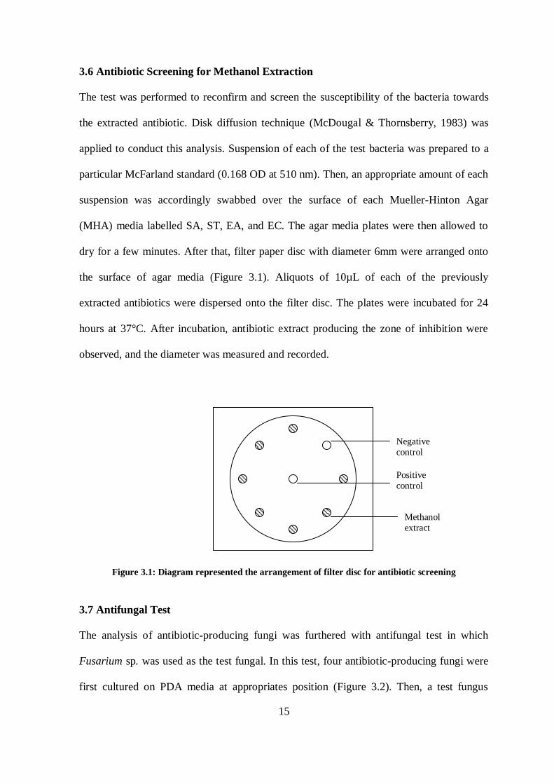
3.6 Antibiotic Screening for Methanol Extraction
The test was performed to reconfirm and screen the susceptibility of the bacteria towards
the extracted antibiotic. Disk diffusion technique (McDougal & Thornsberry, 1983) was
applied to conduct this analysis. Suspension of each of the test bacteria was prepared to a
particular McFarland standard (0.168 OD at 510 nm). Then, an appropriate amount of each
suspension was accordingly swabbed over the surface of each Mueller-Hinton Agar
(MHA) media labelled SA, ST, EA, and EC. The agar media plates were then allowed to
dry for a few minutes. After that, filter paper disc with diameter 6mm were arranged onto
the surface of agar media (Figure 3.1). Aliquots of 10µL of each of the previously
extracted antibiotics were dispersed onto the filter disc. The plates were incubated for 24
hours at 37°C. After incubation, antibiotic extract producing the zone of inhibition were
observed, and the diameter was measured and recorded.
Figure 3.1: Diagram represented the arrangement of filter disc for antibiotic screening
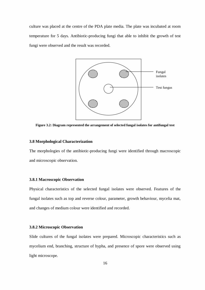
3.7 Antifungal Test
The analysis of antibiotic-producing fungi was furthered with antifungal test in which
Fusarium sp. was used as the test fungal. In this test, four antibiotic-producing fungi were
first cultured on PDA media at appropriates position (Figure 3.2). Then, a test fungus
Negative
control
Positive
control
Methanol
extract
16
culture was placed at the centre of the PDA plate media. The plate was incubated at room
temperature for 5 days. Antibiotic-producing fungi that able to inhibit the growth of test
fungi were observed and the result was recorded.
Figure 3.2: Diagram represented the arrangement of selected fungal isolates for antifungal test
3.8 Morphological Characterization
The morphologies of the antibiotic-producing fungi were identified through macroscopic
and microscopic observation.
3.8.1 Macroscopic Observation
Physical characteristics of the selected fungal isolates were observed. Features of the
fungal isolates such as top and reverse colour, parameter, growth behaviour, mycelia mat,
and changes of medium colour were identified and recorded.
3.8.2 Microscopic Observation
Slide cultures of the fungal isolates were prepared. Microscopic characteristics such as
mycelium end, branching, structure of hypha, and presence of spore were observed using
light microscope.
Fungal
isolates
Test fungus