Embed Size (px)
Citation preview

Faculty of Computer Science
© 2007
Information Theoretic Measures: Object Segmentation and Tracking
CMPUT 615
Nilanjan Ray

2
Information Theory
• Information theory refers to quantification of “information”
• Gained prominence and attention by Shannon in 1948
• Modern computers and communication systems have their deep roots in information theory
• Found numerous applications – coding, wireless, statistical inference, natural language processing, cryptography, image analysis, and many other areas

3
Information Entropy
• One of the basic notions in information theory
i
ii pppH )(log)( 2
pi is a probability mass function

4
Information Divergence Measures
• Compares two probability mass/density functions
• Various divergence measures exists– KL divergence, J-divergence, Bhattacharya coefficient, etc.
• Found applications in– Image registration– Object segmentation– Object classification– Object tracking– …
5
Automated Tumor Segmentation from MRI
• Automated and accurate tumor boundary delineation is an extremely complex, non-trivial task that should use wealth of information, not just images!
• Significant for several reasons, among them:– Clinical applications: diagnosis, drug response,
informatics, MR image database indexing according to location, size etc.
– Medical research: tumor growth prediction, drug discovery,…
MRI Scan

6
Toward Tumor Segmentation: Locating A Fast Bounding Box
• As an important step toward automated tumor boundary delineation, we propose a real-time and convenient method to find a bounding box around an anomalous region (e.g., tumor or edema) on MRI scan
• An initial step toward tumor boundary delineation
• Can help medical informatics: real-time image database indexing based on tumor position, size, shape, etc.

7
Locating a Bounding Box…
D
Image I Reference Image R
Problem definition: Finding an anomalous region D in image I usingreference image R
To detect D from I using R:
Straightforward, fast techniques such as edge detection, point-wise subtractionoften do not work for brain MRI
Unsupervised techniques, such as snakes, level set methods may be insufficient, typically require a lot of tuning parameters, and can be slow
Supervised techniques, such as pixel classifications, require training sets, often need image registration (non-trivial), and can be slow
We propose a fast method, where the only tuning parameter is the number of feature histogram bins. No training data needed. No registration required. Uses single MRI slice, so independent of intensity variation among MR slices.
8
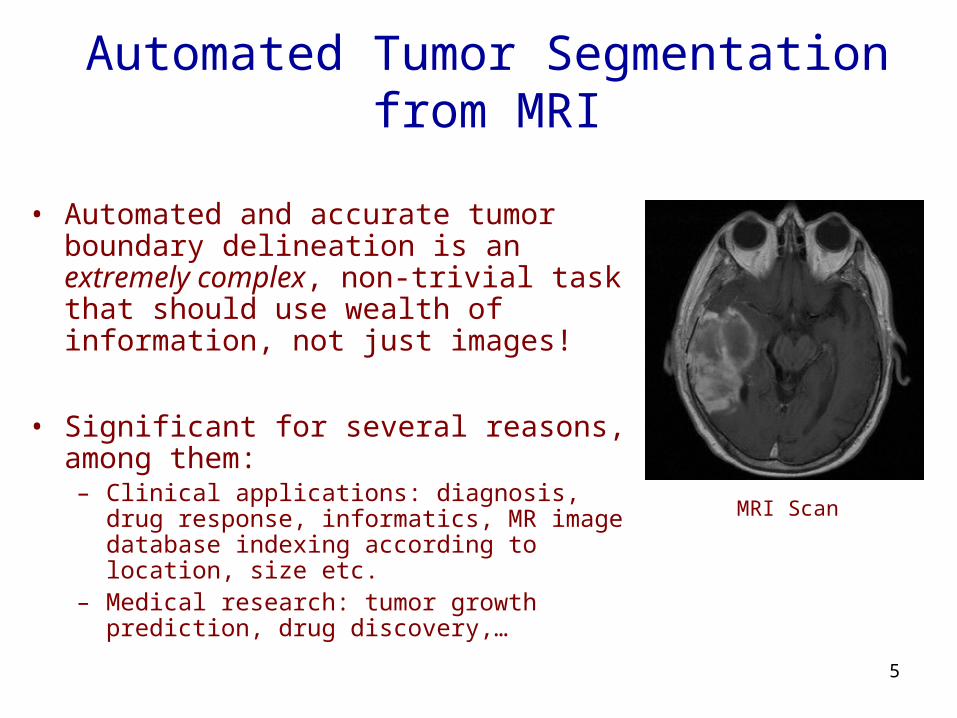
Our Approach: Image Analysis Perspective
,,,)( )()()()( sBR
sBI
sTR
sTI PPPPsE ],[],0[)(],,0[],0[)( hswsBswsT
l
D
I R
Claim: We can show that if the feature histogram within D is sufficiently different from that outside Dthe plot of E(s) vs. s would look like:
s
0, 0 w
h
0 h
Incr. Decr. Incr.
We need two 1D score metric plots to find the dissimilar region D
We propose a score metric based on Bhattacharya coefficient betweenfeature probabilities (normalized histograms) of I and R:
where
T = top portion
B = bottom portion
l
u
u
E(s)
s
9
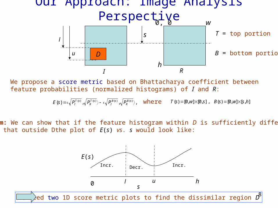
FBB: Locating Brain Tumor
MR Image
(a)50 100 150 200 250
50
100
150
200
250
Boundary and Line of Symmetry
(b)50 100 150 200 250
50
100
150
200
250
0 100 200 300-0.05
0
0.05
0.1
0.15Score plot for vertical direction
(c)0 50 100 150
-0.3
-0.2
-0.1
0
0.1
0.2Score plot for horizontal direction
(d)Fig. 2: Locating brain anomaly.
Use (approximate) symmetry of brain to get reference image and test image
The method can tolerate a reasonable shift of line of symmetry and rotation of the head,because it makes use of region-based symmetry, rather than point-wise symmetry
10
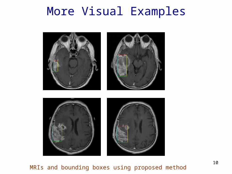
More Visual Examples
MRIs and bounding boxes using proposed method

11
Visual Comparison
Fig. 3: Top row: results of the proposed FBB technique. Bottom row: results of Chan – Vese bounding box algorithm (CVBB).
12
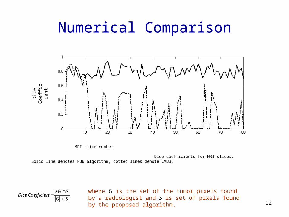
Numerical ComparisonD
ice
Coe
ffic
ient
MRI slice number
Dice coefficients for MRI slices. Solid line denotes FBB algorithm, dotted lines denote CVBB.
,2
SG
SGtCoefficienDice
where G is the set of the tumor pixels found by a radiologist and S is set of pixels found by the proposed algorithm.

13
From Bounding Box toward Boundary: An Example
Chan-Vese level set method without bounding box
Chan-Vese level set method within bounding box
Bounding box by Proposed Method

14
Future Plans
• Extensive testing on Alberta Cross Cancer Institute image database
• Incorporation as a pre-processing step at in-house segmentation algorithms (visit: http://www.cs.ualberta.ca/~btgp/)
• Extension to 3D: should need 3 score metric plots
• A general image analysis technique: can be applied to suitable applications, such as background subtraction with limited number of frames, jittery environment
• Instead of rectangular boxes, can work with general boundaries: level set based framework

15
Summary
• Proposed a bounding box locating method around anomaly
• Uses region-based left-right symmetry, rather than point-wise symmetry
• Uses single MR image
• No training data required
• No image registration needed
• No effect of variability in image intensity across MR images
16
Thanks!
A Short Demo
Visit brain tumor project page:http://www.cs.ualberta.ca/~btgp/