Embed Size (px)
Citation preview
Egyptian Journal of Chest Diseases and Tuberculosis (2013) 62, 59–63
The Egyptian Society of Chest Diseases and Tuberculosis
Egyptian Journal of Chest Diseases and Tuberculosis
www.elsevier.com/locate/ejcdtwww.sciencedirect.com
ORIGINAL ARTICLE
Factors predicting exercise-induced oxygen desaturation
in stable COPD
Khaled Hussein a, Atef Farouk Alkarn a, Raafat El-Sokkary a, Samiaa Hamdy a,*,
Hamdy Shams b
a Department of Chest Diseases, Faculty of Medicine, Assiut University, Egyptb Cardiology Department, Faculty of Medicine, Assiut University, Egypt
Received 20 November 2012; accepted 18 February 2013Available online 18 March 2013
*
Fa
A
E-
@
(R
Pe
D
04
ht
KEYWORDS
COPD;
Exercise;
Desaturation;
Base-line SaO2
Corresponding author. Add
culty of Medicine, Assiut U
ssuit 71111, Egypt. Tel.: +20
mail addresses: khaldhussein
yahoo.com (A. Farouk A
. El-Sokkary), samiaa_sadek
er review under responsibil
iseases and Tuberculosis.
Production an
22-7638 ª 2013 The Egyptia
tp://dx.doi.org/10.1016/j.ejcd
ress: De
niversity
8821805
@yahoo
lkarn),
@yahoo
ity of Th
d hostin
n Society
t.2013.02
Abstract Background: Exercise induced oxygen desaturation (EID) is highly prevalent among
patients with COPD. We suggested that, some resting pulmonary functions and blood gas param-
eters might be used as screening test to predict exercise-induced oxygen desaturation in COPD.
Objective: The aim of this work was to evaluate different parameters that might predict (EID) in
COPD patients.
Design: Randomized, double blind, prospective study.
Methods: This study included 55 patients with stable COPD, resting pulmonary functions; arte-
rial blood gases, echocardiography, and incremental cardiopulmonary exercise testing were done
for all patients. We considered the patient to be desaturated if O2 saturation decreased P4 mmHg
with exercise. We compared desaturated (DS) and nondesaturated (NDS) patients.
Results: Oxygen desaturation was found in 28 subjects after CPET, while 27 subjects were non-
desaturated (NDS). FEV1% was significantly lower in DS (P = 0.001), DLCO was significantly
lower in DS (P = 0.001), and resting oxygen saturation (base-line) was significantly lower in DS
(P = 0.000). Resting PaCO2 was significantly higher in DS (P = 0.000), pulmonary artery systolic
pressure PAP was significantly higher in DS (P = 0.016), and mMRC score was significantly higher
in DS (P = 0.000), while there were no statistically significant differences of age, FEV1/FVC,
(TLC), and (RV).
partment of Chest Diseases,
Hospital, Assiut University,
03; fax: +20 882333327.
.com (K. Hussein), afaroukeg
.com (S. Hamdy).
e Egyptian Society of Chest
g by Elsevier
of Chest Diseases and Tuberculosis. Production and hosting by Elsevier B.V. All rights reserved.
.009
60 K. Hussein et al.
On performing regression analysis the most sensitive predictor for EID was base line SaO2 (rest-
ing SaO2).
Conclusion: Although multiple factors can predict EID in stable COPD (FEV1%, DLCO, rest-
ing SaO2, PaCO2, mMRC score, and PAP), base line SaO2 is the most sensitive factor.
ª 2013 The Egyptian Society of Chest Diseases and Tuberculosis. Production and hosting by Elsevier
B.V. All rights reserved.
Table 1 Clinical and functional parameters.
Parameters Desaturated Nondesaturated P value
Mean ± SD (28) Mean ± SD (27)
Age 55.86 ± 9.35 58.00 ± 8.788 0.386
FEV1/FVC 53.87 ± 10.12 54.77 ± 9.89 0.358
FEV1 (%) 33.75 ± 9.28 49.49 ± 19.86 0.001*
TLC (%) 107.25 ± 33.237 116.52 ± 29.494 0.280
RV 201.46 ± 74.524 202.11 ± 63.634 0.973
DLCO 47.54 ± 20.25 67.35 ± 19.62 0.001*
mMRC score 3.54 ± 0.69 2.44 ± 0.97 0.000*
* means significant < 0.05.
Table 2 Haemogasometric parameters.
Parameters Desaturated Nondesaturated P value
Mean ± SD (28) Mean ± SD (27)
Base-line SaO2 91.92 ± 2.88 95.94 ± 2.9 0.000*
Resting PaCO2 63.46 ± 11.58 38.97 ± 6.38 0.000*
PAP 42.21 ± 11.90 34.15 ± 12.14 0.016*
* means significant < 0.05.
Introduction
The most common functional impairment in patients with alltypes of lung diseases is impaired gas exchange. In the earlystages of many lung diseases, oxygen saturation is maintained
at rest, but when the lung is challenged with increasing demand(exercise), oxygen desaturation may occur [1]. Many previousinvestigators have studied the predictors of oxygen desatura-
tion during maximal exercise in patients with COPD [1].Exercise induced oxygen desaturation (EID) is highly pre-
valent among patients with COPD. EID seems to be associated
with impaired daily physical activity which supports its clinicalimportance [2]. Earlier studies have suggested early mortalityin patients with COPD who are normoxic at rest but experi-ence exercise desaturation [3,4].
Oxygen supplementation with exertion can increase peakexercise level, decrease minute ventilation, improve exercisetolerance, decrease dyspnea, and prevent transitory increases
in pulmonary arterial pressure and pulmonary vascular resis-tance at sub-maximal workloads [5,6].
Previous studies have determined that baseline forced expi-
ratory volume in 1 s (FEV1) or diffusing capacity for carbonmonoxide (DLCO) can serve as screening tests to predictwhich COPD patients will desaturate with exercise [7,8].
Objective
The aim of this work is to study different parameters that maypredict EID in COPD patients, as regards pulmonary function
tests and haemogasometric parameters.
Patients and methods
The present study was conducted in the Department of ChestDiseases, Faculty of Medicine, Assiut University Hospitalfrom March, 2008 to January, 2011. The study included 55 pa-
tients with chronic obstructive pulmonary diseases (COPD),diagnosis of COPD was done according to Global Initiativefor chronic obstructive lung disease guideline, 2008.
All patients were subjected to full medical history, wheredyspnea was graded according to modified Medical ResearchCouncil (mMRC), general and local chest examination, Chest
X-ray PA view, spirometry, and incremental CPET using (Cos-med SrL, Quark PFTs ergo, P/N Co9035-12-99, made inItaly). Lung volumes and diffusion capacity of the lung for car-bon monoxide were determined using single breath method (D
97723; Zan 300, Oberthulba, Germany).Arterial blood gases in room air were obtained both at rest
and at the end of exercise by blood sample from radial artery
and analyzed using blood gases analyzer (Rapid lab 850;CHIRON/Diagnostics; critical care systems). Two dimen-sional Doppler echocardiography was done on all patients.
Exclusion criteria
(1) Patients with primary cardiac diseases include ischemicheart diseases, cardiomyopathy, unstable angina, valvu-
lar heart diseases, and myocardial infarction.(2) Respiratory failure.(3) Decompensated cor-pulmonale.
(4) Acute pulmonary embolism.(5) Uncontrolled cardiac arrhythmia.(6) Severe arterial hypertension.
We considered that a patient would develop exercise-in-duced oxygen desaturation if O2 saturation decreased P4%with exercise. We compared desaturated (DS) with non-desat-
urated (NDS) patients.
Results
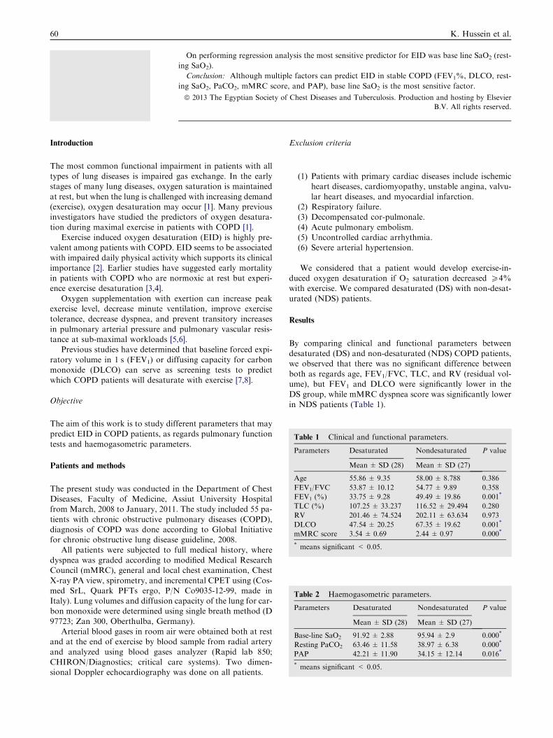
By comparing clinical and functional parameters betweendesaturated (DS) and non-desaturated (NDS) COPD patients,we observed that there was no significant difference between
both as regards age, FEV1/FVC, TLC, and RV (residual vol-ume), but FEV1 and DLCO were significantly lower in theDS group, while mMRC dyspnea score was significantly lower
in NDS patients (Table 1).
0
10
20
30
40
50
60
70
80
DLCOFEV1%
DS
NDS
Figure 1 Comparison between FEV1 & DLCO in both groups.
0
20
40
60
80
100
120
PaCO2SaO2
DS
NDS
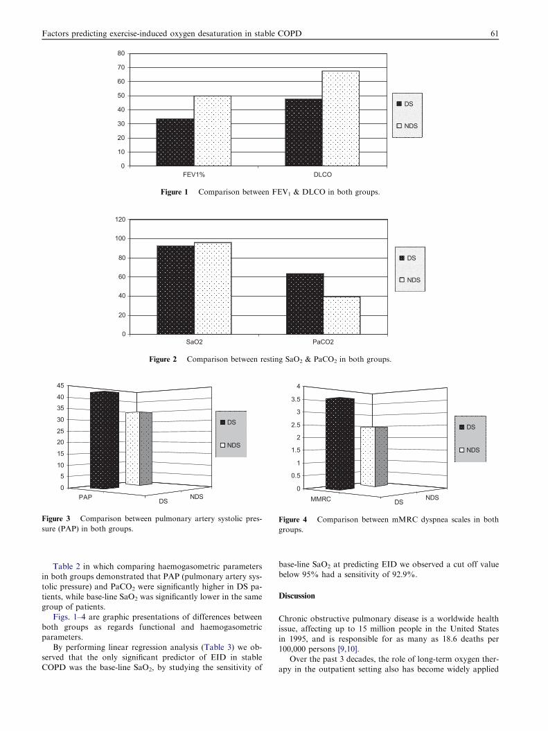
Figure 2 Comparison between resting SaO2 & PaCO2 in both groups.
PAP DSNDS
0
510
15
2025
30
3540
45
DS
NDS
Figure 3 Comparison between pulmonary artery systolic pres-
sure (PAP) in both groups.
MMRC DSNDS
0
0.5
1
1.5
2
2.5
3
3.5
4
DS
NDS
Figure 4 Comparison between mMRC dyspnea scales in both
groups.
Factors predicting exercise-induced oxygen desaturation in stable COPD 61
Table 2 in which comparing haemogasometric parametersin both groups demonstrated that PAP (pulmonary artery sys-
tolic pressure) and PaCO2 were significantly higher in DS pa-tients, while base-line SaO2 was significantly lower in the samegroup of patients.
Figs. 1–4 are graphic presentations of differences betweenboth groups as regards functional and haemogasometricparameters.
By performing linear regression analysis (Table 3) we ob-served that the only significant predictor of EID in stableCOPD was the base-line SaO2, by studying the sensitivity of
base-line SaO2 at predicting EID we observed a cut off valuebelow 95% had a sensitivity of 92.9%.
Discussion
Chronic obstructive pulmonary disease is a worldwide healthissue, affecting up to 15 million people in the United States
in 1995, and is responsible for as many as 18.6 deaths per100,000 persons [9,10].
Over the past 3 decades, the role of long-term oxygen ther-apy in the outpatient setting also has become widely applied
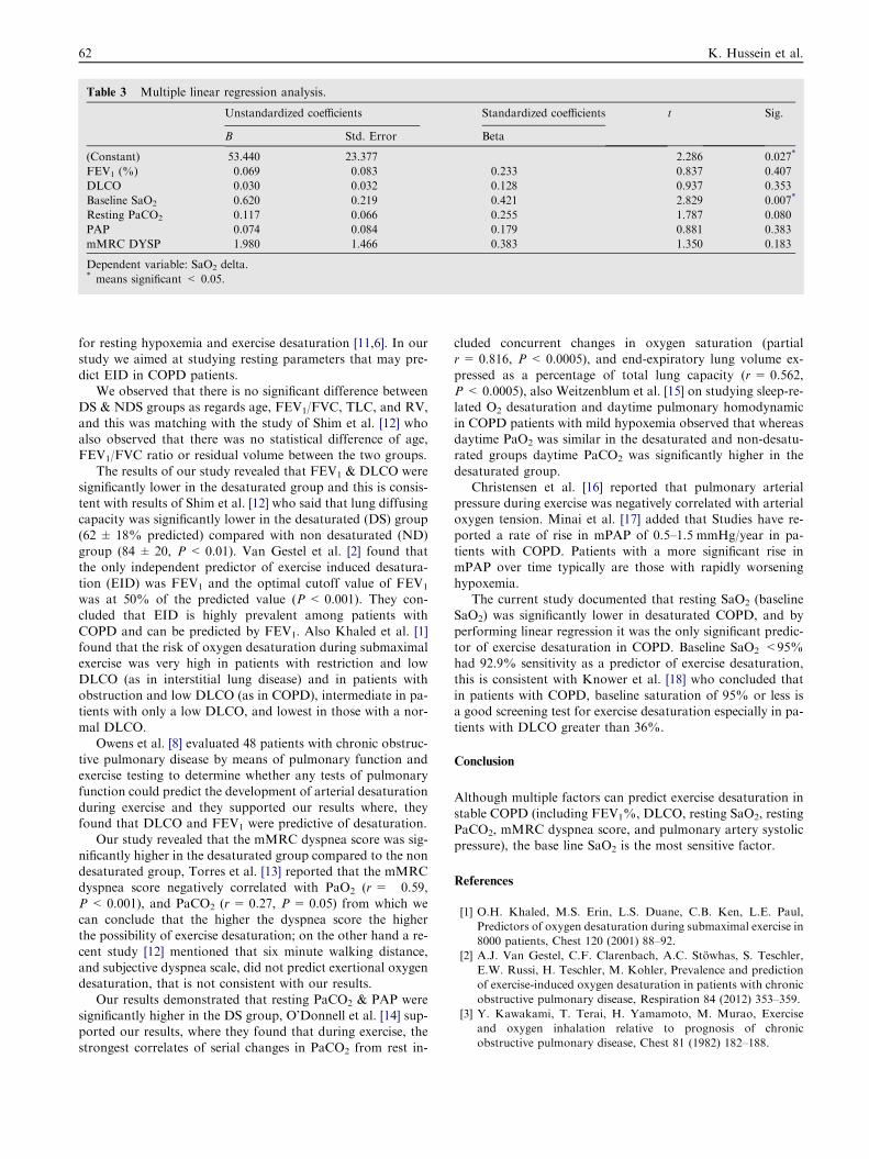
Table 3 Multiple linear regression analysis.
Unstandardized coefficients Standardized coefficients t Sig.
B Std. Error Beta
(Constant) 53.440 23.377 2.286 0.027*
FEV1 (%) 0.069 0.083 0.233 0.837 0.407
DLCO �0.030 0.032 �0.128 �0.937 0.353
Baseline SaO2 �0.620 0.219 �0.421 �2.829 0.007*
Resting PaCO2 0.117 0.066 0.255 1.787 0.080
PAP �0.074 0.084 �0.179 �0.881 0.383
mMRC DYSP 1.980 1.466 0.383 1.350 0.183
Dependent variable: SaO2 delta.* means significant < 0.05.
62 K. Hussein et al.
for resting hypoxemia and exercise desaturation [11,6]. In ourstudy we aimed at studying resting parameters that may pre-
dict EID in COPD patients.We observed that there is no significant difference between
DS & NDS groups as regards age, FEV1/FVC, TLC, and RV,
and this was matching with the study of Shim et al. [12] whoalso observed that there was no statistical difference of age,FEV1/FVC ratio or residual volume between the two groups.
The results of our study revealed that FEV1 & DLCO weresignificantly lower in the desaturated group and this is consis-tent with results of Shim et al. [12] who said that lung diffusingcapacity was significantly lower in the desaturated (DS) group
(62 ± 18% predicted) compared with non desaturated (ND)group (84 ± 20, P < 0.01). Van Gestel et al. [2] found thatthe only independent predictor of exercise induced desatura-
tion (EID) was FEV1 and the optimal cutoff value of FEV1
was at 50% of the predicted value (P < 0.001). They con-cluded that EID is highly prevalent among patients with
COPD and can be predicted by FEV1. Also Khaled et al. [1]found that the risk of oxygen desaturation during submaximalexercise was very high in patients with restriction and lowDLCO (as in interstitial lung disease) and in patients with
obstruction and low DLCO (as in COPD), intermediate in pa-tients with only a low DLCO, and lowest in those with a nor-mal DLCO.
Owens et al. [8] evaluated 48 patients with chronic obstruc-tive pulmonary disease by means of pulmonary function andexercise testing to determine whether any tests of pulmonary
function could predict the development of arterial desaturationduring exercise and they supported our results where, theyfound that DLCO and FEV1 were predictive of desaturation.
Our study revealed that the mMRC dyspnea score was sig-nificantly higher in the desaturated group compared to the nondesaturated group, Torres et al. [13] reported that the mMRCdyspnea score negatively correlated with PaO2 (r = �0.59,P < 0.001), and PaCO2 (r= 0.27, P = 0.05) from which wecan conclude that the higher the dyspnea score the higherthe possibility of exercise desaturation; on the other hand a re-
cent study [12] mentioned that six minute walking distance,and subjective dyspnea scale, did not predict exertional oxygendesaturation, that is not consistent with our results.
Our results demonstrated that resting PaCO2 & PAP weresignificantly higher in the DS group, O’Donnell et al. [14] sup-ported our results, where they found that during exercise, the
strongest correlates of serial changes in PaCO2 from rest in-
cluded concurrent changes in oxygen saturation (partialr= 0.816, P < 0.0005), and end-expiratory lung volume ex-
pressed as a percentage of total lung capacity (r= 0.562,P < 0.0005), also Weitzenblum et al. [15] on studying sleep-re-lated O2 desaturation and daytime pulmonary homodynamic
in COPD patients with mild hypoxemia observed that whereasdaytime PaO2 was similar in the desaturated and non-desatu-rated groups daytime PaCO2 was significantly higher in the
desaturated group.Christensen et al. [16] reported that pulmonary arterial
pressure during exercise was negatively correlated with arterialoxygen tension. Minai et al. [17] added that Studies have re-
ported a rate of rise in mPAP of 0.5–1.5 mmHg/year in pa-tients with COPD. Patients with a more significant rise inmPAP over time typically are those with rapidly worsening
hypoxemia.The current study documented that resting SaO2 (baseline
SaO2) was significantly lower in desaturated COPD, and by
performing linear regression it was the only significant predic-tor of exercise desaturation in COPD. Baseline SaO2 <95%had 92.9% sensitivity as a predictor of exercise desaturation,this is consistent with Knower et al. [18] who concluded that
in patients with COPD, baseline saturation of 95% or less isa good screening test for exercise desaturation especially in pa-tients with DLCO greater than 36%.
Conclusion
Although multiple factors can predict exercise desaturation in
stable COPD (including FEV1%, DLCO, resting SaO2, restingPaCO2, mMRC dyspnea score, and pulmonary artery systolicpressure), the base line SaO2 is the most sensitive factor.
References
[1] O.H. Khaled, M.S. Erin, L.S. Duane, C.B. Ken, L.E. Paul,
Predictors of oxygen desaturation during submaximal exercise in
8000 patients, Chest 120 (2001) 88–92.
[2] A.J. Van Gestel, C.F. Clarenbach, A.C. Stowhas, S. Teschler,
E.W. Russi, H. Teschler, M. Kohler, Prevalence and prediction
of exercise-induced oxygen desaturation in patients with chronic
obstructive pulmonary disease, Respiration 84 (2012) 353–359.
[3] Y. Kawakami, T. Terai, H. Yamamoto, M. Murao, Exercise
and oxygen inhalation relative to prognosis of chronic
obstructive pulmonary disease, Chest 81 (1982) 182–188.
Factors predicting exercise-induced oxygen desaturation in stable COPD 63
[4] V. Vandenbergh, J. Clement, K.P. van de Woestijne, Course and
prognosis of patients with advanced chronic obstructive
pulmonary disease: evaluation by means of functional indices,
Am. J. Med. 55 (1973) 736–746.
[5] J.A. Dempsey, E.H. Vidruk, S.M. Mastenbrook, Pulmonary
control systems in exercise, Fed. Proc. 39 (1980) 1498–1505.
[6] W. Mitlehner, W. Kerb, Exercise hypoxemia and the effects of
increased inspiratory oxygen concentration in severe chronic
obstructive pulmonary disease, Respiration 61 (1994) 255–262.
[7] M.A. Kelley, R.A. Panettieri, A.V. Krupinski, Resting single-
breath diffusing capacity as a screening test for exercise-induced
hypoxemia, Am. J. Med. 80 (1986) 807–812.
[8] G.R. Owens, R.M. Rogers, B.E. Pennock, D. Levin, The
diffusing capacity as a predictor of arterial oxygen desaturation
during exercise in patients with chronic obstructive pulmonary
disease, N. Engl. J. Med. 310 (1984) 1218–1221.
[9] M.S. Niederman, Mechanisms and management of COPD,
Chest 113 (1998) 233S–234S.
[10] American Thoracic Society, Standards for the diagnosis and
care of patients with chronic obstructive pulmonary disease,
Am. J. Respir. Crit. Care Med. 152 (1995) 77S–121S.
[11] Nocturnal Oxygen Therapy Trial Group, Continuous or
nocturnal oxygen therapy in hypoxemic chronic obstructive
lung disease: a clinical trial, Ann. Intern. Med. 93 (1980) 391–
398.
[12] S.W. Shim, Y.J. Jun, S.K. Yong, N.C. Jin, H.P. Jie, Mi-Young
Lee, H.R. Byung, Won-II Choi, Factors related to exertional
oxygen desaturation in patients with COPD, Tuberc. Respir.
Dis. 70 (2011) 498–503.
[13] J.P. Torres, C. Casanova, A.M. Garcini, A. Aguirre-Jaime, B.R.
Celli, Gender and respiratory factors associated with dyspnea in
chronic obstructive pulmonary disease, Respir. Res. 8 (2007) 18.
[14] D.E. O’Donnell, Christine D’Arsigny, Michael Fitzpatrick,
Katherine A. Webb, Exercise hypercapnia in advanced chronic
obstructive pulmonary disease the role of lung hyperinflation,
Am. J. Respir. Crit. Care Med. 166 (2002) 663–668.
[15] C.E. Weitzenblum, C.M. Ehrhart, P. Levi-Valensi, J. Zielinski,
D.R. Cornudella, J.S. Moutinho, Sleep-related O2 desaturation
and daytime pulmonary haemodynamics in COPD patients with
mild hypoxaemia, Eur. Respir. J. 10 (1997) 1730–1735.
[16] C.C. Christensen, M.S. Ryg, A. Edvardsen, O.H. Skjønsberg,
Relationship between exercise desaturation and pulmonary
haemodynamics in COPD patients, Eur. Respir. J. 25 (2005)
1126–1127.
[17] O.A. Minai, C. Ari, A. Serge, Pulmonary hypertension in
COPD: epidemiology, significance, and management:
pulmonary vascular disease: the global perspective, Chest 137
(2010) 39S–51S.
[18] M.T. Knower, D.P. Dunagan, N.E. Adair, R. Chin, Baseline
oxygen saturation predicts exercise desaturation below
prescription threshold in patients with chronic obstructive
pulmonary disease, Arch. Intern. Med. 161 (2001) 732–736.