Embed Size (px)
Citation preview

American Mineralogist, Volume 75, pages 791-800, 1990
Factors influencing shortJived blue cathodoluminescence of a-quartz
K,c.RL RavrsrvnnGeologisches Institut, Universitiit Bern, 3012 Bern, Switzerland
JosBr Mur,r,rsMineralogisch-Petrographisches Institut, Universitiit Basel, 4056 Basel, Switzerland
Ansrucr
Crystals of a-quartz from fissures in the Swiss Alps and from the island of Grand Canarywere investigated to determine the factors influencing their cathodoluminescence charac-tenstlcs.
The cathodoluminescence of a-quartz is commonly a short-lived blue, blue-green, oryellow color with nonluminescent areas. Long-lived violet and a brown luminescence ofincreasing intensity are uncommon for a-quartz.
Cathodoluminescence observations, fluid-inclusion studies, microprobe analysis, heat-and electrodiffusion treatments and morphological data revealed that the cathodolumi-nescence ofa-quartz is influenced by the uptake ofcharge balancing single charged cationsassociated with the substitution of Al for silica. Electrodifusion studies indicate that Hcan cause short-lived cathodoluminescence. The amount and distribution of the traceelements in a-quara are controlled by the growth dynamics, growth direction, twinning,fracturing, and natural a- or 7-irradiation.
Physicochemical growth parameters, such as crystallization temperature, pressure, com-position, and concentrations of volatiles in crystallizing fluids, do not influence the short-lived cathodoluminescence of a-quartz.
INrnonucrroN
Electron-excited luminescence, or cathodolumines-cence, in naturally grown d-quartz was first reported byGoldstein (1907). Later, Smith and Stenstrom (1965),Long and Agrell (1965), Sippel (1968), Sprunt et al. (1978),Zinkernagel (1978), and Matter and Ramseyer (1985)demonstrated the ability of this technique to distinguisho.-quarIz precipitated from an aqueous chloride solution(i.e, diagenetic or hydrothermal origin) from quartz crys-tallized in a melt (i.e., igneous or metamorphic origin).
Few data exist on the cathodoluminescence behaviorof diagenetic or hydrothermally formed a-quartz, whichhas generally been thought to be nonluminescent (Sippel,1965; Smith and Stenstrom,1965;' Zinkernagel, 1978) orto possess nondetectable shortlived luminescence (Zin-kernagel, 1978). Improvements in the detection limits ofvery faint luminescence intensities by a new type of cath-odoluminescence microscope enabled Ramseyer et al.(1988) to record a variety of short-lived luminescencecolors in a-quartz precipitated from an aqueous chloridesolution.
The purpose of this paper is to determine crystallo-graphic, physical, and chemical parameters that influencethe cathodoluminescence of diagenetically or hydrother-mally formed a-quafiz. More than 75 quartz crystals fromnine localities in the Swiss Alps (Mullis, l9'15,1976,1980,1982), from cavities in a Miocene basalt (Canary Islands;Mullis and Sigl, 1982) and a quartz crystal used in elec-
0003-o04x/90/0708-079 I $02.00 79r
trodifusion experiments (kindly provided by the Miner-alogisches Institut ETH-Ziirich) were selected to coverthe physicochemical conditions present in natural dia-genetic and hydrothermal systems (Table l) and to assessthe consistency of cathodoluminescence distributions.
Mnrnons
An improved cathodoluminescence microscope (Ram-seyer et al., 1989) was used to examine the faint andshort-lived luminescence colors in fissure quartz. The ap-plied beam current density for the cathodoluminescencewas 0.2-0.4 pA/mm2 at 30 keV electron energy. Lumi-nescence characteristics were recorded on Ektachrome 400color slide film with l3l+-minutes exposure time and de-veloped at 800 ASA. Changes in the luminescence duringelectron bombardment due to the relatively long expo-sure time (Ramseyer et al., 1989) dictate that comparableresults can only be obtained by using identical operatingconditions and areas of no previous electron bombard-ment. When these requirements are fulfilled, then theconsistency ofresults obtained is excellent. Since lengthyelectron bombardment leads to modification of the emis-sion spectra, cathodoluminescence characteristics arecompared by means of qualitative observations of pre-viously unbombarded areas on color slides. The photo-graphic results obtained are therefore a time-integratedfunction of the continuously changing luminescence char-acteristics. The luminescence spectra were also recorded
792 RAMSEYER AND MULLIS: CATHODOLUMINESCENCE OF a-QUARTZ
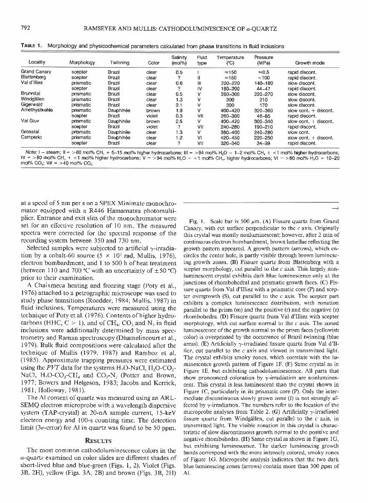
TABLE 1. Morphology and physicochemical parameters calculated from phase transitions in fluid inclusions
Locality Morphology Twinning ColorSalinity(mol%)
Fluid Temperature Pressuretype (rC) (MPa) Growth mode
Grand CanaryBlattenbergVal d'lll iez
BrunnitalWindgallenGigerwaldAmethystkehle
Val Giuv
GrosstalCamperio
scepterscepterprismaticsceprerprismaticprismaticprismaticprismaticsceprerprismaticscepterprismaticprismaticscepter
BrazilBrazilBrazilBtazilBrazilBrazilBrazilDauphin6eBrazilDauphin6eBrazilDauphin6eDauphin6eBrazil
*150c150
200-2201 80-200280-300
300300
400-420260-300400-420240-280380-400420-450320-340
€0.5<100
140-18044-47
220-270210170
320J6046-65
300J401 90-21 0240-280220-25034-39
clearclearclearclearclearclearclearbrownvioletbrownvioletctearcrearcrear
0.57
0.62
0.51 . 32.11 . 90.32.57
1 . 31 . 27
It lil lIV
V
v t l
v t l
VIv t l
rapid discont.rapid discont.slow discont.rapid discont.slow discont.slow discont.slow discont.slow cont. + discont.rapid discont.slow cont. + discont.rapid discont.slow cont.slow cont. + discont.rapid discont.
Note: | : steam; ll : >80 mol7" CH4 + 5-15 molo/" higher hydrocarbons; lll :lV : >80 molo/" CHo + <1 molyo higher hydrocarbons; V : >94 mol7" HrO +molo/" CO,; Vll : >40 md% CO,
>94 mofo/" H2O + 1-2 molo/" CHo * <1 molo/" higher hydrocarbons;<1 molo/" CH4, higher hydrocarbons; Vl : >80 mol7" HrO + 10-20
at a speed of 5 nm per s on a SPEX Minimate monochro-mator equipped with a R446 Hamamatsu photomulti-plier. Entrance and exit slits of the monochromator wereset for an effective resolution of l0 nm. The measuredspectra were corrected for the spectral response of therecording system between 350 and 730 nm.
Selected samples were subjected to artificial 7-irradia-tion by a cobalt-60 source (5 x 10⁷ rad, Mullis, 1976),electron bombardment, and I to 500 h of heat treatment(between I l0 and 700 "C with an uncertainty of +50 "C)prior to their examination.
A Chaixmeca heating and freezing stage (Poty et a1.,1976) attached to a petrographic microscope was used tostudy phase transitions (Roedder, 1984; Mullis, 1987) influid inclusions. Temperatures were measured using thetechnique ofPoty et al. (1976). Contents ofhigherhydro-carbons (HHC, C > l), and of CHo, CO, and N, in fluidinclusions were additionally determined by mass spec-trometry and Raman spectroscopy (Dhamelincourt et al.,1979). Bulk fluid compositions were calculated after thetechnique of Mullis (1919, 1987) and Ramboz et al.(1985). Approximate trapping pressures were estimatedusing the PW data for the systems HrO-NaCl, HrO-COr-NaCl, HrO-COr-CHo and COr-N, (Potter and Brown,1977; Bowers and Helgeson, 1983; Jacobs and Kerrick,l98l ; Hol loway, l98 l ) .
The Al content of qtartz was measured using an ARL-SEMQ electron microprobe with a wavelength dispersivesystem (TAP-crystal) at 20-nA sample current, l5-keVelectron energy and 100-s counting time. The detectionlimit (3o-error) for Al in quartz was found to be 50 ppm.
Rrsur,rsThe most common cathodoluminescence colors in the
a-quartz examined on color slides are different shades ofshort-lived blue and blue-green (Figs. 1, 2). Violet (Figs.38, 2H), yellow (Figs. 3,{, 28) and brown (Figs. 3B, 2H)
Fig. l. Scale bar is 500 pm. (A) Fissure quartz from GrandCanary, with cut surface perpendicular to the c axis. Originallythis crystal was mostly nonluminescent; however, after 2 min ofcontinuous electron bombardment, brown lamellae reflecting thegrowth pattern appeared. A growth pattern (arrows), which en-circles the center hole, is partly visible through brown luminesc-ing growth zones. (B) Fissure quartz from Blattenberg with ascepter morphology, cut parallel to the c axis. This largely non-luminescent crystal exhibits dark blue luminescence only at thejunctions of rhombohedral and prismatic growth faces. (C) Fis-sure quartz from Val d'Illiez with a prismatic core (P) and scep-ter oyergrowth (S), cut parallel to the c axis. The scepter partexhibits a complex luminescence distribution, with zonationparallel to the prism (m) and the positive (r) and the negative (z)rhombohedra. (D) Fissure quartz from Val d'Illiez with sceptermorphology, with cut surface normal to the c axis. The zonedluminescence of the gowth normal to the prism faces (yellowishcolor) is overprinted by the occurrence ofBrazil twinning (blueareas). (E) Artificially 7-irradiated fissure quartz from Val d'Il-liez, iut parallel to the c axis and viewed in transmitted light.The crystal exhibits smoky zones, which correlate with the lu-minescence growth pattern of Figure lF. (F) Same crystal as inFigure lE, but exhibiting cathodoluminescence. All parts thatshow pronounced coloration by z-irradiation are nonlumines-cent. This crystal is less luminescent than the crystal shown inFigure lC, particularly in its prismatic core (P). Only the inter-mediate discontinuous slowly grown zone (I) is not strongly af-fected by .y-irradiation. The numbers refer to the location of themicroprobe analyses from Table 2. (G) Artificially 7-irradiatedfissure quartz from Windgiillen, cut parallel to the c axis, intransmitted light. The visible zonation in this crystal is charac-teristic of slow discontinuous grolvth normal to the positive andnegative rhombohedra. (H) Same crystal as shown in Figure lG,but exhibiting luminescence. The darker luminescing growthbands correspond with the more intensely colored, smoky zonesof Figure lG. Microprobe analysis indicates that the two darkblue luminescing zones (arrows) contain more than 300 ppm ofAl.

RAMSEYER AND MULLIS: CATHODOLUMINESCENCE OF A-QUARTZ 793
B

794
A
RAMSEYER AND MULLIS: CATHODOLUMINESCENCE OF a-QUARTZ

RAMSEYER AND MULLIS: CATHODOLUMINESCENCE OF A-QUARTZ 795
luminescence colors also occur but are less common.Nonluminescent quartz was detected in a few crystals withskeletal growth (Figs. lA-D, lF; Kleber, 1970).
With the exception of the brown luminescence, all theabove colors faded during electron bombardment. Theinduced luminescence color is a bluish or reddish brown(Ramseyer et al., 1988; Figs. 38, 2H).
The spectral response of these various luminescence
(-
Fig. 2. Scale bar is 500 pm. (A) Fissure quartz from Brun-nital, cut parallel to the c axis. The part of the crystal luminescingblue represents growth normal to the rhombohedral faces (r),whereas the area with dark blue luminescence (m) contains car-bonate luminescing yellow and has growth normal to the prismfaces. (B) Fissure quartz from Amethystkehle, cut parallel to thec axis. In this crystal, the three gowth types (continuous slow,cyclic slow, and cyclic rapid) are represented by the core lumi-nescing dark blue (C), the zoned part luminescing lighter blue (I)and the scepter luminescing yellow (S), respectively. Significantgrowth normal to the prism faces (m) is present in the areasluminescing yellow. (C) Fissure quartz from Gigerwald, with cutsurface normal to the c axis. With luminescence, $owth afterthe positive rhombohedra (r) and the negative rhombohedra (z)are distinguishable, as are both prisms (m., m,), the trapezohe-dron (s), and Brazil twins (B) in the zone with growth normal tothe positive rhombohedra. A-A' and B-B' refer to the locationofthe traverses from Figures 4aand 4b, respectively. (D) Fissurequartz from Gigerwald, cut parallel to the c axis. The majorityof the cyclic slow growth in this crystal was normal to bothrhombohedra (r, z) and only a few areas show growth normal tothe prisms. The pronounced zonation is typical of this slow cy-clic growth. In addition, microprobe analyses show that the outerzone luminescing darker blue contains up to 1410 ppm Al (Fig.4c; Table 2). The numbers refer to the location of the microprobeanalyses from Table 2, and C-C' to the location of the traversefrom Figure 4c. (E) Fissure quartz from Grosstal, cut parallel tothe c axis. The homogeneous cathodoluminescence in this crystalis typical ofslow continuous growth. (F) Fissure quartz from ValGiuv, cut parallel to the c axis. The homogeneous core of thecrystal luminescing blue is overprinted by nonluminescent frac-tures (f). Minute solid impurities on the boundary between thecore and the scepter part ofthe crystal (black arrows) lead to theformation of growth defects visible as nonluminescent thin bandswith an orientation approximately normal to the growh faces(white arrows). (G) Section of a quartz crystal from Camperio,cut parallel to the c axis. Two gowth types (e.g., cyclic slow andcyclic rapid) are present in this crysral. In the scepter part (S) ofthe crystal, the fading of the luminescence along growth layers(white arrows) is directly related to the decrease of Al contentfrom 500 to < 50 ppm. The numbers refer to the location of themicroprobe analyses from Table 2. (H) Section of a quartz crys-tal from electrodiffusion experiments, cut parallel to the c axis.The oblique thin line (r) crossing the whole plate represents growthzonation parallel to a rhombohedral face. The unaltered bottlegreen luminescing region (G) is characterized by a bottle greenluminescence color, whereas the altered region is characterizedby streaks ofblue luminescence (B) aligned parallel to the c axis.The boundary region between the two zones is bell-shaped. Thebrown luminescence (b) visible in the composite photograph isdue to an earlier electron bombardment of this area.
400 500 600 700
WAVELENGTH (nm)400 500 600 700
WAVELENGTH (nm)
Fig. 3. Spectra of cathodoluminescence colors from quartzcrystals. All spectra shown are corrected for instrumental re-sponse. (A) Luminescence spectra of yellow (S), lieht blue (I),and dark blue (C) color from the three grouth types distin-guished in the crystal from Amethystkehle (Fig. 2B). @) Lumi-nescence spectra of blue (B), bottle green (G), brown-violet (V),and artificial brown (b) color from the crystal used in electrodif-fusion experiments (Fig. 2H).
colors is characterized by broad peaks and combinationsofpeaks in the violet, blue, green, and yellow-orange re-gion of the visible spectra (Fig. 3). During measurement,continuous change in cathodoluminescence characteris-tics results from electron bombardment, which leads toa detectable increase in the red region of the spectra. Thisincrease is recognizable in the two spectra on Figure 3 forthe blue and blue-green colors. After a few minutes ofelectron bombardment, the spectral response of these areasof previously blue or blue-green luminescence color (Figs.38, 2H) takes the form ofa broad peak centered around650 nm (Ramseyer et al., 1988; Fig. 3B).
The cathodoluminescence pattern in quartz crystals iscommonly heterogeneous and related to gowth history(Fie. 2). Only ordinary colorless (Bambauer et a1., l96l)and smoky quartz exhibit a homogeneous luminescenceintensity and color (Figs. 2E, 2F). The cathodolumines-cence patterns observed were different for each locality,but crystals taken from the same fissure (i.e., Gigerwald,Figs. 2C, 2D) show similar patterns. Quartz crystals fromclosely spaced fissures in different rock types (e.9., dolo-mitic limestone and granite shown by fluid inclusions tohave similar thermal histories) also revealed similar cath-odoluminescence patterns. The color photographs of Fig-ures I and 2 represent the typical cathodoluminescencepattern for a given locality.
Physicochemical growth parameters
The main growh parameters are temperature, pres-sure, composition, and the concentration of volatiles inthe fluids from which quartz precipitated. The effects ofeach parameter on the cathodoluminescence of quartzwere investigated by comparison of the cathodolumi-
z
z
u
fr

o.
796
1 500
RAMSEYER AND MULLIS: CATHODOLUMINESCENCE OF a-QUARTZ
r 500 1 500
eEo_-9 rooozo
t
z qoolrl
z
0
aEo-9 rooozF
tFz 6nnl-rl
z
0
aEg iooozoF
E
7 500U
z
0 100 200
A DTSTANCE (am) A'
nescence patterns obtained from crystals grown under dif-ferent conditions.
Fissure quartz precipitated at similar temperatures andpressures can exhibit variable luminescence intensities.For example, the crystals from Grosstal and the core ofthe crystals from Val Giuv have a homogeneous blueluminescence (Figs. 2E, 2F), whereas the crystals fromAmethystkehle have a dark blue to nonluminescent core(Fig. 2B). In addition, quartz precipitated at differenttemperatures and pressures (e.g., 120 vs. 200 "C and 40vs. 150 MPa, Figs. lC, lH, 2A^,2C,2G) can display asimilar luminescence color and distribution pattern.
Variations in the amount of CO, in the fluid inclusions(e.g., Brunnital: 0.0 molo/o; Gigerwald: 3.1 molo/o) and inthe major volatile species (e.g., the skeletal part of thecrystals from Blattenberg: higher hydrocarbons; from Vald'Illiez: CHo; and from Camperio: COr) have no visibleeffect on the cathodoluminescence color and distribution(Figs. 1B, lC, 2G).
These examples demonstrate that the temperature andpressure during crystal growth do not influence the cath-
Treue 2. Al content of quartz crvstals
Val d'lll iez(Fig. 1F)
Number ppm Al
odoluminescence ofa-quartz. In addition, neither a changeof the major species (e.g., higher hydrocarbons, CHo orCOr) nor the total amount of volatiles in the fluids havean influence on the cathodoluminescence of a-quarlz.
Growth dynamics
In addition to temporature, pressure, and volatile con-tent of the fluid, the mode of growth (e.g., continuous,cyclic, slow, or rapid) is another important parameterpermitting characteization of these crystals. Three typesof crystal growth are distinguishable (Mullis,1976, 1979,1 9 8 0 . 1 9 8 3 ) .
1. Quartz crystals precipitated slowly and continuouslyfrom an aqueous chloride solution that was undersatu-rated in dissolved volatiles. This type of fluid composi-tion is commonly found in fracture systems in tectoni-cally quiescent regions. Quartz crystals from Grosstal (Fig.2E) and the prismatic cores of the Amethystkehle (Fig.28) and Val Giuv (Fig. 2D crystals are examples of thisslow and continuous glowth. Homogeneous lumines-cence is common in this type of quartz.
2. Crystals grown in a slow cyclic manner are charac-terizedby prismatic forms and usually display Dauphin6ehabits with Brazil twinning and horizontal grooves (Weil,I 930; Friedliinder, I 95 I ; Frondel, 1962:- Gigoiev, I 96 5)but without sutures on the prism faces. The outermostzone of the prismatic part of the crystals from Val d'Illiez(Fig. lC), from Amethystkehle (Fig. 28) and from Cam-perio (Fig. 2G), as well as the crystals from Gigerwaldand Windgiillen (Figs. lI{, 2C), all exhibit finely zonedluminescence consistent with cyclic growth.
3. Crystals characterized by cyclic rapid growth wereprobably precipitated during boiling (pure HrO phase) orduring fractionation ofthe fluid-phase into water-rich andvolatile-rich components. Phase separations are mostlycaused by a sudden pressure drop during fissure enlarge-ments in regions with active tectonism (Mullis, 1987).
0 100 200 300 400
B DISTANCE (pm) B'
O 100 2gg 300 499
Q DISTANCE (pm) C'
Fig. 4. Electron microprobe traverses across crystals from Gigerwald showing cathodoluminescence zonation. (a) Traverse acrosscrystal in Figure 2C (A-A') over regions with growth normal to the trigonal prism, bipyramid, and positive rhombohedra. (b) Traverseacross crystal in Figure 2C (B-B') over regions with growth normal to the trigonal prism and negative rhombohedra. (c) Traverseacross crystal in Figure 2D (C-C') over regions with growth normal to the positive rhombohedra.
Camperio(Fig. 2G)ppm Al
Gigerwald(Fig. 2D)ppm Al
122
456
8I
1 01 11 21 31 4
506070
1 940280
1 5 1 043060
9201 960280990
1370170
605050
2750<50
31 501220<50<50260
21 0052050
<50
370<50
9090
1 1 0130100130
1 4 1 01 070

The luminescence color and distribution in these rapidlycyclic grown parts of the crystals from Amethystkehle,Val d'Illiez, Camperio, Val Giuv, and Grand Canary arecomplex, but lack of luminescence in particular growthzones is related to the low Al content ofthese areas (Figs.l, 2, 4, Table 2).
The growth dynamics (i.e., continuous vs. cyclic) andthe growth speed are thus important controls on the dis-tribution of short-lived cathodoluminescence in a-quartz.
Growth directions
The intensity of short-lived luminescence in quartzcrystals from Gigerwald (Fig. 2C, Table 3) decreases fromthe growth normal to the positive rhombohedra (r)through the negative rhombohedra (z), bipyramid (s), tothe trigonal prisms (m., m"). Trends of decreasing inten-sity are similar both for shortlived blue and inducedbrown luminescence.
Thus, the intensity of the cathodoluminescence of quartzcrystallized in a slow cyclic manner depends primarily onthe growth direction.
Density and type of twinning
Crystals showing Brazil twinning (Table l) may benonluminescent or else exhibit variable luminescencecolors (Fig. lD). The luminescence pattern generated inthe case of Brazil twinning is similar to that produced by7-irradiation of quartz with a mimetic structure (Fried-li inder, l95l).
The effect of twinning is also visible on Figure 2C, aquartz crystal from Gigerwald cut perpendicular to the caxis. The difference in luminescence intensity in the samegrowth sector of the positive rhombohedra (r) and thezigzag boundaries ofthese sectors are evidence for Braziltwinning (Mclaren and Pitkethly, 1982).
Crystal fractures
Large, macro- to microscopic cracks are rare in well-crystallized fissure quartz (Mullis, 1976). If fractures arepresent, as in the case of the crystal from Val Giuv (Fig.2E), then the luminescence intensity or color generallydiffers between the fracture filling and the host quartz.
A good example of submicroscopic lattice defects in-duced on a discontinuity surface covered with solid par-ticles is visible in the skeletal part of the crystal from ValGiuv (Fig. 2F). The luminescence color in this rapidlygrown part of the crystal exhibits defect clusters growingoutward similar to the structures observed by Moriya andOgawa ( I 978) using light-scattering tomography.
Trace elements
Measurements of Al content were carried out on crys-tals from Grand Canary, Val d'Illiez, Gigerwald, Gros-stal, and Camperio (Figs. lA, lF, 2C,2D, 28,2G, 4).The detected Al content varies considerably in most ofthe crystals exhibiting rapid or discontinuous growth (Ta-ble 2). Areas with a high Al content (>1000 ppm Al)always exhibit blue luminescence; but the luminescence
797
Tlele 3, Al concentration in different growth zones of a quartzcrystal from Gigerwald (Fig. 2C)
Growth normal to
RAMSEYER AND MULLIS: CATHODOLUMINESCENCE OF a-QUARTZ
n ppm Al
Positive rhombohedra (r)Negative rhombohedra (z)Bipyramid (s)Trigonal prism (m)Trigonal prism (m,)
1 84
1 21 21 3
181 + 20140 + 208 1 + 2 5
12O + 22130 + 21
intensity ofthese zones is not always higher than ofareaswith a lower Al content (e.g., Gigerwald, Camperio).
Analyses of the sample from Gigerwald (Fig. lD) re-vealed a significant difference in the Al content betweenareas with growh normal to the positive rhombohedra(r), the negative rhombohedra (z), the bipyramid (s), andboth trigonal prisms (Table 3). The higher Al content inthe part with growth normal to the positive rhombohedracorresponds with the higher luminescence intensity in thisarea. Differences in the Al content of the other areas (i.e.,z, mt, m., s) show no relation to the observed lumines-cence intensity (Table 3, Fig. lC).
Microprobe measurements in crystals with nonlumi-nescent regions (Grand Canary, Camperio, Val d'Illiez)revealed that short-lived luminescence is absent wherethe Al content is less than 50 ppm but is generally presentin areas with more than 50 ppm Al content (Table 2). Acorrelation between the Al content and the intensity ofthe short-lived blue luminescence seems unlikely, sinceareas with high amounts of Al (> 1000 ppm Al) com-monly exhibit the same or a darker luminescence thanareas of the same crystal with a low (<300 ppm Al) Alcontent. Charge-balancing cations such as H+, Li+, Na+,or K+ located in the channels parallel to the c axis, maybe the causes of the short-lived luminescence in o-quartz.
Artificial and natural'y-irradiation
When the Al-Li pair substitutes for Si and the H con-tent is less than 0.7 ppm, artificial and natural 7-irradia-tion results in a color change in quartz from transparentto smoky (Bambauer, 196l; Poty, 1969; Nassau, 1978;Fig. lE). The effect of artificial 7-irradiation on lumines-cence colors is shown by crystals from Val d'Illiez andWindgiillen (Figs. lE-lH). Slowly grown prismatic parts,areas ofthe rapidly grown skeletal part, and strongly dis-colored smoky parts of the artificially irradiated crystalsexhibit either no or only low luminescence intensity com-pared with crystals from the same locality not subjectedto artificial "y-irradiation (Fig. lC). The crystal fromWindg?illen (Figs. lG, lH), which was also artificially ir-radiated, again shows a lower luminescence intensity inthose growth zones with an intense smoky coloration. Adecrease in luminescence intensity is also observed in thenaturally 7-irradiated quartz (Fig. 2B), with zones ofsmoky and violet colorations from the Amethystkehle(Nassau, 1978), for which heat treatment resulted in in-creased intensity of dark blue luminescence in the core.
These examples show that both natural and artificial

798 RAMSEYER AND MULLIS: CATHODOLUMINESCENCE OF A-QUARTZ
@
Eo
TEMPERATURE ("C)
rooo 800 400 200
0 5 1 1 5 2 2 5 3 3 5 4
1 / T ( 1 / 1 0 3 K )
Fig. 5. Arrhenius plot of heat-treated quartz crystals with cutsurface normal to the c axis. Dots represent conditions wherethe shortJived luminescence could be restored, whereas circlesare for experiments where luminescence was not restored by heattreatment. The dashed boundary between the two areas repre-sents the temperature dependence of the diffusion coefficientcausing the shortlived luminescence. Also included are pub-lished temperature dependences ofthe diffirsion coefrcients par-allel to the c axis for different ions. (1) D*-H* (Kats et al., 1962);(2) H* (Kronenberg et al., 1986); (3) Na* (Rybach and Laves,1967); @) Li+, Na+, K+ (Verhoogen, 1952); (5) Na* (Frischat,l97oa); (6) Ca'?* (Frischat, 1970b); (7) O'? (Giletti and Yund,198a); (8) H,O, Si4* (Freer, 1981).
7=irradiation result in local decrease in the intensity ofshortlived blue luminescence (although this may be re-stored through heat treatment). Furthermore, the smokyareas show generally lower luminescence intensities thantransparent parts of the same crystal (Figs. lD-lH). Sub-stitution of Al + Li for Si, which at low H contents causesthe smoky coloration, does not itself seem to control theluminescence in a- quartz.
Effect of electrodiffusion
Application of an electric field at elevated temperatures(250-900 "C; Warburg and Tegetmeier, 1888; Verhoogen,1952; Pfenninger and Laves, 1960; Brunner et al., l96l)is a well-known technique promoting exchange of inter-stitial cations in quartz. This type of treatment is thus anexcellent method for assessing the effects of charged in-terstitial particles on the color and distribution of theluminescence.
Cathodoluminescence investigation of a quattz crystalused in electrodiffusion experiments (Pfenninger, 196l;Fig. 2H) revealed that the unaltered part of the crystal(on the side of the negative potential) has a homogeneousshortlived bottle-green luminescence color, whereas theremaining altered region (which was located at the posi-tive potential) is characterized by streaks of blue lumi-nescence aligned parallel to the c axis. These streaks oflinearly polarized blue luminescence originate from theside of the positive potential, where H+-ions were takenup (Brunner et al., 196l; Pfenninger, 196l). The bound-ary between the unaltered, bottle-green luminescent zoneand the nonluminescent region is gradational and convexin the direction of the position of the negative potential(Fig. 2H). The boundary of the area of stronger lumines-cence represents the zone ofincreased cation concentra-tion due to diffusion of cations toward the negative po-tential (Pfenninger, 196l; Schindler, 1964). Microprobeanalyses of each luminescence zone showed no correla-tion between the Al content, which varies between 90 and190 ppm Al (mean vafue 122 a 35 ppm Al, n: l0), andthe observed luminescence color. Heating this crystal forone hour at 600 "C generated a more homogeneous short-lived blue to blue-green luminescence color in the area ofbottle green luminescence and restored a blue lumines-cence in the altered regions. These electrodiffusion ex-periments indicate that the blue, blue-green, and bottlegreen luminescence is related to the presence of positivelycharged interstitial cations.
Thermal treatments
Fissure quartz from Grand Canary, Gigerwald, andGrosstal in which luminescence had been destroyed byelectron bombardment in the uppermost 5 to l0 pm (at30 keV, Ramseyer et al., 1989) was thermally treatedbetween I l0 and 700 'C for a maximum of 500 h. Theoriginal shortlived blue luminescence color and distri-bution were restored in samples that were heated to hightemperatures or kept at moderate temperatures for sig-nificantly longer periods of time (e.g., 500'C for 600 s or250.C for 1.8 x 10" s) .
Figure 5 is an Arrhenius plot for these data using themeasured alteration depth (5-10 pm) to calculate the dif-fusion coefficient. Time-temperature conditions that re-generated the short-lived luminescence are representedby dots, and conditions with no regeneration of the lu-minescence are marked as open circles. Thus the first set

RAMSEYER AND MULLIS: CATHODOLUMINESCENCE OF A-QUARTZ 799
of data points (dots) represents time-temperature condi-tions where the diffusion distance of the species causingthe luminescence is equal to or larger than the thicknessof the altered zone. The second set of data points (opencircles) represents conditions where the diffusion distanceis considerably smaller than the alteration depth. Theboundary between the two sets ofdata points representsthe time-temperature conditions where the diffusion dis-tance is equal to the alteration depth. These boundaryconditions characterize the temperature dependence ofthe diffusion coefficient for the species causing the short-lived luminescence in d.-quartz. A comparison with pub-lished data concerning temperature dependences of thediffusion coefficients parallel to the c axis for deuterium-hydrogen exchange, H*, Li*, Na+, K+, Ca2+, Sia*, andO2 ions (Kats et al., 1962; Kronenberg et al., 1986; Ry-bach and Laves, I 967; Verhoogen, 19521, Frischat, L970a,1970b; Giletti and Yund, 1984; Freer, 1981) shows thatthe temperature dependence of the diffusion coefficientobtained here is similar to data for Na+, or K* ions andthe deuterium-hydrogen exchange (Fig. 5). The great dif-ference in the published data for the diffusion of Nat ions(see Freer, 1981) and the large experimental error onlypermit identification of the diffusing species as a singlecharged cation (Fig. 5).
DrscussroN
Since the uptake of trace elements into the quartz struc-ture is controlled by crystal-growth parameters and so-lution chemistry, neither the growth temperature nor theformation pressure should influence the luminescence.Short-lived luminescence in a-quartz is thus controlledby the crystal-groWh parameters, groWh dynamics (e.g.,continuous vs. cyclic and slow vs. rapid), and growth di-rection. The uptake of Al with a charge-balancing cationas a substitution for Si seems to be the cause of the short-lived blue luminescence colors in d-quartz, since onlyquartz with more than 50 ppm Al exhibits this type ofluminescence.
Observations of luminescence of artificially treatedquartz crystals (e.g., electron bombardment, 7-irradia-tion, electrodifusion, and thermal treatment) provide ad-ditional insights into the nature and origin of the short-lived luminescence in a-qtnrtz. The rapid decrease inluminescence intensity during excitation by highly accel-erated electrons, the regeneration of this luminescencethrough heating, and the redistribution of the lumines-cence colors after electrodiffirsion (Ramseyer et al., 1988)indicate the close relationship between luminescence andthe presence of positively charged interstitial ions inchannels parallel to the c axis. The observations of,y-irra-diated crystals and from thermal experiments restrict thepossible range ofresponsible species to the singly chargedcations H+, Na+, and K*. In addition, the electrodiffu-sion experiment reveals that luminescence can result fromthe uptake of hydrogen (H+) and the diffusion of cations.
CoNcr-usroNs
The most important factor influencing the short-livedcathodoluminescence in a-quartz during crystallization isthe uptake ofcharge balancing, singly charged cations as-sociated with the substitution of Si by Al. The concen-tration and distribution of these trace elements are con-trolled by (l) the growth dynamics (continuous slow, cyclicslow, or cyclic rapid), (2) the growth direction, and (3)the presence of Brazil twinning. The temperature andpressure conditions of crystal growth do not appear toaffect a-quartz luminescence.
After crystallization, natural irradiation by ^y-rays ora-particles may modify the luminescence characteristics.In addition, later filled fractures in the crystals may ex-hibit luminescence colors or intensities that are differentfrom those of the host crystal.
Luminescence in a-quartz is destroyed by electronbombardment, a- and 7-irradiation, and electrodiffusionbut is regenerated by heat treatment.
AcxNowr,rcMENTS
This research was funded by the Swiss National Science Foundation
Grant no. 2000-5.287), which we gratefully acknowledge We thank Dr.
G.A. Waychunas and Dr. Ian Steele for their helpfirl cornrnents. Thanksare also due to Dr. J. Zimmermann, Centre de Recherches P6trog;ra-phiques et G6ochimiques (Nancy, France) for mass spectrometric analy-
ses, to Dr. J. Dubessy, Centre de Recherches sur la G6ologie de I'Uranium(Nancy, France) for Raman spectroscopic investigations, to Dr' J. Bau-
mann, Institut fiir anorganische und physikalische Chemie (Bern) for the
analyses of luminescence spectra, and to Dr. T' Armbruster, Dr. M. Frey,
Dr. N.H. Platt, and Dr. P. Mozley for reviewing and polishing the English
rcxt.
RnrnnnNcns crrED
Bambauer, H.U. (1961) Spurenelementgehalte und 7-Farbzentren in
Quarzen aus Zerrkliiften der Schweizer Alpen. Schweizerische miner-
alogische und petrographische Miueilungen, 41, 335-369.Bambauer, H.U., Brunner, G.O., and Iaves, F. (1961) Beobachtungen
iiber den l-amellenbau an Bergkristallen. Zeitschrift fiir Kristallogra-ph ie , l l 6 , 173 -181 .
Bowers, T.S., and Helgeson, H.C. (1983) Calculation of the thermody-namic and geochemical consequences ofnonideal mixing in the systemHrO-COr-NaCl on phase relations in geologic systems: Metamorphicequilibria at high pressures and temperatures. American Mineralogist,68, 1059-1075.
Brunner, G.O., Wondratscheck, H., and Iaves, F. (1961) Ultrarot-unter-suchungen iiber den Einbau von H in natiirtchem Quarz. Zeitschriftfiir Elektrochemie. 65. 7 !5-7 5O.
Dhamelincourt, P., Beny, J.M., Dubessy, J., and Poty, B.P. (1979) Ana-lyse d'inclusions fluides i la microsonde MOLE i effet Raman. Bulletinde Min6ralogie, 102, 600-610.
Freer, R. (1981) Diffi.rsion in silicate minerals and glasses: A data digestand guide to the literature. Contributions to Mineralogy and Petrology,76,440-454.
Friedliinder, C. (195 l) Untersuchungen iiber die Eignung alpiner Quarzefiir piezoelektrische Zwecke. Beitriige zur Geologie der Schweiz, Geo-technische Serie 29.
Frischat, G.H. (1970a) Kationentransport in Quarzkristallen: I. Natrium-diffirsion parallel zur c-Achse. Bericht Deutsche Keramische Gesell-schaft,. 47. 218-243.
- (1970b) Kationentransport in Quarzkristallen: III. C-alciumdiftr-sion parallel zur c-Achse. Bericht Deutsche Keramische Gesellschaft,47,36+i68.

800 RAMSEYER AND MULLIS: CATHODOLUMINESCENCE OF a-QUARTZ
Frondel, C. (l 962) The system ofmineralogy, silica minerals (7th edition),334 p. Wiley, New York.
Giletti, B.J , and Yund, R.A. (1984) Oxygen diffi-rsion in quarrz. Journalof Geophysical Research, 89, 4039-4046.
Goldstein, E. (1907) Uber das Auftreten roten Phosphoreszenzlichtes anGeissler' schen R6hren: Bericht der Deutschen Physikalischen Gesell-schaft,598-605.
Grigoriev, D.P. (1965) Ontogeny ofrninerals. Israel Program ofScientificTranslations, p. 250. Jerusalem.
Holloway, J.R. (1981) Compositions and volumes of supercritical fluidsin the earth's crust. In L.S. Hollister and M.L. Crawford, Eds., Shortcourse in fluid inclusions. Mineralogical Association of Canada ShortCourse.6. l3-38.
Jacobs, G.K., and Kerrik, D.M. (1981) Methane: An equation of statewith application to the temary system H'O-CO,-CH.. Geochimica etCosmochimica Acta. 45. 607 -61 4
Kats, A., Haven, Y., and Stevels, J.M. (1962) Hydroxyl groups ind-quarIz. Physics and Chemistry of Glasses, 3 , 69-7 5 .
Kleber, W. (1970) An introduction to crystallography, 366 p. VEB VerlagTechnik, Berlin.
Kronenberg, A.K., Kirby, S.H., Aines, R.D., and Rossman, G.R. (1986)Solubility and diffusional uptake of hydrogen in quartz at high waterpressures: Implications for hydrolytic weakening. Journal of Geophys-ical Research. 91. 127 23-127 44.
I-ong, J.V.P., and Agrell, S.O. (1965) The cathodoluminescence of min-erals in thin section. Mineralogical Magazine, 34,318-326.
Matter, A., and Ramseyer, K. (1985) Cathodoluminescence microscopyas a tool for provenance studies ofsandstones NATO ASI Series, 148,r9t-zr1.
Mclaren, A.C., and Pitkethly, D.R. (1982) The twinning microstrucrureand growth of amethyst quartz. Physics and Chemistry of Minerals, 8,128- l 35.
Moriya, K., and Ogawa, T. (1978) Observation of growth defects in syn-thetic quartz crystals by light-scattering tomography. Journal of CrystalGrowth. 44.53-60.
Mullis, J. (1975) Growth conditions of quartz crystals from Val d'Illiez(Valais, Switzerland). Schweizerische mineralogische und petroga-phische Mitteilungen, 55, 41 9-430.
-(1976) Das Wachstumsmilieu der Quarzkristalle im Val d'Illiez.Schweizerische mineralogische und petrographische Mitteilungen, 56,219J68.
- (1979) The system methane-water as a geologic thermometer andbarometer from the external part ofthe Central Alps. Bulletin de Mi-n6ralogie, 102, 526-536.
-(1980) Quarzkristalle aus den Maderanertal. Iapis, 5, 19-21.-(1982) Sternquarz. Schweizer Strahler, 6, 125-l4O-(1983) Einschliisse in Quarzkristallen der Schweizer Alpen und
ihre mineralogisch-geologische Bedeutung. Bulletin de la Societ6 fri-bourgeoise des Sciences naturelles, 72,5-19.
-(1987) Fluid inclusion studies during very low-grade metamor-phism. In M. Frey, Ed., Low temperature metamorphism, p. 162-199.Backie, Glasgow.
Mullis, J., and Sigl, F. (1982) Die Entstehungsgeschichte von Opal, Chal-cedon und Quarz von Gran Canaria. Schweizer Strahler, 6,155-176.
Nassau, K. (1978) The origins of color in minerals. American Mineralo-
eist, 63 , 219-229 .Pfenninger, H.H. (1961) Diftrsion von Kationen und Abscheidung von
Metallen in Quarz unter elektrischer Feldeinwirkung. Ph.D. thesis,University of Ziirich, I 30 p.
Pfenninger, H.H., and Laves, F. (1960) Kationenwanderung und Farb-zentrenbildung bei der Elektrolyse von Quarzplatten senkrecht zur op-tischen Achse. Naturwissenschaften. 47. 276.
Potter, R.W., and Brown, D.L. (1977) The volumetric properties of aqueoussodium chloride solutions from 0'to 500'C and pressures up to 2000bars based on a regression ofavailable data in the literature. U.S. Geo-logical Survey Open-file Report 75-636, 3 I p.
Poty, B.P. (1969\La croissance des cristaux de quartz dans les filons surI'example du filon de I-a Gardette (Bourg d'Oisans) et des filons dumassif du Mont Blanc. Thdse Universit6 de Nancy, Science de la Terre,M6moir 17.
Poty, B.P , Leroy, J., and Jachimowicz, L. (1976) Un nouvel appareil pourla mesure des temp6ratures sous la microscope: I'installation de mi-crothermometrie Chaixmeca. Bulletin de Min6ralogie, 99, 182-186.
Ramboz, C., Schnapper, D., and Dubessy, J. (1985) The P-V-T-X-fO,evolution of H,O-CO,-CH. bearing fluid in a wolframite vein: Recon-struction from fluid inclusion studies Geochimica et CosmochimicaActA, 49,205-2t9.
Ramseyer, K., Baumann, J., Matter, A., and Mullis, J. (1988) Cathodolu-minescence colours of a-quartz. Mineralogical Magazinq 52, 669477.
Ramseyer, K., Fischer, J., Matter, A, Eberhardt, P., and Geiss, J. (1989)A cathodoluminescence microscope for low intensity luminescence.Journal of Sedimentary Petrology, 59, 619422.
Roedder, E. (1984) Fluid inclusions. Mineralogical Society ofArnericaReviews in Mineralogy, 12,646 p.
Rybach, L., and Laves, F. (1967) Sodium diffusion experiments in quartzcrystals. Geochimica et cosmochimica Acta, 31, 539-546.
Schindler, P. (1 964) Untersuchungen eines paramagnetischen Defektes in
Quarz. Ph.D. thesis, University of Ziirich, 53 p.Sippel, R.F. (1965) Simple device for luminescence peuog&phy. Review
of Scientific Instruments, 36, I 556-1 558.-(1968) Sandstone petrology, evidence from luminescence micros-
copy. Joumal of Sedimentary Perology, 38, 530-554.Smith, J.V., and Stenstrom, R.C. (1965) Electron-excited luminescence
as a petrologic tool. Journal of Geology, '13,
627435.Sprunt, E.S., Dengler, L.A., and Sloan, D. (1978) Effects of metamor-
phism on quartz cathodoluminescence. Geology, 6, 305-308.Yerhoogen, J. (1952) Ionic diffi:sion and electrical conductivity in quartz.
American Mineralogist, 37, 637 455.Warburg, E., and Tegetmeier, F. (1888) flber die electrolytische I-eitung
des Bergkrystalls. Annalen der Physik und Chemie, 35, 455-467.Weil, R. (1930) Observations sur le quartz. Comptes rendus hebdoma-
daires des seancos de I'Academie des sciences Paris sciences naturelles,r9r, 270-272,380-382, 93s-937 .
Zinkernagel, U. (1978) Cathodoluminescence ofquartz and its applicationto sandstone petrology. Contributions to Sedimentology, 8, l-69.
Mnm,ncnrm RECEMD Drceusen 14, 1988MnNuscnrp'r AcCEPTED Mav 14, 1990