Embed Size (px)
Citation preview

King Saud University
College of Nursing
Medical Surgical Department
Application of Adult Health Nursing Skills
(NUR 317)
Prepared by
Ms: Alwah M. Alkathiri
BSN, RN, MS
0
Care for patients with fluid and electrolytes imbalance

Outline of lecture; Introduction Fluid and electrolytes balance Fluid and electrolytes imbalance Assessment of Edema, Dehydration Measuring intake and output IVF ( intravenous fluids)
Care for patients with fluid and electrolytes imbalance
Normal anatomy and physiology
1

Water comprises 60% of the body weight of an average adult, the total body water is divided functionally into the extracellular (ECF = 20% of body weight) and the intracellular fluid spaces (ICF = 40% of body weight) separated by the cell membrane.
The ECF is further divided into the intravascular (within the circulation) and the interstitial (extravascular fluid surrounding the cells) fluid space.
Fluid Functions:
Help regulate body temperature Transport nutrients and gases throughout the body Carry cellular waste products to excretion sites
Electrolytes :
Electrolytes are a major component of body fluids that play important roles in maintaining chemical balance, there are six major electrolytes; sodium, potassium,calcium, chloride, phosphorus, and magnesium.
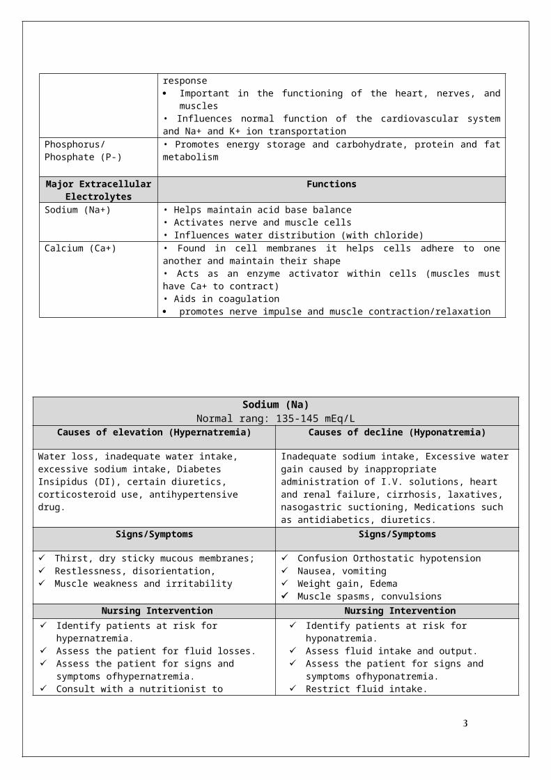
Major Intracellular Electrolytes
Functions
Potassium (K+) • Regulates cell excitability & nerve impulse conduction • Permeates cell membranes, thereby affecting the cell’s electrical status (resting membrane potential)• Regulates muscle contraction and myocardial membrane responsiveness
Magnesium (Mg+) • Modifies nerve impulse transmission and skeletal muscle response Important in the functioning of the heart, nerves, and muscles • Influences normal function of the cardiovascular system and Na+ and K+ ion transportation
Phosphorus/Phosphate (P-) • Promotes energy storage and carbohydrate, protein and fat metabolism
Major Extracellular Electrolytes
Functions
Sodium (Na+) • Helps maintain acid base balance • Activates nerve and muscle cells • Influences water distribution (with chloride)
Calcium (Ca+) • Found in cell membranes it helps cells adhere to one another and maintain their shape • Acts as an enzyme activator within cells (muscles must have Ca+ to contract) • Aids in coagulation promotes nerve impulse and muscle contraction/relaxation
Sodium (Na)Normal rang: 135-145 mEq/L
Causes of elevation (Hypernatremia) Causes of decline (Hyponatremia)
Water loss, inadequate water intake, excessive sodium intake, Diabetes Insipidus (DI), certain diuretics, corticosteroid use, antihypertensive drug.
Inadequate sodium intake, Excessive water gain caused by inappropriate administration of I.V. solutions, heart and renal failure, cirrhosis, laxatives, nasogastric suctioning,
2
Medications such as antidiabetics, diuretics.Signs/Symptoms Signs/Symptoms
Thirst, dry sticky mucous membranes; Restlessness, disorientation, Muscle weakness and irritability
Confusion Orthostatic hypotension Nausea, vomiting Weight gain, Edema Muscle spasms, convulsions
Nursing Intervention Nursing Intervention
Identify patients at risk for hypernatremia. Assess the patient for fluid losses. Assess the patient for signs and symptoms
ofhypernatremia. Consult with a nutritionist to determine Encourage the patient to increase his fluidintake but
decrease his sodium intake. Teach the patient and his family how to
prevent,recognize, and treat hypernatremia
Identify patients at risk for hyponatremia. Assess fluid intake and output. Assess the patient for signs and symptoms
ofhyponatremia. Restrict fluid intake. Administerisotonic I.V. fluids. that ensure appropriate fluid and sodium intake.
Potassium ( K) Normal Level 3.5 - 5 mEq/L
Causes of elevation (Hyperkalemia) Causes of decline (Hypokalemia)
High potassium intake related to the improper use of oralsupplements, excessive use of salt substitutes, or rapid infusion of potassium solutions.
GI losses from diarrhea, laxative abuse, prolonged gastricsuctioning, prolonged vomiting.
Signs/Symptoms Signs/Symptoms
arrhythmias, decreased strength of contraction,and cardiac arrest Nausea, vomiting, diarrhea, intestinal colic, uremic enteritis, decreased bowel sounds, abdominal distention.
fatigue, muscle weakness orthostatic hypotension cardiac arrest Suppressed insulin release and aldosterone secretion Respiratory muscle weakness slightly elevated glucose
level Nursing Intervention Nursing Intervention
Identify patients at risk for hyperkalemia. Assess for signs and symptoms of hyperkalemia. Have emergency equipment available. Administer calcium gluconate to decrease
myocardial irritability. Administer insulin and I.V. glucose to move
potassium back into cells. Carefully monitor serum glucose levels. Administer sodium polystyrene sulfonate(Kayexalate) with 70% sorbitol to exchange sodium ions for potassium ions in the intestine
Identify patients at risk for hypokalemia. Assess the patient’s diet for a lack of
potassium. Assess the patient for signs and symptoms of
hypokalemia. Administer a potassium replacement asprescribed. Encourage intake of high-potassium foods,such as
bananas, dried fruit, and orange juice. Monitor the patient for complications. Have emergency equipment available for cardiopulmonary
resuscitation and cardiac defibrillation.
CalciumNormal Level 4.5 – 5.5 mEq/L
Causes of elevation (hypercalcemia) Causes of decline (hypocalcemia)
Metastatic bone cancer, hyperparathyroidism,High calcium intake, Hyperthyroidism or hypothyroidism
acute pancreatitis, inadequate dietary intake of vitamin D, longterm use of laxatives, thyroid carcinoma, loop diuretics.
Signs/Symptoms Signs/Symptoms Muscle weakness and lack of coordination Anorexia, constipation, abdominal pain, nausea,
Tingling around the mouth and in the fingertips and feet, numbness,
3

vomiting, peptic ulcers, and abdominal distention Confusion, impaired memory,slurred speech, and coma Cardiac arrest
painful muscle spasms. Positive Chvostek’s signs or Positive trousseau's sings Seizures confusion, and hallucinations Skeletal fractures resulting from osteoporosis
Nursing Intervention Nursing Intervention Assess the patient for signs and symptoms of
hypercalcemia. Encourage ambulation. Move the patient carefully to prevent fractures. Administer phosphate to inhibit GI absorptionof calcium. Administer a loop diuretic to promote calcium excretion. Reduce dietary calcium.
Assess the patient for signs and symptoms of hypocalcemia, especially changes in cardiovascularand neurologic status and in vital signs.
Administer I.V. calcium as prescribed. Administer a phosphate-binding antacid. Take seizure or emergency precautions as
needed. Encourage the patient to increase his intake of foods that
are rich in calcium and vitamin D.Magnesium ( Mg)
Normal level 1.5 - 2.5 mEq/LCauses of elevation (Hypermagnesemia) Causes of decline (Hypomagnesemia)
Renal failure, adrenal insufficiency, or diuretic abuseExcessive magnesium replacement or excessive useof milk of magnesia .
malnutrition, malabsorption anorexia, intestinal bypass for obesity, diarrhea, diuretics or antibiotics, such as gentamicin, Overdose of vitamin D or calcium, burns, pancreatitis, or diabetic ketoacidosis
Signs/Symptoms Signs/Symptoms
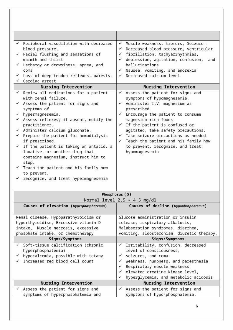
Peripheral vasodilation with decreased blood pressure, Facial flushing and sensations of warmth and thirst Lethargy or drowsiness, apnea, and coma Loss of deep tendon reflexes, paresis. Cardiac arrest
Muscle weakness, tremors, Seizure . Decreased blood pressure, ventricular fibrillation, tachyarrhythmias, depression, agitation, confusion, and hallucinations Nausea, vomiting, and anorexia Decreased calcium level
Nursing Intervention Nursing Intervention Review all medications for a patient with renal failure. Assess the patient for signs and symptoms of hypermagnesemia. Assess reflexes; if absent, notify the practitioner. Administer calcium gluconate. Prepare the patient for hemodialysis if prescribed. If the patient is taking an antacid, a laxative, or another
drug that contains magnesium, instruct him to stop. Teach the patient and his family how to prevent, recognize, and treat hypermagnesemia
Assess the patient for signs and symptoms of hypomagnesemia.
Administer I.V. magnesium as prescribed. Encourage the patient to consume magnesium-rich
foods. If the patient is confused or agitated, take safety
precautions. Take seizure precautions as needed. Teach the patient and his family how to prevent,
recognize, and treat hypomagnesemia
Phosphorus (p)Normal level 2.5 - 4.5 mg/dl
Causes of elevation (Hyperphosphatemia) Causes of decline (Hypophosphatemia)
4

Renal disease, Hypoparathyroidism or hyperthyroidism, Excessive vitamin D intake, Muscle necrosis, excessivephosphate intake, or chemotherapy
Glucose administration or insulin release, respiratory alkalosis, Malabsorption syndromes, diarrhea, vomiting, aldosteronism, diuretic therapy.
Signs/Symptoms Signs/Symptoms Soft-tissue calcification (chronic hyperphosphatemia) Hypocalcemia, possible with tetany Increased red blood cell count
Irritability, confusion, decreased level of consciousness, seizures, and coma Weakness, numbness, and paresthesia Respiratory muscle weakness elevated creatine kinase level, hyperglycemia, and metabolic acidosis
Nursing Intervention Nursing Intervention Assess the patient for signs and symptoms of
hyperphosphatemia and hypocalcemia, including tetany and muscle twitching. Advise the patient to avoid foods and medications that
contain phosphorus. Administer phosphorus-binding antacids. Prepare the patient for possible dialysis.
Assess the patient for signs and symptoms of hypo-phosphatemia, especially neurologic.
Administer phosphate supplements as prescribed. Note calcium and phosphorus levels because calcium and
phosphorus have an inverse relationship.
Fluid and electrolyte imbalances
Fluid and electrolyte balance is essential for health. Many factors, such as illness, injury, surgery, and treatments, can disrupt a patient’s fluid and electrolyte balance. Even a patient with a minor illness is at risk for fluid and electrolyte imbalance.
Fluid Volume Deficit (Hypovolemia) Fluid Volume Excess (Hypervolemia)
The body loses water all the time. A person responds to the thirst reflex by drinking fluids and eating foods that contain water. However, if water isn’t adequately replaced, the body’s cells can lose water. This causes dehydration, or fluid volume deficit. Dehydration refers to a fluid loss of 1% or more of body weight
Hypervolemia refers to an excess of fluid (water andsodium) in ECF. The body has compensatory mechanismsto deal with hypervolemia. However, if these fail, signs andsymptoms develop.
Etiology/Cause• Hemorrhage• Vomiting • Diarrhea• Burns• Diuretic therapy• Fever• Impaired thirst
Etiology/Cause Congestive Heart Failure Early renal failure IV therapy Excessive sodium ingestion Corticosteroid
Fluid Volume Deficit (Hypovolemia) Fluid Volume Excess (Hypervolemia)
Signs/Symptoms ;
Mild Fluid Loss: Orthostatic hypotension, Increased heart rate Restlessness, anxiety Weight loss
Moderate Fluid Loss:
Signs/Symptoms ;
Tachypnea ,Dyspnea, crackles Rapid or bounding pulse Hypertension (unless in heart failure) Distended neck and hand veins Acute weight gain
5
Confusion, dizziness, irritability Extreme thirst Nausea -Cool, clammy skin Rapid Pulse Decreased urine output (10-30 ml/hr)
Severe Fluid Loss: Decreased cardiac output Unconsciousness Hypotension Weak or absent peripheral pulses
Edema Pulmonary edema
- Dyspnea -Orthopnea (diff. breathing when supine) -crackles
Assessing fluid balance
There are three elements to assessing fluid balance and hydration status:
Review of fluid balance charts; Clinical assessment; Review of blood chemistry.
1- Review of fluid balance charts; Fluid balance means the amount of fluid intake equal the amount of fluid excreted .
Intake include; water, juice, tea and coffe, IV fluid , NG feeding Output include; urine, emesis, NG drainage, and blood drainage.
Record all fluid intake in the sheet and calculate the total at the end of each shift Record all fluid output remember if patients on urine catheter each shift
empty urine from catheter. IF Intake ( I ) more than Output (O) look for signs of edema IF Intake ( I ) less than Output (O) look for signs of dehydration
2- Nursing assessment for dehydration Observations Vital signs, such as pulse, blood pressure
and respiratory rate, will change when a patient becomes dehydrated
Skin elasticity The elasticity of skin, or turgor, is an indicator of fluid status in most patients. However, this assessment can be an unreliable indicator of dehydration in older people as skin elasticity reduces with age
2- Nursing assessment for edema
6

Medical treatment
Treatment involves determining the cause(such as diarrhea or decreased fluid intake) and replacing lost fluids either orally or I.V.
Most patients receive hypotonic, low sodiumfluids such as dextrose 5% in water (D5W).
Medical treatment
Treatment involves determining the cause and treating the underlying condition.
Typically, patients require fluid and sodium restrictions
Diuretics therapy may be ordered if renal failure is not the cause.
I.V. fluid replacement
The doctor may order I.V. fluid to maintain or restore fluid balance. I.V. fluid replacement fall into the broad categories of crystalloids and colloids;
A. Colloids - contain larger insoluble molecules (blood, albumin, plasma) used to increase the blood volume following severe loss of blood (haemorrhage) or loss of plasma ( severe burns).
B. Crystalloids – contains aqueous solutions of mineral salts or other water-soluble molecules ( salts and sugar.) to correct body fluids and electrolyte deficit
Isotonic A solution that has the same salt concentration as the normal cells of the body and the blood.
Examples:
Ringer Lactate . 0.9% NaCl (0.9% NSS ) D5W. Normal saline same tonicity as body
Indication: Hypotension )increases
BP(, Hypovolemia
Complications of Isotonic IV fluid overload
7

Hypertonic A solution with a higher salts concentration than in normal cells of the body and the blood.Examples :
D5W in normal Saline solution , D5W in half normal Saline D10W. 5% normal saline D5 Ringers Lactate
Indication: low BP slight edema but not w/CHF
Complications ; circulatory overload.
Hypotonic
A solution with a lower salts concentration than in normal cells of the body and the blood.Examples :
0.45% NaCl . 0.33% NaCl . 45% sodium chloride 5%dextrose water (becomes hypotonic in body)
Indication: Dehydration
Complications ; May cause edema
Types of IV lines ;
1. Peripheral (hands)2. Central Venous Catheter (big veins)
- PICC (Peripherally inserted Central Catheter)- CVC ( Central venous catheter )
Advantages of IVI
• Immediate effect • Patient cannot tolerate drugs / fluids orally• Some drugs cannot be absorbed by any other route • Pain and irritation is avoided compared to some substances when given SC/IM
Disadvantages/Complications of IVI • Phlebitis; is inflammation of a vein
• Thrombophlebitis; is an irritation of the vein along with the formation of a clot; it’s usually more painful than phlebitis. Look for pain, redness, swelling, or a red line streaking along the vein
8

• Infiltration; fluid may leak from the vein into surrounding Tissue, If you see infiltration, stop the infusion, elevate the extremity, and apply warm soaks.
• Infection ; Adhering to aseptic technique is vital in the prevention of intravenous related infections. Swab the site for culture and remove the catheter as ordered.
• Anaphylaxis / Allergic reactions (Itching, rash, shortness of breath)
What the Nurse should do? • STOP INFUSION and treat as indicated by Pharmacy, Medication package insert or drug reference
book.• Notify MD and document
9


![L-14 Fluids [3] Fluids at rest Fluids at rest Why things float Archimedes’ Principle Fluids in Motion Fluid Dynamics Fluids in Motion Fluid Dynamics](https://img.pdfslide.us/doc/110x75/56649d845503460f94a6ab30/l-14-fluids-3-fluids-at-rest-fluids-at-rest-why-things-float-archimedes.jpg)