Embed Size (px)
Citation preview
Immunolabelling artifactsImperial CollegeImperial CollegeLondonLondon
So th KensingtonSo th Kensington
“Immunolabeling artifacts and
–– South KensingtonSouth Kensington
Facility for Imagingby Light Microscopy
the need for live-cell imaging”
ClubUlrike Schnell, Freark Dijk, Klaas A Sjollema &
Ben N G Giepmans (Groningen, NL)
Observing Life As It Happens
Martin Spitaler, FILM
Nature Methods 9/2: 152-158


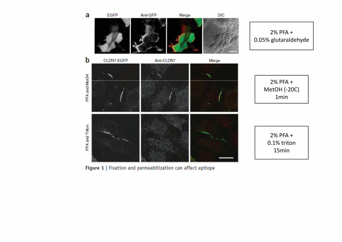
2% PFA +0.05% glutaraldehyde
2% PFA2% PFA +MetOH (‐20C)
1min
2% PFA +2% PFA +0.1% triton15min

Why fixation artifacts?What happens during fixation and permabilisation?
Fixation:• arrests the sample in a live‐like state• ideally should fix everything (proteins, lipids,
nucleic acids )
Common fixatives:• dehydrating:
• methanol (precipitates proteins)ld h d ( li ki )nucleic acids, …)
• must not block access for labels (antibodies, lipid dyes, DNA dyes, …)
• aldehydes (crosslinking):• formaldehyde • glutaraldehyde
Permeabilisation:• opens cell boundaries (plasma membrane,
organelles) to antibodies and other labels• should not affect the shape of the cell or organelles• should leave localisation of proteins in membranes
Permeabilising agents:• saponin• non‐ionic detergents• methanol• should leave localisation of proteins in membranes
intact• methanol

dehydrating fixatives:methanol, ethanol, acetone
• for microscopy, usually methanol is used• replaces water from the proteins surface, thereby inducing precipitation• low temperature (usually ‐20C) and short incubation avoid denaturation of proteins
( ti f t i i )(preserves antigens for staining)• also solubilises membrane lipids (permeabilisation)
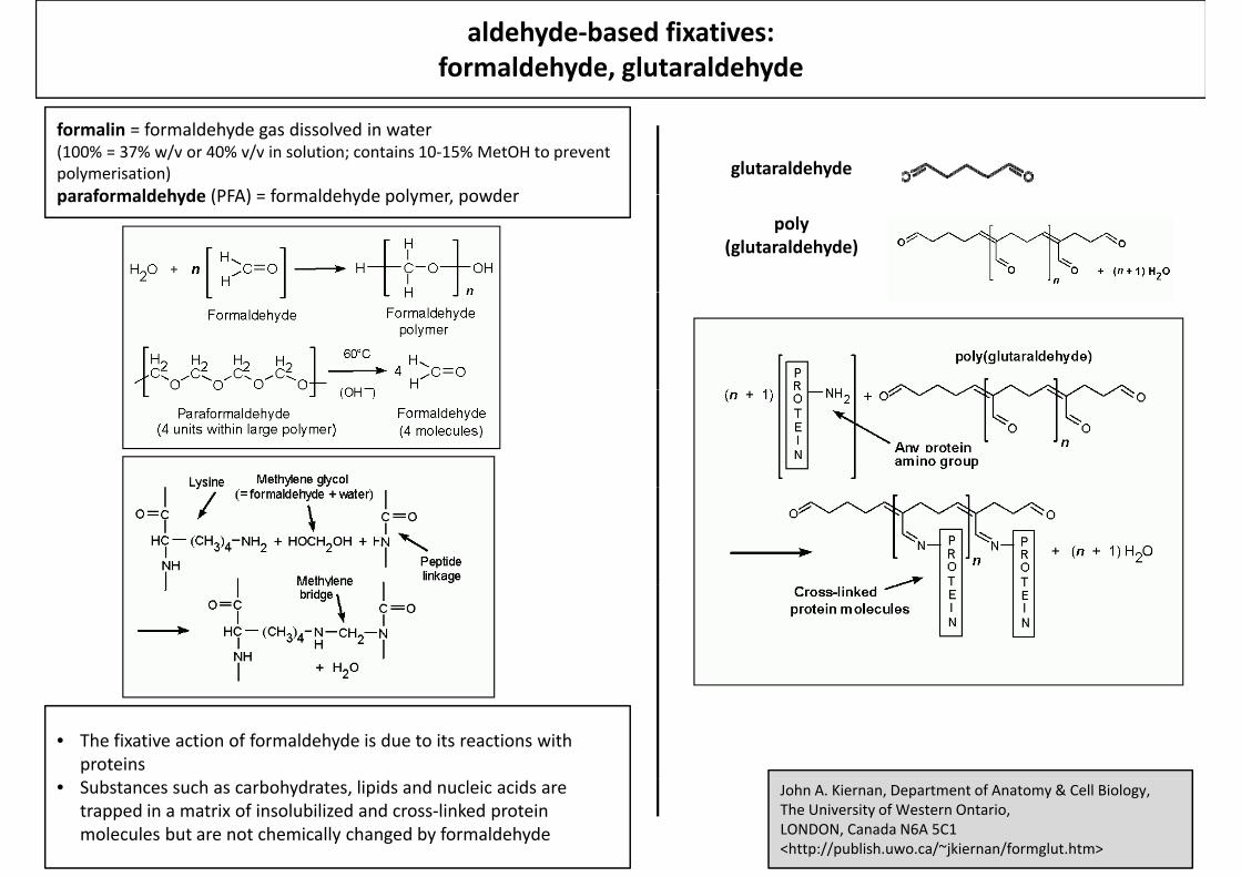
aldehyde‐based fixatives:formaldehyde, glutaraldehyde
formalin = formaldehyde gas dissolved in water(100% = 37% w/v or 40% v/v in solution; contains 10‐15% MetOH to prevent polymerisation)paraformaldehyde (PFA) = formaldehyde polymer powder
glutaraldehydeparaformaldehyde (PFA) = formaldehyde polymer, powder
poly (glutaraldehyde)
• The fixative action of formaldehyde is due to its reactions with proteins
John A. Kiernan, Department of Anatomy & Cell Biology,The University of Western Ontario,LONDON, Canada N6A 5C1 <http://publish.uwo.ca/~jkiernan/formglut.htm>
• Substances such as carbohydrates, lipids and nucleic acids are trapped in a matrix of insolubilized and cross‐linked protein molecules but are not chemically changed by formaldehyde

aldehyde‐based fixatives:formaldehyde, glutaraldehyde
Advantages:
• strong crosslinking and fixation of proteins
• also fixes DNA (crosslinking histones to DNA)
Possible side effects and considerations
• destruction or shielding of epitopes for antibody staining
(worse with glutaraldehyde)also fixes DNA (crosslinking histones to DNA)
• largely irreversible
(worse with glutaraldehyde)
• most proteins are fixed within seconds, but some very
dense or loose complexes can take hours
• changed protein conformation (loss of fluorescence of
fluorescent proteins)
• left‐over aldehyde groups react with any amine group, i.e.
also the lysins of the antibodies high background
staining (worse with glutaraldehyde)
• fluorescent side products (aldehyde reactions)
(worse with glutaraldehyde)
• lipids are not crosslinked
Remedies
• blocking of free aldehyde groups after fixation with
sodium borohydride or ammonium chloride
• good blocking with protein‐rich solutions (fish skin
gelatine skimmed milk BSA)gelatine, skimmed milk, BSA)sodium borohydride
NaBH4
ammonium chlorideNH4Cl
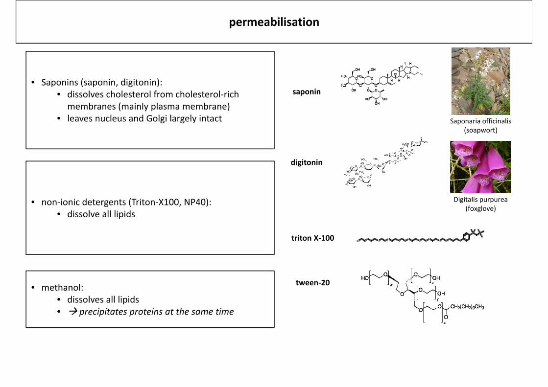
permeabilisation
• Saponins (saponin, digitonin):
Saponaria officinalis(soapwort)
p ( p , g )• dissolves cholesterol from cholesterol‐rich
membranes (mainly plasma membrane)• leaves nucleus and Golgi largely intact
saponin
(soapwort)
digitonin
Digitalis purpurea(foxglove)
• non‐ionic detergents (Triton‐X100, NP40):• dissolve all lipidsdissolve all lipids
triton X‐100
tween‐20• methanol:• dissolves all lipidsdissolves all lipids• precipitates proteins at the same time
2% PFA +0.05% glutaraldehyde
2% PFA2% PFA +MetOH (‐20C)
1min
2% PFA +2% PFA +0.1% triton15min
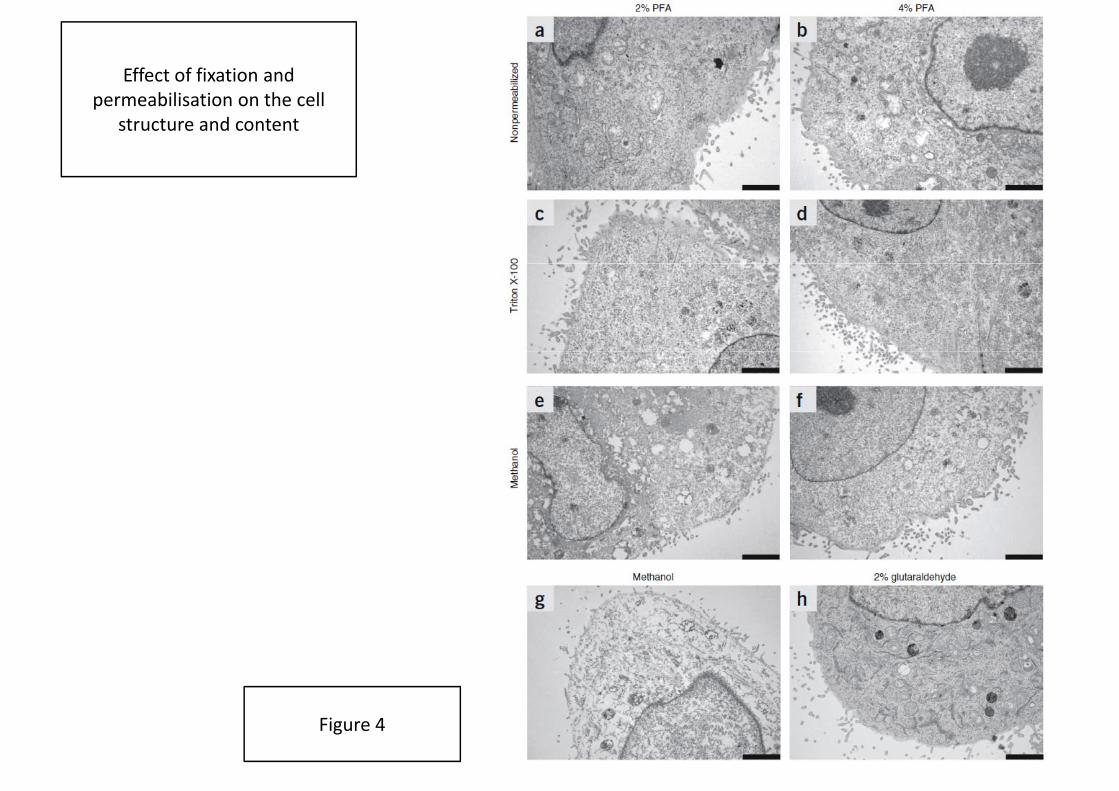
Effect of fixation and permeabilisation on the cell
structure and content
Figure 4

Fig 3
Fig 2b
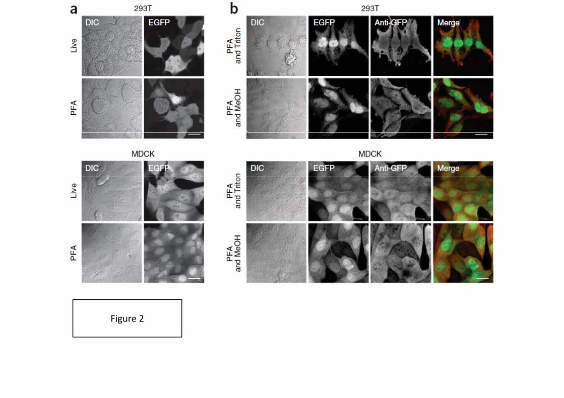
Figure 2

Figure 2
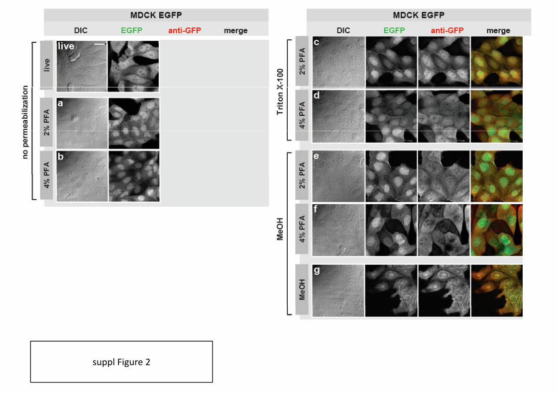
suppl Figure 2
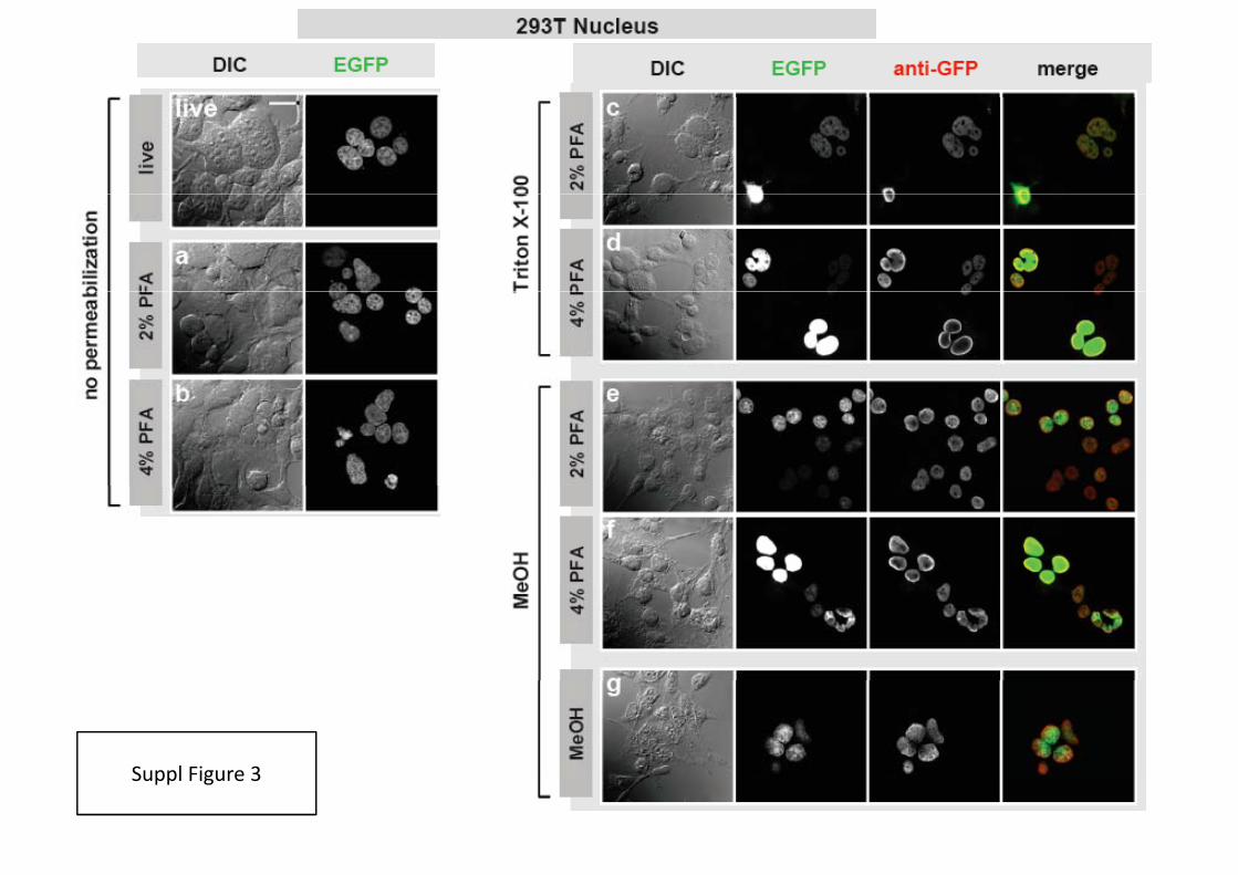
Suppl Figure 3Suppl Figure 3
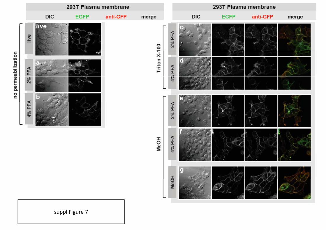
suppl Figure 7

Gerald R. V. Hammond et al.Biochem J. 2009 :422(Pt 1): 23–35.
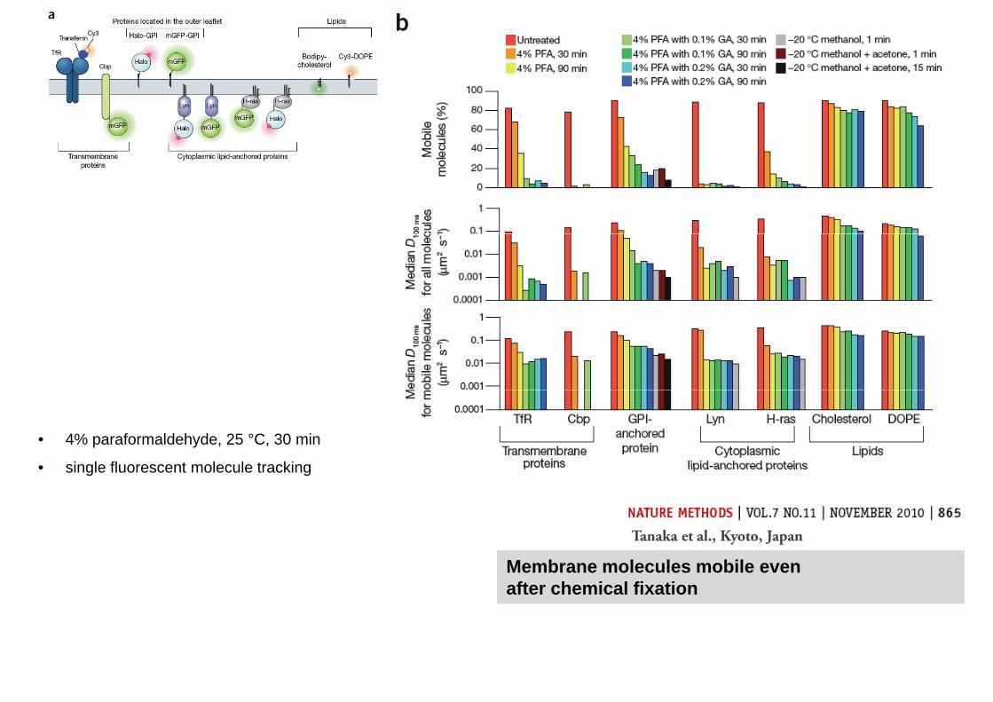
• 4% paraformaldehyde, 25 °C, 30 min
• single fluorescent molecule tracking• single fluorescent molecule tracking
Tanaka et al., Kyoto, Japan
Membrane molecules mobile evenafter chemical fixation
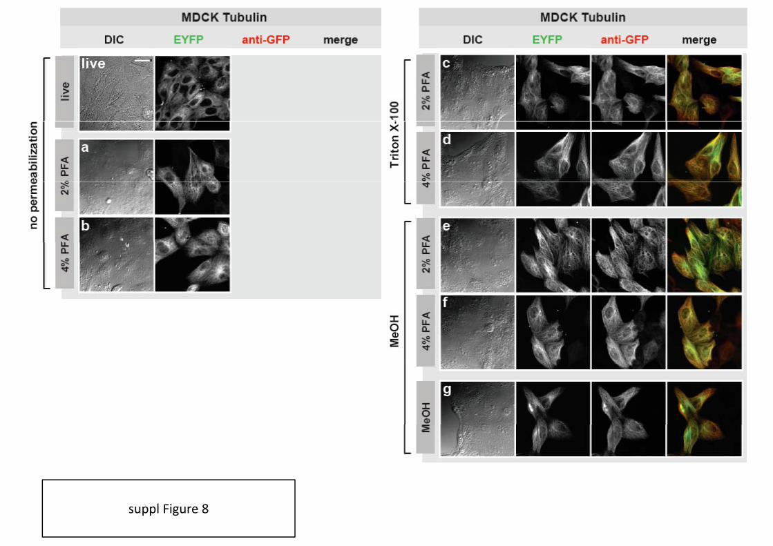
suppl Figure 8

suppl Figure 9

How to avoid artefacts?
• use fresh chemicals:
• prepare PFA freshly, adjust the pH carefully
• ready‐to‐use fixatives (‘Fix&Perm’) contain preservatives (avoiding polymerisation) that can induce
artefatcs; work for some samples but not other
h t’ d h f FACS i ’t il d h f i• what’s good enough for FACS isn’t necessarily good enough for microscopy
• Controls:
• comparison live (fluorescent protein) / fixed (antibody staining)
caution: fluorescent proteins tend to dimerise / oligomerise and can interfere with protein functions / interactions
• comparison various fixation / permeabilisation chemicals, protocols and conditions
• comparison various cell typesp yp
• positive control: known physiological effect (change in expression and / or localisation of protein of
interest)
• Observation:
• optimise cell viability before fixation
• observe samples throughout fixation / permeabilisation process
• good brightfield images! (DIC, phase contrast, darkfield)

good brightfield images!
DIC images of Jurkat cells

control: physiological effect
GFPanti‐GFPAlx‐633
neg control(unstimulated)
stimulation(PKD translocation)
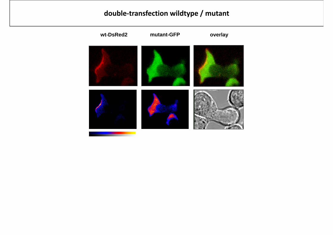
double‐transfection wildtype / mutant
wt-DsRed2 mutant-GFP overlay

A
BC
D
Imperial CollegeImperial CollegeLondonLondon
–– South KensingtonSouth KensingtonSouth KensingtonSouth Kensington
Facility for Imagingby Light Microscopy
Observing Life As It Happens
ClubObserving Life As It Happens
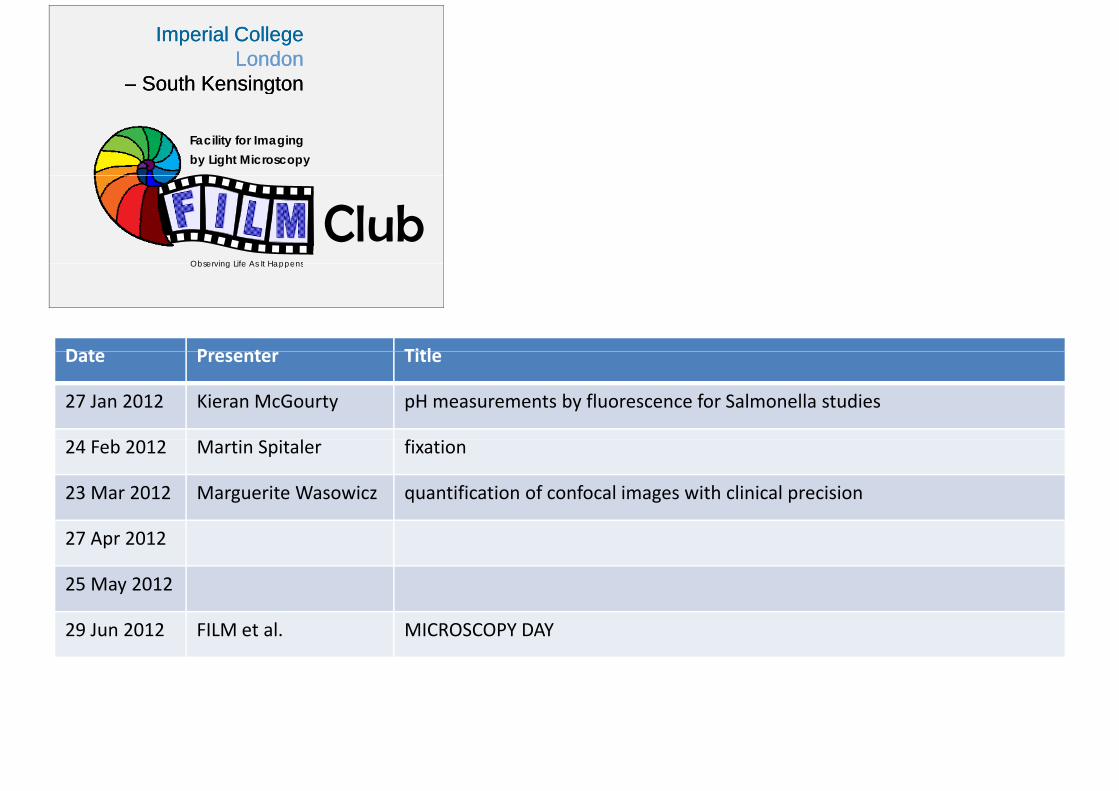
D t P t TitlDate Presenter Title
27 Jan 2012 Kieran McGourty pH measurements by fluorescence for Salmonella studies
24 Feb 2012 Martin Spitaler fixation
23 Mar 2012 Marguerite Wasowicz quantification of confocal images with clinical precision
27 Apr 2012
25 May 2012
29 Jun 2012 FILM et al. MICROSCOPY DAY