Embed Size (px)
Citation preview

Facial Chemical Peels
Gordon Shields, MD
The University of Texas Medical Branch
Department of Otolaryngology
Grand Rounds Presentation
May 12, 2004

History
Ebers papyrus 3,500 years ago mentions keratolytic formulas
Egyptians used particles of alabaster mixed with milk and honey
1882 German dermatologist P.G Unna described resorcinol, salicylic acid, phenol, trichloroacetic acid
Frenchman La Gasse World War I – treated soldiers with powder burns to face with phenol
daughter Antoinette brought to Los Angeles 1930’s

American lay peelers 1920’s-1950’s
1961 Litton presented 50 patients treated
with formula he bought from Coopersmith,
a lay peeler in Fort Lauderdale
1961 Baker and Gordon presented a peel
formula with one patient with a 3 month
follow up, became the standard formula
1966 Baker published results in 250
patients

Application of chemical exfoliant to
wound the dermis and epidermis
Creates a superficial burn
Classified by depth of injury
Superficial/Very superficial
Medium
Deep


Post Peel

72 hours post-peel

Post Peel Complete

Sun exposure and aging
Coarse rhytids
Rougher skin texture
Pigmentary mottling
Solar lentigines/actinic keratoses
Epidermal dysplasia
Epidermal atrophy
Dermoelastosis

Elastosis- irregular
formation of
connective tissue
14 years post peel
with more orderly,
parallel collagen
fibers

Patient evaluation
Skin type
Complexion
Skin texture
Thickness
Degree of photoaging
Severity of facial rhytids
Age related gravitational changes
Expectations

Patient evaluation
History of skin disorders like rosacea,
seborrheic dermatitis, psoriasis, contact
dermatitis
History of radiation to the face (facial hair?)
History of Accutane use (12-24 months)
General medical condition including renal,
liver and cardiac function if phenol peel is
considered


Fitzpatrick types I and II are best
canidates for chemical peel
Types III-VI have greater risk of
pigmentary dyschromia

Pretreatments
Tretinoin (thins stratum corneum,
thickens epidermis, disperses melanin
throughout epidermis)
Hydroquinone 4-8% -blocks
tyrosinase from forming precursors for
melanin (use when treating
dyschromias or Type III-VI)
Topical steroids

Pretreatments
Herpes simplex outbreak can be caused by chemical peels
Must be considered in all patients – especially those with a history
Heralded by unusually sever facial pain that is unexpected and delayed
Can cause scarring
Acyclovir from 1-3 days preoperatively to 14 days postoperatively
Some start antibiotics 1-2 days preoperatively (cephalexin)

Superficial peels
Indicated for: Comedonal acne
Melasma
Skin refresher
Nonfacial peeling
Repetitive peels may be required to gain maximum benefit (usually 6-8 every 1-4 weeks)
Low-risk, rapid recovery
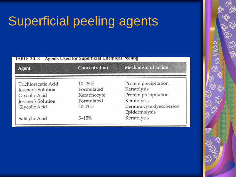
Superficial peeling agents

Heals in 1-5 days
Very superficial agents include
glycolic acid 20-70%, 10% TCA,
salicylic acid
Multiple superficial peels do not equal
a medium depth peel

Medium and deep peel indications
Actinic changes and preneoplasia
Rhytides (fine)
Pigmentary dyschromias
Woods lamp helpful
Superficial scarring
Radiation dermatitis
Acne vulgaris and rosacea


Specific chemicals
Alpha-hydroxyacids
Jessner’s solution
Trichloroacetic acid
Baker-Gordon peel formula

Alpha-hydroxy acids
Present in foods
Glycolic acid, Lactic acid most common
Low concentrations - reduction of cohesiveness of stratum corneum cells resulting in thinning
High concentrations – complete epidermolysis
Action is stopped by water dilution
No known systemic toxicity
Can be combined with TCA to produce medium peel

Jessner’s solution

Jessner’s solution
Standardized solution
Salicylic acid – keratolytic
Resorcinol – benzene derivative, keratolysis
Lactic acid – AHA, keratolysis
Used alone causes superficial epidermal peel
Number of applications controls depth
No neutralization is necessary
Can be combined with TCA for medium peel
Possible to have salicylate toxicity (tinnitus, headache, nausea) or resorcinol toxicity (methemoglobinemia)

Trichloroacetic acid
Dissolves keratin, coagulates skin proteins, causes precipitation of salts
Neutralized by tissue fluids
Causes frosting
10-30% superficial peel
35-50% superficial-medium depth peel
50% should be avoided due to scarring
Proper technique to mix is weight-to-volume ratio, i.e. 50% solution is 50 g TCA crystals in enough distilled water to make 100cc of solution

Phenol
88% is standard concentration
Carbolic acid or hydroxybenzene
Used alone causes medium depth peel
Causes keratin protein coagulation
Rapidly absorbed through skin, metabolized in the liver, excreted renally
Can lead to renal failure, hepatotoxicity, directly irritates myocardium causing arrythmias
Requires cardiac monitoring and testing of kidney/liver/cardiac function
Use hydration to prevent problems
Peel in subunits with 15-20 minutes per site

Deep Peel
Baker-Gordon formula
3cc of 88% phenol
2cc distilled water
8 drops Septisol (emulsifying agent)
3 drops croton oil (lysis epidermal cells, causes
inflammation from plant Croton tiglium)
Some authors have used occlusion to increase
penetration
Requires anesthesia and monitoring

TCA Peeling
Depth of peel depends on multiple
factors
Skin type
How skin was primed
How acid is applied
How many layers of acid are applied
How wet the applicator is with acid

TCA peeling steps
Skin priming – increase
reepithelialization and decreased risk
of hyperpigmentation
Cleaning – alcohol, acetone,
Hibiclens, etc.

Application
Patient is inclined at 45 degrees
Use cotton-tipped applicators or
folded 2x2 gauze
Peel in aesthetic units
Feather edges
Observe for signs of peel depth


Peel depth
Level 0 – no frost, minimal erythema, removes stratum corneum
Level I – irregular light frost, some erythema, superficial epidermal peel, 2-4 days healing
Level 2 – white frost with pink showing through, full thickness epidermal peel, about 5 days to heal
Level 3 – solid white frost, no pink background, extends into papillary dermis and takes 5-7 days to heal
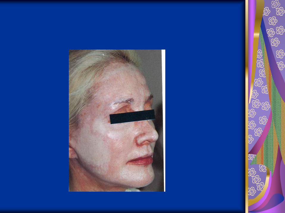

30% TCA peel

30% TCA

Deep Peel, Baker-Gordon

Application

Tape occlusion

6 months
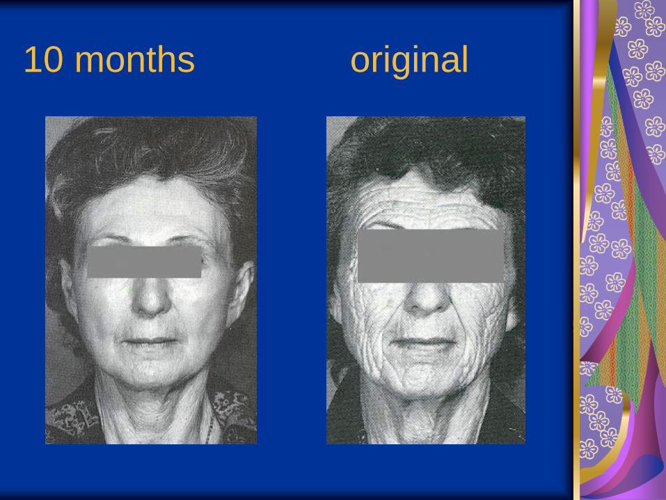
10 months original

Baker-Gordon/CO2-35%TCA

Baker-Gordon peel

Post peel care
Occlusive ointment: Eucerin cream, Elta,
bacitracin ointment, A&D ointment, Crisco
until re-epthelialization occurs
Lubriderm or Eucerin cream
May wear makeup
Topical steroids
Antivirals and antibiotics
Sunscreen

Wound healing when dry

Wound healing when moist

Complications
Hyperpigmentation
Hypopigmentation
Scarring
Persistent erythema
Herpes outbreak
Milia
Infection
Lines of demarcation

Hyperpigmentation

Hypopigmentation

Persistent erythema

Lines of demarcation

Herpes outbreak

Candida infection

Pseudomonal infection

Scarring

Laser versus chemical peel
CO2 laser produces injury to depth of
0.14-0.22mm compared with 0.60-
0.80mm with deep chemical peel
Chemical peel requires some user
expertise, mixing chemicals
Laser requires having a laser,
computer controlled handpiece with
pattern generator makes much easier

Laser versus chemical peel

Immediate

2 weeks later

One year later

Authors conclude that peel is equally
effective in thin-skinned areas
Laser better in thick glandular skin but
had more hypopigmentation, longer
discomfort, longer postop erythema

Conclusions
Chemical peels remain an important
tool
Knowledge of chemicals used,
methods and indications important
Make sure the treatment matches the
patient’s problem and expectations

Facial Chemical Peels
Gordon Shields, MD
The University of Texas Medical Branch
Department of Otolaryngology
Grand Rounds Presentation
May 12, 2004