Embed Size (px)
Citation preview
IJCA Special Issue on “Recent Trends in Pattern Recognition and Image Analysis" RTPRIA 2013
27
Face Detection Algorithm for Skintone Images using
Robust Feature Extraction in HSV Color Space
H C VijayLakshmi Dept of Computer Science & Engineering
S J College of Engineering, Mysore,INDIA
S. Patilkulkarni, Ph.D
Associate Professor
S J College of Engineering, Mysore,INDIA
ABSTRACT
A robust feature extraction method in HSV space is proposed
for face detection problem in skin toned images using
biorthogonal wavelet detail coefficients. It is demonstrated
that followed with neural network classifier, proposed method
is robust under varying conditions.
General Terms
Feature extraction, threshold, wavelet, color space, skin tone
Keywords
Biorthogonal, HSV color space,
1. INTRODUCTION
1.1 Face Detection Face detection and localization is the task of checking
whether the given input image or video sequence contains any
human face, and if human faces are present, returning the
location of the human face in the image. The face detection
problem involves segmentation, feature extraction, and
classification of the segmented region as face or non face
irrespective of the background and occlusion. Several
researchers are at this task with different approaches, so that
the machine detects and locates the faces efficiently as we
human beings do in any complex scenarios.
The faces play a major role in identifying and recognizing
people. The area of face detection has gained considerable
importance with the advancement of human-machine
interaction as it provides a natural and efficient way to
communicate between humans and machines. Face detection
and localization in image sequences has become a popular
area of research due to emerging applications in intelligent
human-computer interface, surveillance systems, content-
based image retrieval, video conferencing, financial
transaction, forensic applications, and many other fields. Face
detection is essentially localizing and extracting a face region
from the background. This may seem like an easy task but the
human face is a dynamic object and has a high degree of
variability in its appearance. A large number of factors that
govern the problem of face detection [5, 6].The long list of
these factors include the pose, orientation, facial expressions,
facial sizes found in the image, luminance conditions,
occlusion, structural components, gender, ethnicity of the
subject, the scene and complexity of image‟s background.
Faces appear totally different under different lighting
conditions. A thorough survey of face detection research work
is available in [5, 6]. In terms of applications, face detection
and good localization is an important pre-processing step in
online face recognition and surveillance systems. In the recent
years lots of progress has been made in detecting faces with
slight variation in illumination, pose, expression and
background. A number of techniques have been developed by
researchers in order to efficiently detect human faces in any
given input image or video sequence.
A face detection algorithm with a novel and robust feature
extraction method is proposed, which is invariant to
illumination, background and expression. Each face image
contains wavelet decomposed detail coefficient extracted in
hue, saturation, value channels, eye, nostril and mouth blobs
are dilated and threshold to obtain facial features.
Experiments are conducted using the personal database
developed by the authors as well as Bao Face Database
available on the internet.
1.2 Wavelets Wavelets are mathematical functions that initially cut up the
data into different frequency components and then study each
component with a resolution matched to its scale. Thus
wavelets analyze according to scale and self-similarity caused
by scales and dilations. Wavelet algorithms process data at
different scales or resolutions. The wavelet analysis
procedures adopt a wavelet prototype function called an
analyzing wavelet or mother wavelet [3]. The Discrete
Wavelet Transform (DWT) has become a very versatile signal
processing tool over the last decade. In fact, it has been
effectively used in signal and image processing applications
ever since Mallat [12] proposed the multiresolution
representation of signals based on wavelet decomposition.
Wavelet transform is a representation of a signal in terms of a
set of basis functions, which is obtained by dilation and
translation of a basis wavelet. The advantage of DWT over
other traditional transformations is that it performs
multiresolution analysis of signals with localization both in
time and frequency. Orthogonal or biorthogonal wavelet
transform has often been used in many image processing
applications, because it makes possible multiresolution
analysis and does not yield redundant information, refer
Mallat [12]. The wavelet consists of two components, the
scaling function which describes the low-pass filter for the
wavelet transform, and the wavelet function which describes
the band-pass filter for the transform. For orthogonal
wavelets, the scaling function Φ and mother wavelet 𝛹 are
given by the recursion relations
𝛷 𝑥 = 2 𝑘𝛷 2𝑥 − 𝑘 𝑘 (1)
𝛹 𝑥 = 2 𝑔𝑘𝛷 2𝑥 − 𝑘 𝑘 (2)
Their scaled translates are denoted by
𝛷𝑘𝑛 𝑥 = 2
𝑛
2𝛷(2𝑛 𝑥 − 𝑘) (3)
𝛹𝑘𝑛 𝑥 = 2
𝑛
2𝛹(2𝑛 𝑥 − 𝑘) (4)
IJCA Special Issue on “Recent Trends in Pattern Recognition and Image Analysis" RTPRIA 2013
28
The translates of scaling function form the basis set for the
approximation subspace, where as those of wavelet function
form the basis set of detail subspace respectively, at the n-th
level resolution. Wavelet transforms can be applied in a
number of scientific research areas such as feature extraction,
edge and corner detection, partial differential equation
solving, transient detection, filter design, electrocardiogram
(ECG) analysis, texture analysis, business information
analysis and gait analysis. Transforms in image processing are
two-dimensional. Discrete wavelet transform is calculated by
applying the corresponding one-dimensional transform to the
columns first, and then to the rows. When filtering, we have
four possibilities
low-pass filter to rows, followed by low-pass filter
to columns (LL coefficients)
low-pass filter to rows, followed by high-pass filter
to columns (HL coefficients)
high-pass filter to rows, followed by low-pass filter
to columns (LH coefficients)
high-pass filter to rows, followed by high-pass filter
to columns (HH coefficients)
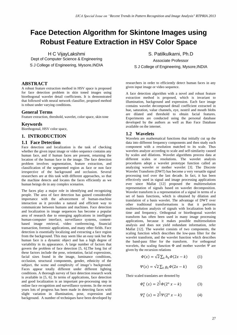
In wavelet decomposition, the image is split into an
approximation and details images. Approximation image is
obtained by low pass filtering and detail images are obtained
by high pass filtering. Further decomposing the approximation
image (LL1 subband), we will get second level LL2, HL2,
LH2 and HH2 coefficients as shown in the Fig. 1a. The high
pass or detail component characterizes the images‟ high
frequency information and the low pass or approximation
component contains its low frequency information.
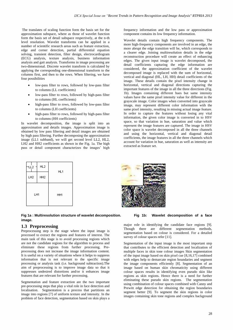
Wavelet details contain high frequency components. The
more high-frequency components are involved in an edge, the
more abrupt the edge transition will be, which corresponds to
a clearer edge. Joining multiresolution details in the edge
reconstruction procedure will create an effect of enhancing
edges. The given input image is wavelet decomposed, the
detail coefficients capturing the edge information are
considered, the approximation coefficient of the wavelet
decomposed image is replaced with the sum of horizontal,
vertical and diagonal (HL, LH, HH) detail coefficients of the
image. These details contain the pixel intensity change in
horizontal, vertical and diagonal directions capturing the
important features of the image in all the three directions (Fig.
1b). Images containing different hues but same intensity
values have the same pixel intensity value for different in the
grayscale image. Color images when converted into grayscale
image, may represent different color information with the
same pixel intensity, resulting in missing actual image feature.
In order to capture the features without losing any vital
information, the given color image is converted in to HSV
space, so that variation in hue, saturation and value which
represent the image features are captured. The image in HSV
color space is wavelet decomposed in all the three channels
and using the horizontal, vertical and diagonal detail
coefficients, the image features in all the three channels which
account for variation in hue, saturation as well as intensity are
extracted as feature set.
Fig 1a : Multiresolution structure of wavelet decomposition. Fig 1b: Wavelet decomposition of a face
image.
1.3 Preprocessing Preprocessing step is the stage where the input image is
processed to extract the regions and features of interest. The
main task of this stage is to avoid processing regions which
are not the candidate regions for the algorithm to process and
eliminate these regions from further processing. Pre-
processing does not increase the image information content.
It is useful on a variety of situations where it helps to suppress
information that is not relevant to the specific image
processing or analysis task (i.e. background subtraction).The
aim of preprocessing is to improve image data so that it
suppresses undesired distortions and/or it enhances image
features that are relevant for further processing.
Segmentation and feature extraction are the two important
pre-processing steps that play a vital role in face detection and
localisation. Segmentation is a process that partitions an
image into regions [7] of uniform texture and intensity. In the
problem of face detection, segmentation based on skin plays a
major role in identifying the candidate face regions [9].
Though there are different segmentation methods,
segmentation based on colour is considered. For a detailed
survey of colour spaces refer [11].
Segmentation of the input image is the most important step
that contributes to the efficient detection and localization of
multiple faces in skin tone colour images Skin segmentation
of the input image based on skin pixel cue [8,16,17] combined
with edges help to demarcate region boundaries and segment
the image components efficiently [8]. Segmentation of an
image based on human skin chromaticity using different
colour spaces results in identifying even pseudo skin like
regions as skin regions. Hence there is a need for further
eliminating these pseudo skin regions. The segmentation
using combination of colour spaces combined with Canny and
Prewitt edge detection for obtaining the region boundaries
segment better [9]. To segment the skin regions in color
images containing skin tone regions and complex background
IJCA Special Issue on “Recent Trends in Pattern Recognition and Image Analysis" RTPRIA 2013
29
refer [10]. In this approach, a wavelet texture energy based
illumination compensation for segmenting the outdoor bright
sunlight skin tone images even with complex background is
used. This methodology generates a texture energy image for
wavelet approximation image, addresses illumination
compensation and results in smooth segmentation. The
blurred wavelet texture energy image generated removes
small details and bridges small gaps in lines and curves.
Random noise typically consists of sharp transitions in pixel
intensity level are minimized and thus results in a better object
boundary. For each channel approximation image, using a
3X3 image window a texture energy image is generated using
the following equation
𝑡 𝑥, 𝑦 =1
∗
𝑎
𝑠=−𝑎
𝑓 𝑥 + 𝑠, 𝑦 + 𝑡 ∗ 𝑓(𝑥 + 𝑠, 𝑦 + 𝑡)
𝑏
𝑡=−𝑏
Here f(x, y) is the input image, s and t the size of the window,
in our case 3X3 kernel is considered and t(x, y) is the
weighted average texture image generated and „h‟ is the
threshold. Using the approximation texture energy images
generated, the image in each channel is reconstructed and the
entire three channel images are combined to obtain the
illumination compensated RGB image.
2. FEATURE EXTRACTION Feature extraction is the method of capturing information
about the object in compact way, using these features one
should be able to retrieve and hence recognize the object
under consideration. Features should contain vital information
about the object of interest; the feature extraction should be
easy to compute and should be robust and compact enough to
represent the object under consideration uniquely. The
features extracted should well depict the human perception
about the object. Image features can refer to global
properties of an image i.e. average gray level, shape of
intensity histogram etc. and local properties of an image such
as edges, textures, important features uniquely representing
the object and also shape of contours etc. From object
recognition purpose these image features should be local,
meaningful, detectable parts of an image. Meaningful
features are associated to unique information in the image
(e.g., like eyes, eyebrows, mouth and nose in a human face).
They should be invariant to some variations in the image
formation process (i.e. invariance to viewpoint and
illumination for images captured with digital cameras). These
features extracted should be detectable, they can be located or
detected from images via algorithms and features should
robustly capture the salient features of the object. The features
extracted should be represented using a feature vector with
less complexity. Robust and compact feature set [13, 14, 15]
yield good detection rates. In this paper in order to extract
facial features without losing any information, color face
image is converted in to HSV color space and the variation in
hue, saturation and value are also considered.
Edges occur in images due to sudden change in pixel intensity
values. Edges are the most important feature of an image.
Edge detection is an important task in image processing. It is a
main tool in pattern recognition, image segmentation, and
scene analysis. An edge detector is basically a high pass filter
that can be applied to extract the edge points in an image.
Object recognition, image segmentation and image coding
applications require robust and smooth edges clearly
highlighting the regions of interest. For a detail survey of
edges in grayscale images refer [2]. Important features can be
extracted from the edges of an image (e.g., corners, lines,
curves). These features are used by higher-level computer
vision algorithms (e.g., recognition). In gray scale images, the
edges correspond to change in illumination, pixel intensity
values. This situation is different in case of color images,
which gives more detailed edge information. Color plays a
very important role in determining object boundaries in color
images. In color images, intensity, hue and saturation of a
color all play a part in determining object boundaries. Objects
having different hues but same intensity values represent the
same pixel intensity value in the grayscale image. Color
images containing such hues, when converted into grayscale
image, may represent different color information in to a single
region, resulting in missing actual object feature and
boundary. This is very vital for object segmentation, detection
and recognition in color images. Edge extraction in color
images provide vital clue about the object‟s hue variation,
intensity variation and also the variation in the amount of
saturation in hue. In case of grayscale images, the object
boundary and also object features are captured when there is a
sharp change in the intensity values in a single channel. In
case of color images, more information regarding the object
can be captured in more than one channel. For a
comprehensive analysis refer [1].
3. PROPOSED APPROACH A holistic approach is used, instead of looking for the
presence of facial features such as eye, mouth, nose
separately. In the proposed approach, the algorithm uses the
following steps to extract the prominent features.
Step1: Extract the facial features by first converting the
windows containing face image from RGB color space to
HSV color space.
Step2: Obtain the detail coefficients of each of the three
channels (H, S, V) after wavelet decomposition using discrete
biorthogonal wavelet transform, in particular, „bior1.3‟
wavelet filters. The eye and mouth socket regions extracted in
H, S, V channel are dilated in all the three channels using
morphological operation to enhance the features and make it
clearer. Consider only the horizontal, vertical and diagonal
details in all the three channels.
Step3: Replace the LL subband image i.e. approximation
coefficients with the sum of horizontal, vertical and diagonal
details as these detail coefficients capture all the important
features of the face. Reconstruct the image with enhanced
detail coefficients in each channel.
Step4: The reconstructed image in HSV channel which
predominantly contains eyebrows, eyes, nostrils and mouth
regions as shown in Figure 2b is converted back to RGB color
space and then to grayscale image and an image histogram is
generated. Using suitable hard threshold, face images
highlighting only the prominent facial features are obtained.
Step5: Using this procedure features are extracted for the
facial window images considered for training and stacked as
column vectors in a data matrix. Similarly nonface images are
also wavelet decomposed in H, S and V channel and using the
above procedure, the non facial features extracted are
appended to the data matrix. This serves as the input data
matrix for training the neural network.
Step6 : Create an output column vector having the number of
rows equal to the number of columns of input data matrix
created in the previous steps. Initialize the output column
vector values to +1 or 0 indicating face or non-face. Initialize
the neural network parameters and train the network to obtain
appropriate weight sequence w(n) using back propagation
IJCA Special Issue on “Recent Trends in Pattern Recognition and Image Analysis" RTPRIA 2013
30
algorithm. Multilayer perceptrons (MLP), with input layer,
hidden layer and output layer is used for classifying the
window image as face or nonface. Using this approach, the
number of face and non face images required for training is
vey less and training is very fast, when compared with other
approaches which use neural network for classification.
In testing phase, segment the input image containing multiple
faces using the approach proposed by the authors in [10],
using a sliding window across the skin segmented regions and
convert the window image into HSV color space and apply
steps mentioned above to extract the features. Test the
extracted features with the neural network trained earlier
using features extracted using step1 to step5. Classify the
window image as face, if the output is greater than 0.5,
otherwise nonface. If the input image contains faces of
different size, then the sliding window size is scaled up or
down depending on the need and the procedure is repeated to
locate faces of different sizes.
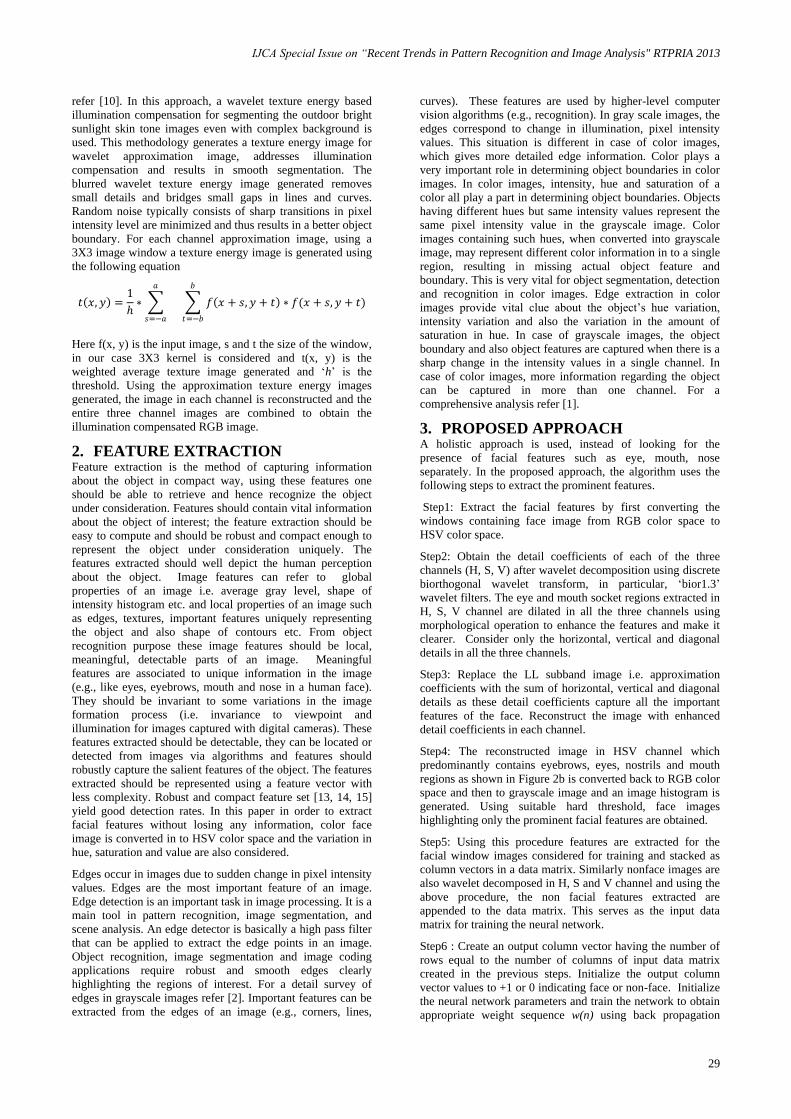
The features extracted are as shown in Figure 2. Sample
features extracted for windows containing faces with
variations in pose and expression used in the experiment are
as shown in Figure 3. The images considered for training
have upright frontal faces, faces with variation in pose,
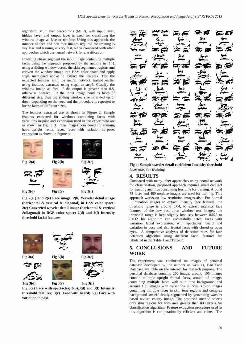
expression as shown in Figure 4.
Fig 2(a) Fig 2(b) Fig 2(c)
Fig 2(d) Fig 2(e) Fig 2(f)
Fig 2(a ) and 2(e) Face image; 2(b) Wavelet detail image
(horizontal & vertical & diagonal) in HSV color space;
2(c) Converted wavelet detail image (horizontal & vertical
&diagonal) in RGB color space; 2(d) and 2(f) Intensity
threshold facial features
Fig 3(a) Fig 3(b) Fig 3(c)
Fig 3(d) Fig 3(e) Fig 3(f)
Fig 3(a) Face with spectacles; 3(b),3(d) and 3(f) Intensity
threshold features; 3(c) Face with beard; 3(e) Face with
variation in pose.
Fig 4: Sample wavelet detail coefficient Intensity threshold
faces used for training.
4. RESULTS Compared with many other approaches using neural network
for classification, proposed approach requires small data set
for training and thus consuming less time for training. Around
75 faces and 450 nonface images are used for training. This
approach works on low resolution images also. For normal
illumination images to extract intensity face features, the
threshold range is around 0.04, to extract intensity face
features of the low resolution window test images, the
threshold range is kept slightly low, say between 0.028 to
0.032.This algorithm can successfully detect faces with
variation facial expression, with spectacles, beard and
variation in pose and also frontal faces with closed or open
eyes. A comparative analysis of detection rates for face
detection algorithm using different facial features are
tabulated in the Table 1 and Table 2.
5. CONCLUSIONS AND FUTURE
WORK
The experiment was conducted on images of personal
database developed by the authors as well as, Bao Face
Database available on the internet for research purpose. The
personal database contains 250 image, around 105 images
contain multiple upright frontal faces, around 45 images
containing multiple faces with skin tone background and
around 100 images with variations in pose. Color images
containing multiple faces in skin tone regions and complex
background are efficiently segmented by generating wavelet
based texture energy image. The proposed method selects
only skin regions for with area greater than 800 pixels for
classification algorithm. Feature extraction procedure used in
this algorithm is computationally efficient and robust. The

IJCA Special Issue on “Recent Trends in Pattern Recognition and Image Analysis" RTPRIA 2013
31
features extracted using this procedure clearly distinguishes a
facial image from a non facial image. We plan to extend this
approach to only profile faces as future work. The result is
also compared with other approaches [18], [19] proposed by
the authors during experimentation and this method is found
to have better false rejection and acceptance values.
Table-1 Comparative result of different methods
Features Used Classification
Method
Windows
Tested
False
Positive
False
Rejection
Nature of Dataset Used
Wavelet Approximation
coefficients
Bhattacharya
Distance
1500
128
20
Frontal Faces Under Normal illumination
condition.
City block
1500
44
38
Frontal Faces Under Normal illumination
condition.
Signature of Wavelet
Approximation
coefficients Image
correlation
2000
50
0
Frontal Faces Under Normal illumination
condition.
Gray Scale Edges of
Wavelet Approximation
coefficients
City block
1700
30
3
Frontal faces with slight variation in pose and
expression
R & G channel Edges of
Wavelet Approximation
coefficients
City block
2800
18
8
Frontal faces with slight variation in pose and
expression
Approximation image
with Gray Scale Edges
Neural
Network
3000
5
0
Frontal faces with variation in pose and
expression and spectacles
Proposed Method Euclidean
Distance
3000 100 25 Frontal faces with variation in pose and
expression and spectacles.
Proposed Method Neural
network
4000 5 10 Frontal faces with sufficient variation in pose
and expressions, faces containing spectacles,
Beard, Partly covered faces. Bright sunlight,
Normal Illumination Low resolution images.
Table 2 Comparison of approximation image edge features Vs HSV detail image features results
Features Used Classification
method
Windows
Tested
False
Positive
False
Rejection
Nature of Dataset Used
Approximation image
with Gray Scale
Edges
Neural
Network
2000
15
25
Frontal faces with sufficient variation in pose and
expressions, faces containing spectacles, Beard,
Partly covered faces. Bright sunlight, Normal
Illumination Low resolution images.
Proposed Method Neural
network
4000 5 10 Frontal faces with sufficient variation in pose and
expressions, faces containing spectacles, Beard,
Partly covered faces. Bright sunlight, Normal
Illumination Low resolution images.
IJCA Special Issue on “Recent Trends in Pattern Recognition and Image Analysis" RTPRIA 2013
32
6. REFERENCES [1] Shu-Yu Zhu, Konstantinos N. Plataniotis, Anastasios N.
Venetsanopoulos, Comprehensive analysis of edge
detection in color image processing, Optical Engineering,
Vol. 38 No. 4, (April 1999), pp 612-625.
[2] Raman Maini, Dr. Himanshu Aggarwal, Study and
Comparison of Various Image Edge Detection
Techniques, International Journal of Image Processing
(IJIP), Vol. 3, Issue 1, pp. 2-12.
[3] L. Feng, C. Y. Suen, Edge extraction of images by
reconstruction using wavelet decomposition details at
different resolution levels, International Journal of
Pattern Recognition and Artificial Intelligence, Vol. 14,
No. 6, pp. 779-793.
[4] S. Anila, N. Devarajan, Simple and Fast Face Detection
System Based on Edges, International Journal of
Universal Computer Sciences, Vol. 1, Issue 2, pp. 54-58.
[5] Y. Ming-Hsuan, D J Kreigman, and N Ahuja “Detecting
Faces in Images: a Survey”, IEEE Transactions on
Pattern Analysis and Machine intelligence, (2002), Vol.
24, pp. 34-58.
[6] W. Zhao, R. Chellappa, A. Rosenfeld, and P. Phillips
“Face recognition:A literature survey”, ACM Computing
Surveys , (2003), pp. 399.458.
[7] R. C. Gonzalez, R. E. Woods Digital Image processing,
Second edition Prentice Hall India.
[8] F Fritsch, S Lang, M Kleinehagenbrock, G A Fink and
Sagerer “Improving Adaptive Skin Colour Segmentation
by incorporating Results from Face Detection”, In
Proceedings of the IEEE International workshop on
Robot and Human Interactive Communication, Berlin,
Germany, (September 2002), pp.337-343.
[9] H.C. Vijaylakshmi, S. PatilKulakarni, Segmentation
Algorithm for Multiple Face Detection for Color Images
with Skin Tone Regions, International Journal of
Computer Theory and Engineering, (August, 2010), Vol.
2, No. 4, pp. 552-558.
[10] H.C. Vijaylakshmi, S. PatilKulakarni “Illumination
Compensation to Segment True Skin and Non-skin
Regions For Skin Tone Images” Springer Computational
Intelligence and Information Technology, (2011), Pune
Volume 250, 2011, pp 494-499.
[11] V. Veznevets, V Sazonov, nad A Andreeva, A survey on
pixel-based skin color detection techniques. In
Proceedings of GRAPHICON conference (2003), pp. 85-
92.
[12] S. Mallat, “A Theory forMultiresolution Signal
Decomposition: TheWavelet Representation,” IEEE
Trans. Pattern Analysis and Machine Intelligence,
(1989), vol. 11, no. 7, pp. 674–693.
[13] P. Viola and M. Jones, "Robust real time object
detection," International Journal of Computer Vision,
vol. 57, no. 2, pp. 137-154, 2001.
[14] Rein-Lein Hsu, Mohamed-Abdel Mottaleb, Anil K jain
Face Detection in Color Images, IEEE Transactions on
Pattern Analysis and Machine Intelligence, Vol. 42, Issue
5, pp.696-706.
[15] C. Garcia, G. Zikos, G. Tziritas, Face Detection in Color
Images using Wavelet Packet Analysis. In Proceedings
of IEEE International Conference on Multimedia
Computing and Systems, (1999) Vol.1, pp. 703 – 708.
[16] Mohamed A. Berbar Hamdy M. Kelash, and Amany A.
Kandeel, “Faces and facial features detection in color
images,” In Proceedings. Geometric Modeling and
Imaging― New Trends (GMAI'06), pp 209 – 214.
[17] Son Lam Phung, Abdesselam Bouzerdoum, and Douglas
Chai, Skin segmentation using colour and edge
information. In Proceedings of the International
Symposium on Signal Processing and its Applications,
Paris, France, (July 2003), pp. 353-356.
[18] H.C. Vijaylakshmi, S. PatilKulakarni, Face Detection in
Skin-Toned Images Through Wavelet Edges and Neural
Network, International Journal of Computer and
Electrical Engineering,Vol. 4, No. 5.
[19] H.C. Vijaylakshmi, S. PatilKulakarni, Face Detection in
Skin-toned Images Using EdgeDetection and Feature
Extraction Using R and G Channels through Wavelet
Approximation, International Journal of Computer
Theory and Engineering, Vol. 5, No. 1.


![Face Recognition Performance: Role of Demographic Information · Subspace Features (4SF) face recognition algorithm [5] (i.e., the trainable face recognition algorithm used in this](https://img.pdfslide.us/doc/110x75/5bfcb1c809d3f297368b9dee/face-recognition-performance-role-of-demographic-subspace-features-4sf-face.jpg)