Embed Size (px)
Citation preview
ORIGINAL
Fabrication of DNA nanotubes using origami-basednanostructures with sticky ends
Mohammad Mousavi-Khattat • Adele Rafati •
Pooria Gill
Received: 30 October 2014 /Accepted: 16 January 2015 / Published online: 5 February 2015
� The Author(s) 2015. This article is published with open access at Springerlink.com
Abstract Described here is a simplified method for fab-
rication of DNA nanotubes using a minimum numbers of
staple oligomers for DNA origami. For this purpose, the
cylindrical nanotemplates with two sticky ends have been
designed using caDNAno software. Then, the nanostruc-
tures were shaped in an optimized experimental condition
via an origami-based self-assembly reaction. Finally, the
produced nanostructures were joined together through their
sticky ends using a ligase enzyme. Transmission electron
microscope confirmed fabrication of these elongated
nanotubes. In addition, high-resolution microscopy of
DNA nanotubes by scanning tunnelling microscope indi-
cated efficient attachments of the primarily DNA nano-
structures via their sticky ends. The results demonstrated
that a ligase treatment of cylindrical DNA nanostructures
with the sticky ends made DNA nanotubes with standard
shapes using minimum numbers of staples.
Keywords DNA nanotube � T4 ligase � Sticky ends �TEM � STM
Introduction
DNA nanotechnology uses DNA as a building block to
create nano-scale structures which acts as a candidate in
multidisciplinary applications such as fabrications of nan-
odevices, nanoarrays, nanodiagnostics, nanotherapeutics,
and the other demanding areas [1–4]. Among DNA nano-
structures, DNA nanotubes have particularly potentials for
functions due to high aspect ratio [5], controllability in
fabrications [6, 7], and some capabilities for applications as
nanocarriers [8], nanoarrays [9], nanowires [10], nanotools
for structural analysis [11], and nanoanalytes for control-
ling some fabrication processes [12].
DNAorigamiwas introduced as a newgrowthmechanism
for DNA assembly for fabrication of various shapes of DNA
nanostructures. In this technique, a long single-stranded
DNA templated the final structure, so-called DNA scaffold
[13]. DNA origami gives excellent control over the size and
shape of objects [14, 15]. However, this level of structural
complexity requires hundreds of staple strands with different
sequence as well as long annealing times [16]. In other
words, obtaining nanotubes with high aspect ratio need more
quantities of staple oligonucleotides within a DNA origami
procedure; however, more staples in fabrication made more
costs in practice. Hence, we aimed here to introduce a simple
procedure for fabrication of DNA nanotubes via an origami-
based technique assisted with a ligase reaction. This fabri-
cation process provides DNA nanotube with minimum
numbers of the staples within a self-assembly reaction.
In this study, computational modelling of DNA nano-
tubes performed using caDNAno software [17] that
superimposed a cylindrical scaffold with staples as essen-
tials in a self-assembly process. The nanotemplates had
sticky ends for jointing together; the nanotubes were made
with high aspect ratio (Fig. 1).
M. Mousavi-Khattat � A. Rafati � P. GillDepartment of Medical Biotechnology, Faculty of Advanced
Medical Technologies, Golestan University of Medical Sciences,
Gorgan, Iran
P. Gill
Nanomedicine Group, Research Center for Immunogenetics,
Mazandaran University of Medical Sciences, Sari, Iran
P. Gill (&)
Department of Physio Pharmacology and NanoBioMedicine,
Faculty of Medicine, Mazandaran University of Medical
Sciences, Sari, Iran
e-mail: [email protected]; [email protected]
123
J Nanostruct Chem (2015) 5:177–183
DOI 10.1007/s40097-015-0148-z
The cylindrical nanostructures were constructed using
M13mp18 DNA as scaffold and staples with specific
sequences. The nanotemplates jointed together to form
DNA nanotubes via a ligase reaction.
Materials and methods
Chemicals and instruments
Thermal condition for self-assembly in origami reaction
was set using thermocycler CA 1000 (Bio-Rad, USA). Gel
electrophoresis experiments were performed using elec-
trophoresis mini set from Bio-Rad (USA). The micro-
graphs were obtained by NAMA-STM SS-3 (Nano System
Pars Corporation, Natsyco, Iran) and TEM (Philips EM028,
USA). Highly ordered pyrolytic graphite (HOPG) was
prepared from Nano System Pars Corporation. M13mp18phage genome was purchased from New England Biolabs
(USA). T4 DNA ligase was provided by Thermo (USA)
and the desired single strand oligonucleotides were syn-
thesised by Sigma. SYBRGold gel staining solution was
purchased from Molecular Probes Inc. (Oregon, USA).
Quantum PrepTM Freeze ‘N Squeeze DNA Gel Extraction
Spin Columns were from Bio-Rad, USA.
Computational modelling of nanotube-based templates
with sticky ends
Recently caDNAno is an attractive software for scientists
to design variable origami shapes. It commonly applies two
inputs, including a lengthy scaffold and a group of crossing
staples. M13mp18 DNA was used as DNA scaffold and
staples were designed based on their complementarities
with special sites of the scaffold sequences for shaping
sticky-ended nanostructures.
Self-assembly of nanotube-based templates
Assembly of DNA origami nanotubes was accomplished
by mixing 10 nM M13mp18 DNA with 100 nM of each
staple oligonucleotide. This self-assembly mixture con-
tained TAE–Mg2? (40 mM Tris, 20 mM acetic acid, 2 mM
EDTA and 12.5 mM magnesium acetate, pH 8.0). Thermal
annealing program was applied via a hybridization pro-
gram from 90 to 25 �C with 1 �C/min ramping rate.
Ligase treatment of the nanotube templates
Ligation was the next step of the fabrication method that
resulted longitudinal binding of primarily short DNA
nanotubes together. For this purpose, 10 ll origami prod-
uct, 2 ll 109 T4 DNA ligation buffer, 2 ll 50 % PEG
4,000 solution, 5 U T4 DNA ligase, and nuclease-free
water were mixed up to 20 ll in a total volume. The
mixture was incubated for 1 h at 22 �C according to the
manufacturer’s instruction.
Gel electrophoresis
The origami product was loaded into the gel electropho-
resis wells after completing the folding phase immediately.
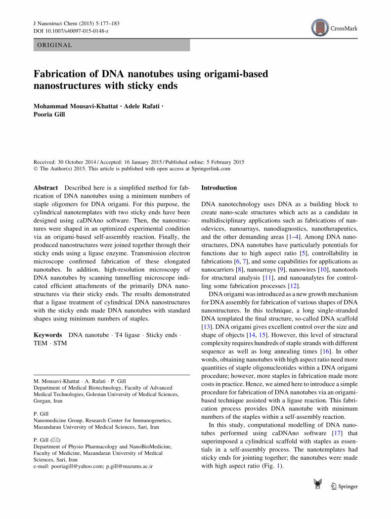
Fig. 1 Schematic fabrication of
DNA nanotubes designed by
caDNAno. a Three-dimensional
graphical view of DNA
nanotemplates with sticky ends;
b DNA nanotemplates jointed
together by ligase treatment;
c the positions of sticky ends in
ligation for shaping DNA
nanotubes with high aspect ratio
178 J Nanostruct Chem (2015) 5:177–183
123
The experiment was performed in 1 % agarose gel at TAE
buffer with a voltage of 100 V applied in 25 min by power
supply. Gel staining was performed by SYBRGold working
solution. In addition, the ligation product was run in the
same experimental condition for the gel electrophoresis.
After electrophoresis, DNA nanotubes were extracted from
the gels using Quantum PrepTM Freeze ‘N Squeeze DNA
Gel Extraction Spin Columns according to the manual
instruction.
Transmission electron microscopy of DNA nanotubes
Transmission electron microscope was used to determine
the size and morphology of DNA nanotubes. For this
purpose, the samples were immobilized by syringe spray-
ing on agar scientific holey carbon film with 300 mesh
Cu(50).
Characterization of DNA nanotubes by STM
The ligation product-contained DNA nanotubes were
diluted 103 folds in TAE–Mg2? buffer (pH 8). Then, 5 lldiluted sample was immobilized on the HOPG by drying
for 3 h at room temperature [18]. The samples were imaged
using topographic mode with STM, with 0.1 nA current set
point and 0.2 V sample bias through a platinum iridium tip.
Rough data were first processed using line adjust, plain
adjust and average filters of the NAMA-STM Nanoana-
lyzer software. Then, the colouring process was tested on
the obtained micrographs for different levels [19].
Results
DNA nanotube templates with sticky ends
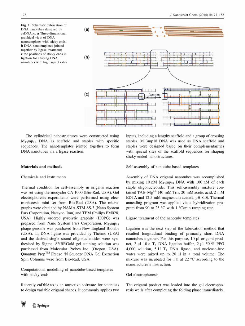
The modelled nanotube obtained from caDNAno software
was demonstrated in Fig. 2. The model consisted of a
cylindrical scaffold assembled with the staples from the
oligonucleotides. The scaffold has been formed from six
single-stranded DNA with the M13mp18 nucleotide con-
tents that were shown with blue colour. In addition,
sequences of the staples were determined as those applied
on the scaffold and eventually 24 staples (Table 1) were
determined with the variable lengths.
The 24 staples were indicated with different colours in
the computational model. The computerized nanotube
contained two sticky ends that those could be jointed to
their complements for shaping DNA nanotubes via a ligase
reaction.
Fig. 2 The caDNAno
decoration of a DNA nanotube-
based template with two sticky
ends. The scaffold DNA was
differentiated from the staples
with blue colour. Not all the 24
staples were hybridized to the
scaffold DNA into double-
stranded DNAs. Many of the
staples enclosed to the scaffold
into crossing-over strands and
holiday junctions
Table 1 Staple oligomers for self-assembly of DNA nanotemplates
Oligomer
name
Sequence (50–30)
S1 CCAACGTGCAGGTCATTCGTA
S2 CACTATTCCGGTTCATGGTCG
S3 TTCCAGTTCCCTTAAGCAGGC
S4 GAGATAGGGTTGACGCGCGGGGAGAGGCGGT
S5 ACGGCCAGTGCCTGTTTCCTG
S6 CATGCCTCAAAGGGGCGCTCA
S7 GAGGATCAAAGAACGTCGGGA
S8 GGCAAAATTGGAACGCTGCAT
S9 ATCATGGGCTCACAAATGAGTGAGCTAACTCAC
S10 GGTACCGACGAGCCAGTGTAA
S11 GAAAATCTTGCCCTCACCAGT
S12 AGCGGTCCACGCTGGTTGAGAGACGCCAGG
S13 TGTGAAATTGTTATCCTCATAGCAAGCTTG
S14 ACAACATAGCTCGAGACTCTA
S15 CAGCTGACTGTTTGCGAAATC
S16 CTGGCCCTTGCCCCTAAATCAAAAGAATAGCCC
S17 AGCCTGGCTTTCCAGTGGACT
S18 GAGACGGCGTGCCAAAGAGTC
S19 GTGGTTTTCGGCCAAGTGTTG
S20 TTGCGTATTGGGGTTGCAGCA
S21 ATTAATTGCGTTCGAAAAACCGTCTATCACG
S22 CTGCCCGGGTGCCTATTCCAC
S23 AACCTGTGCCATAAGGAAGAA
S24 TAATGAATTCTTTTTCACCGC
J Nanostruct Chem (2015) 5:177–183 179
123
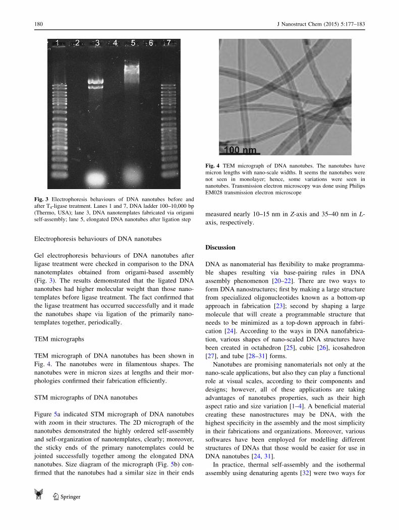
Electrophoresis behaviours of DNA nanotubes
Gel electrophoresis behaviours of DNA nanotubes after
ligase treatment were checked in comparison to the DNA
nanotemplates obtained from origami-based assembly
(Fig. 3). The results demonstrated that the ligated DNA
nanotubes had higher molecular weight than those nano-
templates before ligase treatment. The fact confirmed that
the ligase treatment has occurred successfully and it made
the nanotubes shape via ligation of the primarily nano-
templates together, periodically.
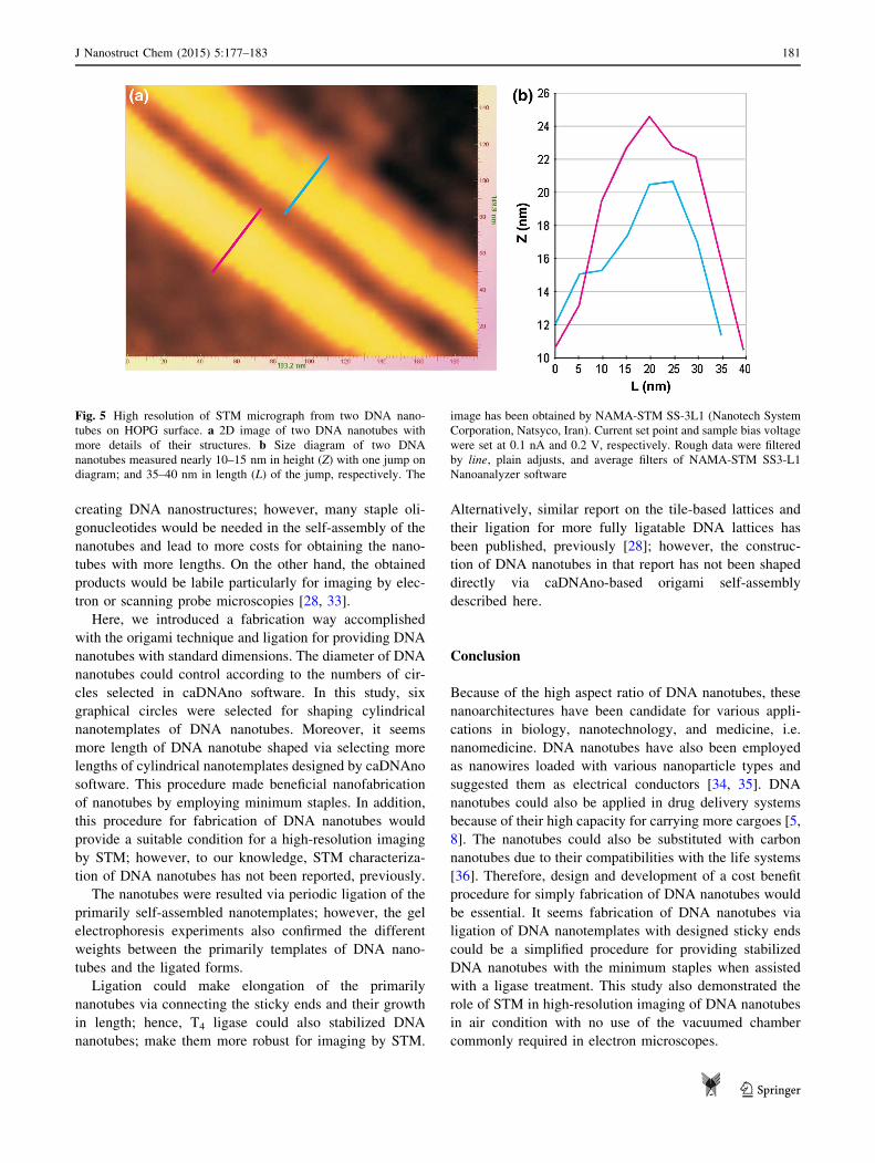
TEM micrographs
TEM micrograph of DNA nanotubes has been shown in
Fig. 4. The nanotubes were in filamentous shapes. The
nanotubes were in micron sizes at lengths and their mor-
phologies confirmed their fabrication efficiently.
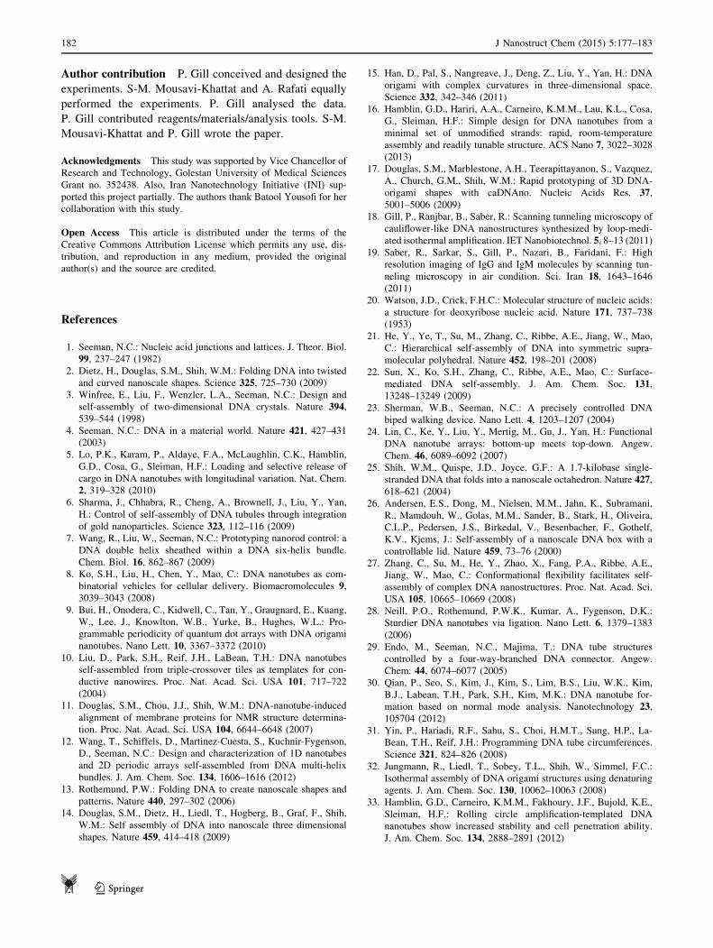
STM micrographs of DNA nanotubes
Figure 5a indicated STM micrograph of DNA nanotubes
with zoom in their structures. The 2D micrograph of the
nanotubes demonstrated the highly ordered self-assembly
and self-organization of nanotemplates, clearly; moreover,
the sticky ends of the primary nanotemplates could be
jointed successfully together among the elongated DNA
nanotubes. Size diagram of the micrograph (Fig. 5b) con-
firmed that the nanotubes had a similar size in their ends
measured nearly 10–15 nm in Z-axis and 35–40 nm in L-
axis, respectively.
Discussion
DNA as nanomaterial has flexibility to make programma-
ble shapes resulting via base-pairing rules in DNA
assembly phenomenon [20–22]. There are two ways to
form DNA nanostructures; first by making a large structure
from specialized oligonucleotides known as a bottom-up
approach in fabrication [23]; second by shaping a large
molecule that will create a programmable structure that
needs to be minimized as a top-down approach in fabri-
cation [24]. According to the ways in DNA nanofabrica-
tion, various shapes of nano-scaled DNA structures have
been created in octahedron [25], cubic [26], icosahedron
[27], and tube [28–31] forms.
Nanotubes are promising nanomaterials not only at the
nano-scale applications, but also they can play a functional
role at visual scales, according to their components and
designs; however, all of these applications are taking
advantages of nanotubes properties, such as their high
aspect ratio and size variation [1–4]. A beneficial material
creating these nanostructures may be DNA, with the
highest specificity in the assembly and the most simplicity
in their fabrications and organizations. Moreover, various
softwares have been employed for modelling different
structures of DNAs that those would be easier for use in
DNA nanotubes [24, 31].
In practice, thermal self-assembly and the isothermal
assembly using denaturing agents [32] were two ways for
Fig. 3 Electrophoresis behaviours of DNA nanotubes before and
after T4-ligase treatment. Lanes 1 and 7, DNA ladder 100–10,000 bp
(Thermo, USA); lane 3, DNA nanotemplates fabricated via origami
self-assembly; lane 5, elongated DNA nanotubes after ligation step
Fig. 4 TEM micrograph of DNA nanotubes. The nanotubes have
micron lengths with nano-scale widths. It seems the nanotubes were
not seen in monolayer; hence, some variations were seen in
nanotubes. Transmission electron microscopy was done using Philips
EM028 transmission electron microscope
180 J Nanostruct Chem (2015) 5:177–183
123
creating DNA nanostructures; however, many staple oli-
gonucleotides would be needed in the self-assembly of the
nanotubes and lead to more costs for obtaining the nano-
tubes with more lengths. On the other hand, the obtained
products would be labile particularly for imaging by elec-
tron or scanning probe microscopies [28, 33].
Here, we introduced a fabrication way accomplished
with the origami technique and ligation for providing DNA
nanotubes with standard dimensions. The diameter of DNA
nanotubes could control according to the numbers of cir-
cles selected in caDNAno software. In this study, six
graphical circles were selected for shaping cylindrical
nanotemplates of DNA nanotubes. Moreover, it seems
more length of DNA nanotube shaped via selecting more
lengths of cylindrical nanotemplates designed by caDNAno
software. This procedure made beneficial nanofabrication
of nanotubes by employing minimum staples. In addition,
this procedure for fabrication of DNA nanotubes would
provide a suitable condition for a high-resolution imaging
by STM; however, to our knowledge, STM characteriza-
tion of DNA nanotubes has not been reported, previously.
The nanotubes were resulted via periodic ligation of the
primarily self-assembled nanotemplates; however, the gel
electrophoresis experiments also confirmed the different
weights between the primarily templates of DNA nano-
tubes and the ligated forms.
Ligation could make elongation of the primarily
nanotubes via connecting the sticky ends and their growth
in length; hence, T4 ligase could also stabilized DNA
nanotubes; make them more robust for imaging by STM.
Alternatively, similar report on the tile-based lattices and
their ligation for more fully ligatable DNA lattices has
been published, previously [28]; however, the construc-
tion of DNA nanotubes in that report has not been shaped
directly via caDNAno-based origami self-assembly
described here.
Conclusion
Because of the high aspect ratio of DNA nanotubes, these
nanoarchitectures have been candidate for various appli-
cations in biology, nanotechnology, and medicine, i.e.
nanomedicine. DNA nanotubes have also been employed
as nanowires loaded with various nanoparticle types and
suggested them as electrical conductors [34, 35]. DNA
nanotubes could also be applied in drug delivery systems
because of their high capacity for carrying more cargoes [5,
8]. The nanotubes could also be substituted with carbon
nanotubes due to their compatibilities with the life systems
[36]. Therefore, design and development of a cost benefit
procedure for simply fabrication of DNA nanotubes would
be essential. It seems fabrication of DNA nanotubes via
ligation of DNA nanotemplates with designed sticky ends
could be a simplified procedure for providing stabilized
DNA nanotubes with the minimum staples when assisted
with a ligase treatment. This study also demonstrated the
role of STM in high-resolution imaging of DNA nanotubes
in air condition with no use of the vacuumed chamber
commonly required in electron microscopes.
Fig. 5 High resolution of STM micrograph from two DNA nano-
tubes on HOPG surface. a 2D image of two DNA nanotubes with
more details of their structures. b Size diagram of two DNA
nanotubes measured nearly 10–15 nm in height (Z) with one jump on
diagram; and 35–40 nm in length (L) of the jump, respectively. The
image has been obtained by NAMA-STM SS-3L1 (Nanotech System
Corporation, Natsyco, Iran). Current set point and sample bias voltage
were set at 0.1 nA and 0.2 V, respectively. Rough data were filtered
by line, plain adjusts, and average filters of NAMA-STM SS3-L1
Nanoanalyzer software
J Nanostruct Chem (2015) 5:177–183 181
123
Author contribution P. Gill conceived and designed the
experiments. S-M. Mousavi-Khattat and A. Rafati equally
performed the experiments. P. Gill analysed the data.
P. Gill contributed reagents/materials/analysis tools. S-M.
Mousavi-Khattat and P. Gill wrote the paper.
Acknowledgments This study was supported by Vice Chancellor of
Research and Technology, Golestan University of Medical Sciences
Grant no. 352438. Also, Iran Nanotechnology Initiative (INI) sup-
ported this project partially. The authors thank Batool Yousofi for her
collaboration with this study.
Open Access This article is distributed under the terms of the
Creative Commons Attribution License which permits any use, dis-
tribution, and reproduction in any medium, provided the original
author(s) and the source are credited.
References
1. Seeman, N.C.: Nucleic acid junctions and lattices. J. Theor. Biol.
99, 237–247 (1982)
2. Dietz, H., Douglas, S.M., Shih, W.M.: Folding DNA into twisted
and curved nanoscale shapes. Science 325, 725–730 (2009)
3. Winfree, E., Liu, F., Wenzler, L.A., Seeman, N.C.: Design and
self-assembly of two-dimensional DNA crystals. Nature 394,539–544 (1998)
4. Seeman, N.C.: DNA in a material world. Nature 421, 427–431(2003)
5. Lo, P.K., Karam, P., Aldaye, F.A., McLaughlin, C.K., Hamblin,
G.D., Cosa, G., Sleiman, H.F.: Loading and selective release of
cargo in DNA nanotubes with longitudinal variation. Nat. Chem.
2, 319–328 (2010)
6. Sharma, J., Chhabra, R., Cheng, A., Brownell, J., Liu, Y., Yan,
H.: Control of self-assembly of DNA tubules through integration
of gold nanoparticles. Science 323, 112–116 (2009)
7. Wang, R., Liu, W., Seeman, N.C.: Prototyping nanorod control: a
DNA double helix sheathed within a DNA six-helix bundle.
Chem. Biol. 16, 862–867 (2009)
8. Ko, S.H., Liu, H., Chen, Y., Mao, C.: DNA nanotubes as com-
binatorial vehicles for cellular delivery. Biomacromolecules 9,3039–3043 (2008)
9. Bui, H., Onodera, C., Kidwell, C., Tan, Y., Graugnard, E., Kuang,
W., Lee, J., Knowlton, W.B., Yurke, B., Hughes, W.L.: Pro-
grammable periodicity of quantum dot arrays with DNA origami
nanotubes. Nano Lett. 10, 3367–3372 (2010)
10. Liu, D., Park, S.H., Reif, J.H., LaBean, T.H.: DNA nanotubes
self-assembled from triple-crossover tiles as templates for con-
ductive nanowires. Proc. Nat. Acad. Sci. USA 101, 717–722
(2004)
11. Douglas, S.M., Chou, J.J., Shih, W.M.: DNA-nanotube-induced
alignment of membrane proteins for NMR structure determina-
tion. Proc. Nat. Acad. Sci. USA 104, 6644–6648 (2007)
12. Wang, T., Schiffels, D., Martinez-Cuesta, S., Kuchnir-Fygenson,
D., Seeman, N.C.: Design and characterization of 1D nanotubes
and 2D periodic arrays self-assembled from DNA multi-helix
bundles. J. Am. Chem. Soc. 134, 1606–1616 (2012)
13. Rothemund, P.W.: Folding DNA to create nanoscale shapes and
patterns. Nature 440, 297–302 (2006)
14. Douglas, S.M., Dietz, H., Liedl, T., Hogberg, B., Graf, F., Shih,
W.M.: Self assembly of DNA into nanoscale three dimensional
shapes. Nature 459, 414–418 (2009)
15. Han, D., Pal, S., Nangreave, J., Deng, Z., Liu, Y., Yan, H.: DNA
origami with complex curvatures in three-dimensional space.
Science 332, 342–346 (2011)
16. Hamblin, G.D., Hariri, A.A., Carneiro, K.M.M., Lau, K.L., Cosa,
G., Sleiman, H.F.: Simple design for DNA nanotubes from a
minimal set of unmodified strands: rapid, room-temperature
assembly and readily tunable structure. ACS Nano 7, 3022–3028(2013)
17. Douglas, S.M., Marblestone, A.H., Teerapittayanon, S., Vazquez,
A., Church, G.M., Shih, W.M.: Rapid prototyping of 3D DNA-
origami shapes with caDNAno. Nucleic Acids Res. 37,5001–5006 (2009)
18. Gill, P., Ranjbar, B., Saber, R.: Scanning tunneling microscopy of
cauliflower-like DNA nanostructures synthesized by loop-medi-
ated isothermal amplification. IETNanobiotechnol. 5, 8–13 (2011)19. Saber, R., Sarkar, S., Gill, P., Nazari, B., Faridani, F.: High
resolution imaging of IgG and IgM molecules by scanning tun-
neling microscopy in air condition. Sci. Iran 18, 1643–1646
(2011)
20. Watson, J.D., Crick, F.H.C.: Molecular structure of nucleic acids:
a structure for deoxyribose nucleic acid. Nature 171, 737–738(1953)
21. He, Y., Ye, T., Su, M., Zhang, C., Ribbe, A.E., Jiang, W., Mao,
C.: Hierarchical self-assembly of DNA into symmetric supra-
molecular polyhedral. Nature 452, 198–201 (2008)
22. Sun, X., Ko, S.H., Zhang, C., Ribbe, A.E., Mao, C.: Surface-
mediated DNA self-assembly. J. Am. Chem. Soc. 131,13248–13249 (2009)
23. Sherman, W.B., Seeman, N.C.: A precisely controlled DNA
biped walking device. Nano Lett. 4, 1203–1207 (2004)
24. Lin, C., Ke, Y., Liu, Y., Mertig, M., Gu, J., Yan, H.: Functional
DNA nanotube arrays: bottom-up meets top-down. Angew.
Chem. 46, 6089–6092 (2007)
25. Shih, W.M., Quispe, J.D., Joyce, G.F.: A 1.7-kilobase single-
stranded DNA that folds into a nanoscale octahedron. Nature 427,618–621 (2004)
26. Andersen, E.S., Dong, M., Nielsen, M.M., Jahn, K., Subramani,
R., Mamdouh, W., Golas, M.M., Sander, B., Stark, H., Oliveira,
C.L.P., Pedersen, J.S., Birkedal, V., Besenbacher, F., Gothelf,
K.V., Kjems, J.: Self-assembly of a nanoscale DNA box with a
controllable lid. Nature 459, 73–76 (2000)
27. Zhang, C., Su, M., He, Y., Zhao, X., Fang, P.A., Ribbe, A.E.,
Jiang, W., Mao, C.: Conformational flexibility facilitates self-
assembly of complex DNA nanostructures. Proc. Nat. Acad. Sci.
USA 105, 10665–10669 (2008)
28. Neill, P.O., Rothemund, P.W.K., Kumar, A., Fygenson, D.K.:
Sturdier DNA nanotubes via ligation. Nano Lett. 6, 1379–1383(2006)
29. Endo, M., Seeman, N.C., Majima, T.: DNA tube structures
controlled by a four-way-branched DNA connector. Angew.
Chem. 44, 6074–6077 (2005)
30. Qian, P., Seo, S., Kim, J., Kim, S., Lim, B.S., Liu, W.K., Kim,
B.J., Labean, T.H., Park, S.H., Kim, M.K.: DNA nanotube for-
mation based on normal mode analysis. Nanotechnology 23,105704 (2012)
31. Yin, P., Hariadi, R.F., Sahu, S., Choi, H.M.T., Sung, H.P., La-
Bean, T.H., Reif, J.H.: Programming DNA tube circumferences.
Science 321, 824–826 (2008)
32. Jungmann, R., Liedl, T., Sobey, T.L., Shih, W., Simmel, F.C.:
Isothermal assembly of DNA origami structures using denaturing
agents. J. Am. Chem. Soc. 130, 10062–10063 (2008)
33. Hamblin, G.D., Carneiro, K.M.M., Fakhoury, J.F., Bujold, K.E.,
Sleiman, H.F.: Rolling circle amplification-templated DNA
nanotubes show increased stability and cell penetration ability.
J. Am. Chem. Soc. 134, 2888–2891 (2012)
182 J Nanostruct Chem (2015) 5:177–183
123

34. Monson, C.F., Woolley, A.T.: DNA-templated construction of
copper nanowires. Nano Lett. 3, 359–363 (2003)
35. Ford, W.E., Harnack, O., Yasuda, A., Wessels, J.M.: Platinated
DNA as precursors to templated chains of metal nanoparticles.
Adv. Mater. 13, 1793–1797 (2001)
36. Bianco, A., Kostarelos, K., Prato, M.: Applications of carbon
nanotubes in drug delivery. Cur. Opin. Chem. Biol. 9, 674–679(2005)
J Nanostruct Chem (2015) 5:177–183 183
123