Embed Size (px)
Citation preview
Canine heartworm disease
1
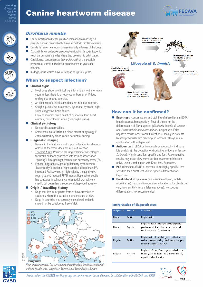
Dirofilaria immitis ! Canine heartworm disease (cardiopulmonary dirofilariosis) is a
parasitic disease caused by the filarial nematode Dirofilaria immitis. ! Despite its name, heartworm disease is mainly a disease of the lungs. ! D. immitis larvae undertake an extensive migration through tissues to
reach the pulmonary arteries where they develop into adult stages. ! Cardiological consequences (cor pulmonale) or the possible
presence of worms in the heart occur months to years after infection.
! In dogs, adult worms have a lifespan of up to 7 years.
When to suspect infection? ! Clinical signs
o Most dogs show no clinical signs for many months or even years unless there is a heavy worm burden or if dogs undergo strenuous exercise.
o An absence of clinical signs does not rule out infection. o Coughing, exercise intolerance, dyspnoea, syncope, right-
sided congestive heart failure. o Caval syndrome: acute onset of dyspnoea, loud heart
murmur, red-coloured urine (haemoglobinuria). ! Clinical pathology
o No specific abnormalities. o Sometimes microfilariae on blood smear or cytology if
contaminated by blood (often accidental finding). ! Diagnostic imaging
o Normal in the first few months post infection. An absence of lesions therefore does not rule out infection.
o Thoracic X-ray: Perivascular lung inflammation; enlarged, torturous pulmonary arteries with loss of arborisation (‘pruning’); Enlarged right ventricle and pulmonary artery (PA).
o Echocardiography: Signs of pulmonary hypertension (hypertrophy/dilatation of right ventricle, dilated main PA, increased PA flow velocity, high-velocity tricuspid valve regurgitation, reduced RPAD index); Hyperechoic double-line structures in pulmonary arteries (adult worms): very specific but dependent on operator skills/probe frequency.
! Origin / travelling history o Dogs that live in, originate from or have travelled to
countries where the parasite is endemic are at risk. o Dogs in countries not currently considered endemic
should not be considered free of risk.
2
How can it be confirmed? ! Knott test (concentration and staining of microfilaria in EDTA
blood): Acceptable sensitivity. Test of choice for the differentiation of filaria species (Dirofilaria immitis, D. repens and Achantochelionema reconditum. Inexpensive. False negative results occur (occult infections), mainly in patients treated previously with macrocyclic lactones. Always run in combination with antigen test.
! Antigen test (ELISA or immunochromatography, in-house kits available): the detection of circulating antigens of female D. immitis. Highly sensitive, specific and fast. False negative results may occur (low worm burden, male worm infection only). Use in combination with Knott test. Expensive.
! PCR (detection of DNA of microfilariae): Highly specific, less sensitive than Knott test. Allows species differentiation.Expensive.
! Fresh blood drop exam (visualisation of living, mobile microfilariae). Fast and inexpensive, educational for clients but very low sensitivity (many false negatives). No species differentiation. Not recommended.
FECAVAFederation of European CompanionAnimal Veterinary Associations
FECAVA FECAVA FECAVA FEC
AVA
WERSJA POPRAWIONA A
© E
SDA
Working Group on
Canine vector borne
diseases
Mean prevalence rates. The current area where Dirofilaria immitis is considered endemic includes most countries in Southern and South-Eastern Europe.
Interpretation of diagnostic tests
Produced by the FECAVA working group on canine vector-borne diseases in collaboration with ESCCAP and ESDA
Canine heartworm disease
FECAVAFederation of European CompanionAnimal Veterinary Associations
FECAVA FECAVA FECAVA FEC
AVA
WERSJA POPRAWIONA A
Working Group on
Canine vector borne
diseases
3
Staging the disease ! Heartworm-positive dogs first need to be staged according to
their risk of pulmonary thromboembolism (PTE). ! Dogs that have at least one of the following signs are at high
risk of having pulmonary thromboembolism: o Disease-related clinical signs o Pathological patterns on thoracic radiography. o High level of circulating antigens o Visualisation of worms in pulmonary artery and/or right
ventricle/atrium o Evidence of pulmonary hypertension o Concurrent disease o No restricted exercise
! If all of the following applies, dogs are considered being at low risk of pulmonary thromboembolism: o No disease-related clinical signs o No pathological patterns on thoracic radiography o Low level of circulating antigens, or negative antigen test
and positive Knott test o No worms visualised by echocardiography o No signs of pulmonary hypertension o No concurrent disease o Restricted exercise
Disease management
o The surgical removal of worms is always recommended in dogs with the caval syndrome (several worms present in the right ventricle and atrium).
o Echographic visualisation of worms in the pulmonary artery allows the use of flexible alligator forceps under fluoroscopic guidance to remove the worms, avoiding pulmonary thromboembolism.
! Adulticide treatment with melarsomine o Day 1: doxycyline 10 mg/kg q12-24h for 30 days;
macrocyclic lactone (heartworm prevention) o Day 15: macrocyclic lactone (heartworm prevention) o Day 30: melarsomine dihydrochloride* 2.5 mg/kg deep IM
Days 60 and 61: melarsomine dihydrochloride* 2.5 mg/kg deep IM
o Coughing dogs should receive prednisolone at anti-inflammatory dose until effect.
*Calcium heparin 50-100 IU should be given during melarsomine treatment, from 1-2 weeks before to several weeks after treatment
! Alternative adulticide treatment (‘slow kill’) o Second-choice treatment o Only if surgery is not possible and melarsomine is unavailable:
" Doxycycline 10 mg/kg q12-24h for 30 days " Ivermectin 6-12µg/kg or spot-on moxidectin 2.5 mg/kg
bimonthly until two consecutive Ag test results are obtained (usually after 12 months).
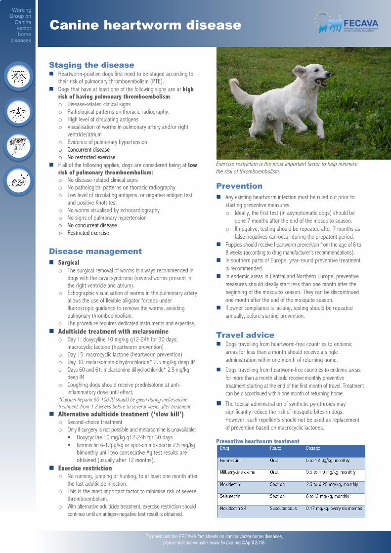
! Exercise restriction o No running, jumping or hunting, to at least one month after
the last adulticide injection. o This is the most important factor to minimise risk of severe
thromboembolism. o With alternative adulticide treatment, exercise restriction should
continue until an antigen-negative test result is obtained.
4
Prevention ! Any existing heartworm infection must be ruled out prior to
starting preventive measures. o Ideally, the first test (in asymptomatic dogs) should be
done 7 months after the end of the mosquito season. o If negative, testing should be repeated after 7 months as
false negatives can occur during the prepatent period. ! Puppies should receive heartworm prevention from the age of 6 to
8 weeks (according to drug manufacturer’s recommendations). ! In southern parts of Europe, year-round preventive treatment
is recommended. ! In endemic areas in Central and Northern Europe, preventive
measures should ideally start less than one month after the beginning of the mosquito season. They can be discontinued one month after the end of the mosquito season.
! If owner compliance is lacking, testing should be repeated annually, before starting prevention.
Travel advice ! Dogs travelling from heartworm-free countries to endemic
areas for less than a month should receive a single administration within one month of returning home.
! Dogs travelling from heartworm-free countries to endemic areas for more than a month should receive monthly preventive treatment starting at the end of the first month of travel. Treatment can be discontinued within one month of returning home.
! The topical administration of synthetic pyrethroids may significantly reduce the risk of mosquito bites in dogs. However, such repellents should not be used as replacement of prevention based on macrocyclic lactones.
Exercise restriction is the most important factor to help minimise the risk of thromboembolism.
Preventive heartworm treatment
To download the FECAVA fact sheets on canine vector-borne diseases, please visit our website: www.fecava.org ©April 2018.
FECAVAFederation of European CompanionAnimal Veterinary Associations
FECAVA FECAVA FECAVA FEC
AVA
WERSJA POPRAWIONA A