Embed Size (px)
Citation preview
Volume 10 • Issue 4 • 1000544J Cytol Histol, an open access journalISSN: 2157-7099
Research Article Open Access
Massoud et al., J Cytol Histol 2019, 10:4
Research Article Open Access
Journal of Cytology & HistologyJour
nal o
f Cytology & Histology
ISSN: 2157-7099
Keywords: Diabetes mellitus; Fermented deglycyrrhizinated liquorice extract; Skeletal muscles; Peripheral nerve; Rats; Histology
IntroductionDiabetes mellitus (DM) is a potentially serious metabolic disorder
with increasing occurrence all over the world. According to the International Diabetes Federation, there was approximately 366 million people suffered from DM (aged 20-79 years) in 2011 and this figure would climb up to 552 million by the year of 2030 [1]. All types of DM are characterized by hyperglycemia and the progression of multiple debilitating complications. In addition to infection and premature death, diabetic complications include cerebrovascular disorders, myocardial infarction, renal failure, blindness, limb amputation and a variety of neuropathies [2].
The skeletal muscle is notably affected by DM. It has been discovered that DM induces skeletal muscle atrophy, fiber-type alteration from oxidative to glycolytic and impaired energy metabolism in skeletal muscle [3]. These changes result in skeletal muscle dysfunction, such as muscle weakness and exercise intolerance [4]. Moreover, the commonest cause of peripheral neuropathy is diabetes, and 30-90% of patients with diabetes have peripheral neuropathy [5]. Diabetic peripheral neuropathy is characterized by pain, paraesthesia and sensory loss [6].
Interest in the use of plant extracts that possess widespread biological functions has increased in recent years. Liquorice (Glycyrrhiza glabra) is an herb of Mediterranean and Asian area. Root of this plant has various useful pharmacological properties
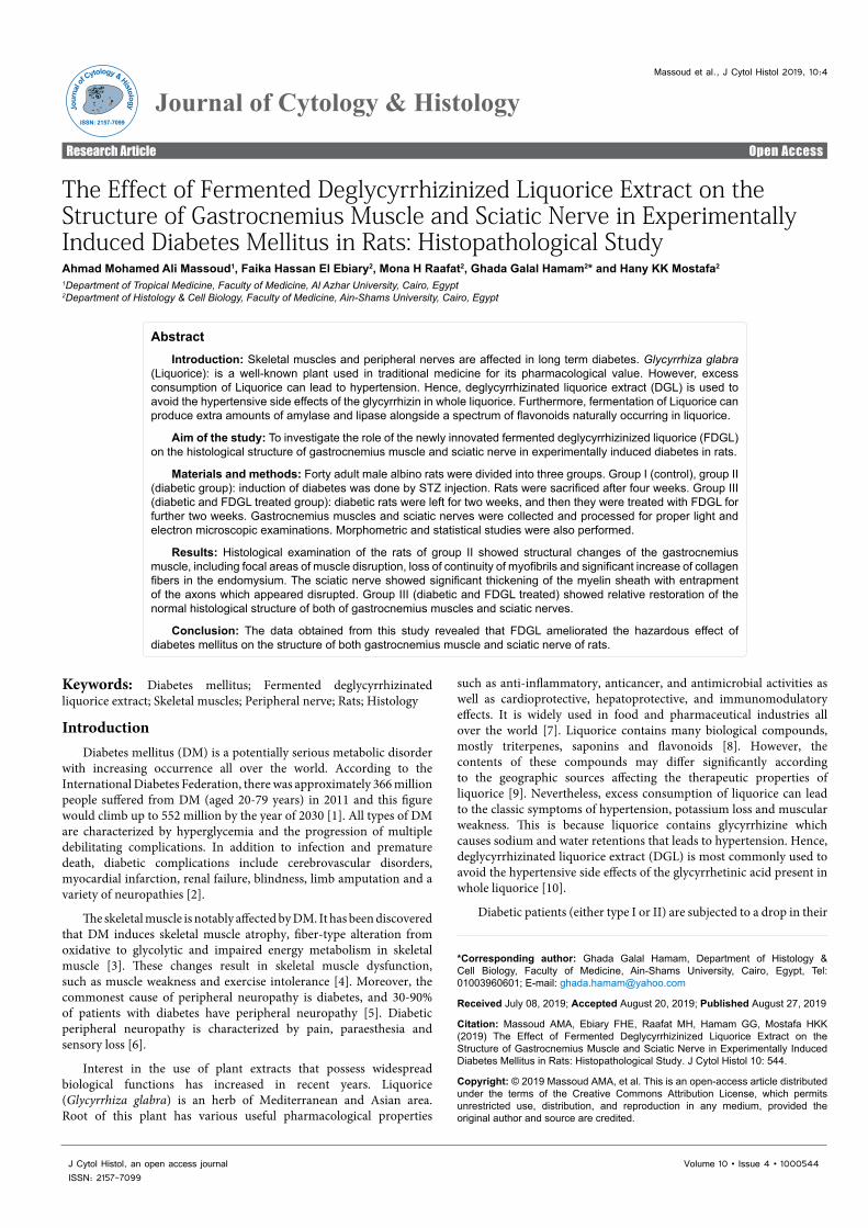
The Effect of Fermented Deglycyrrhizinized Liquorice Extract on the Structure of Gastrocnemius Muscle and Sciatic Nerve in Experimentally Induced Diabetes Mellitus in Rats: Histopathological StudyAhmad Mohamed Ali Massoud1, Faika Hassan El Ebiary2, Mona H Raafat2, Ghada Galal Hamam2* and Hany KK Mostafa2
1Department of Tropical Medicine, Faculty of Medicine, Al Azhar University, Cairo, Egypt2Department of Histology & Cell Biology, Faculty of Medicine, Ain-Shams University, Cairo, Egypt
*Corresponding author: Ghada Galal Hamam, Department of Histology & Cell Biology, Faculty of Medicine, Ain-Shams University, Cairo, Egypt, Tel: 01003960601; E-mail: [email protected]
Received July 08, 2019; Accepted August 20, 2019; Published August 27, 2019
Citation: Massoud AMA, Ebiary FHE, Raafat MH, Hamam GG, Mostafa HKK (2019) The Effect of Fermented Deglycyrrhizinized Liquorice Extract on the Structure of Gastrocnemius Muscle and Sciatic Nerve in Experimentally Induced Diabetes Mellitus in Rats: Histopathological Study. J Cytol Histol 10: 544.
Copyright: © 2019 Massoud AMA, et al. This is an open-access article distributed under the terms of the Creative Commons Attribution License, which permits unrestricted use, distribution, and reproduction in any medium, provided the original author and source are credited.
such as anti-inflammatory, anticancer, and antimicrobial activities as well as cardioprotective, hepatoprotective, and immunomodulatory effects. It is widely used in food and pharmaceutical industries all over the world [7]. Liquorice contains many biological compounds, mostly triterpenes, saponins and flavonoids [8]. However, the contents of these compounds may differ significantly according to the geographic sources affecting the therapeutic properties of liquorice [9]. Nevertheless, excess consumption of liquorice can lead to the classic symptoms of hypertension, potassium loss and muscular weakness. This is because liquorice contains glycyrrhizine which causes sodium and water retentions that leads to hypertension. Hence, deglycyrrhizinated liquorice extract (DGL) is most commonly used to avoid the hypertensive side effects of the glycyrrhetinic acid present in whole liquorice [10].
Diabetic patients (either type I or II) are subjected to a drop in their
AbstractIntroduction: Skeletal muscles and peripheral nerves are affected in long term diabetes. Glycyrrhiza glabra
(Liquorice): is a well-known plant used in traditional medicine for its pharmacological value. However, excess consumption of Liquorice can lead to hypertension. Hence, deglycyrrhizinated liquorice extract (DGL) is used to avoid the hypertensive side effects of the glycyrrhizin in whole liquorice. Furthermore, fermentation of Liquorice can produce extra amounts of amylase and lipase alongside a spectrum of flavonoids naturally occurring in liquorice.
Aim of the study: To investigate the role of the newly innovated fermented deglycyrrhizinized liquorice (FDGL) on the histological structure of gastrocnemius muscle and sciatic nerve in experimentally induced diabetes in rats.
Materials and methods: Forty adult male albino rats were divided into three groups. Group I (control), group II (diabetic group): induction of diabetes was done by STZ injection. Rats were sacrificed after four weeks. Group III (diabetic and FDGL treated group): diabetic rats were left for two weeks, and then they were treated with FDGL for further two weeks. Gastrocnemius muscles and sciatic nerves were collected and processed for proper light and electron microscopic examinations. Morphometric and statistical studies were also performed.
Results: Histological examination of the rats of group II showed structural changes of the gastrocnemius muscle, including focal areas of muscle disruption, loss of continuity of myofibrils and significant increase of collagen fibers in the endomysium. The sciatic nerve showed significant thickening of the myelin sheath with entrapment of the axons which appeared disrupted. Group III (diabetic and FDGL treated) showed relative restoration of the normal histological structure of both of gastrocnemius muscles and sciatic nerves.
Conclusion: The data obtained from this study revealed that FDGL ameliorated the hazardous effect of diabetes mellitus on the structure of both gastrocnemius muscle and sciatic nerve of rats.

Page 2 of 8
Citation: Massoud AMA, Ebiary FHE, Raafat MH, Hamam GG, Mostafa HKK (2019) The Effect of Fermented Deglycyrrhizinized Liquorice Extract on the Structure of Gastrocnemius Muscle and Sciatic Nerve in Experimentally Induced Diabetes Mellitus in Rats: Histopathological Study. J Cytol Histol 10: 544.
Volume 10 • Issue 4 • 1000544J Cytol Histol, an open access journalISSN: 2157-7099
-Subgroup Ia: Rats were sacrificed after four weeks.
-Subgroup Ib: Each rat received freshly prepared FDGL extract at a dose of 0.12 g/kg body weight/day, dissolved in 2 mL distilled water by oral gavage for two weeks.
Group II (Diabetic group): It included fifteen rats in which induction of diabetes was done. After confirmation of diabetes, they were left without treatment and were sacrificed after four weeks.
Group III (Diabetic and FDGL extract treated group): It included fifteen rats in which induction of diabetes was done as in group II. After confirmation of diabetes, rats were left for two weeks then FDGL was given daily for further two weeks as in subgroup Ib, then they were sacrificed (i.e. after four weeks from confirmation of diabetes).
Blood glucose level analysis
A drop of fresh blood was collected from the animal`s tail using a lancet at fasting conditions. Fasting blood glucose levels were measured in all groups twice weekly for the period of the experiment using glucometer instrument One Touch Ultra 2 glucose meter and strips (LifeScan, Inc. USA) and statistical analysis were done.
Sample collection and preparation of tissue
At the end of the experiment, animals were sacrificed by decapitation after ether inhalation anesthesia.
The right gastrocnemius muscle was fixed in 10% formalin and processed to form paraffin blocks for light microscopic study (LM). Longitudinal 5 µm thick sections, were stained with hematoxylin and eosin stain (H&E) and Mallory`s trichrome stain [17].
Small pieces (1-2 mm thick) of left gastrocnemius muscle were processed for transmission electron microscopic study (TEM). They were fixed in 2.5% phosphate buffered glutaraldehyde [17]. Ultra-thin sections (50-60 nm) were examined and photographed by (JEM-1200EXII Tokyo, Japan) transmission electron microscope at at the Regional Center for Mycology and Biotechnology Faculty of Medicine, El Azhar University.
Right sciatic nerves were isolated, cut into 1-2 mm thick segments and fixed in phosphate buffered glutaraldehyde followed by 1% osmium tetroxide, dehydrated, and embedded in epoxy resin. Semi thin sections (350 nm) were stained with 1% toluidine blue and examined by light microscope. Meanwhile, for ultrastructural study, ultrathin sections (60-80 nm) were stained with 1% uranyl acetate and 2% lead citrate, examined and photographed with (JEM-1200EXII Tokyo, Japan) transmission electron microscope at the Regional Center for Mycology and Biotechnology Faculty of Medicine, El Azhar University.
Morphometric and statistical study
Five animals from all groups were subjected for morphometric study. Measurements were taken from five different slides obtained from each animal. Five haphazardly selected non-overlapping fields were examined for each slide. Five different readings from every captured photo were counted and the mean was calculated for each specimen. Measurements were taken by an independent observer blinded to the specimens’ details to perform an unbiased assessment.
An image analyzer Leica Q win V.3 program installed on a computer in the Department of Histology and Cell Biology, Faculty of Medicine, Ain Shams University, was used. The computer was connected to a Leica DM2500 microscope (Wetzlar, Germany).
serum amylase level comparative with the severity of their diabetic complication [11]. Therefore, liquorice was fermented under specific circumstances and was termed fermented deglycyrrhizinized liquorice extract (FDGL) to produce large amounts of enzymes as amylase and lipase. Although boiling is important for formation of DGL by removal of glyceryhizin (a compound responsible for several side effects of liquorice), it also prevents fermentation process which is important for production of lipase and amylase [12].
The rapidly increasing prevalence of DM worldwide leads to a great need for effective and safe functional biomaterials with anti-diabetic criteria. Thus, the aim of the study was to investigate the possible therapeutic role of FDGL on the structural changes in gastrocnemius muscle and sciatic nerve in experimentally induced diabetes mellitus.
Materials and MethodsThe experiment was carried out in the Animal Research Center,
Faculty of medicine, Ain Shams University, Cairo, Egypt.
Animals
Forty adult male albino Wistar rats of average weight 180-250 g and their ages ranged from two to three months were used in this study. They were given food and tap water ad libitum, and were kept under proper conditions of light, temperature and humidity. All animal procedures were carried out according to the guideline of animal care and the scientific research ethical committee of the faculty of Medicine, Ain Shams University.
Induction of diabetes
Before streptozotocin (STZ) injection, blood glucose level was measured to ensure that rats were normoglycemic. Rats were fasted for 18 hours before induction of DM. Diabetes was induced by a single intraperitoneal (IP) injection of freshly prepared solution of STZ (Sigma, Aldrich, USA.) at the dose of 45 mg/kg body weight diluted in 0.5 mL of 0.1 M/L citrate buffer, (pH 4.5) [13]. After three days, fasting blood glucose level was measured from the tail vein using One Touch Ultra 2 glucose meter and strips (LifeScan, Inc. USA). Rats with blood glucose above 250 mg/dL were confirmed to be diabetic and were selected for the experiment [14].
Drug preparation
Fermented deglycyrrhizinized liquorice extract powder was prepared in Pharmacology Research Unit, National Research Center, Cairo, Egypt, according to the EUROPEAN PATENT SPECIFICATION Ep 1 925 312 B1 through three consecutive steps: fermentation of liquorice root, deglycyrrhizination and lyophilization [12]. Preliminary pilot studies were conducted in Pharmacology Research Unit, National Research Center, Cairo, Egypt, on adult rats to detect both the acute toxicity of FDGL extract [15] and the chronic toxicity [16]. It was deduced that DGL extract is safe for oral administration up to 0.12 g/kg body weight/day for two months [12].
Experimental protocol
After seven days of acclimatization, rats were randomly divided into three groups:
Group I (Control group): It included ten rats. Each rat received single IP injection of 0.5 mL of 0.1 M/L citrate buffer (Vehicle of Streptozotocin). Then they were divided into two subgroups five animals each.
Page 3 of 8
Citation: Massoud AMA, Ebiary FHE, Raafat MH, Hamam GG, Mostafa HKK (2019) The Effect of Fermented Deglycyrrhizinized Liquorice Extract on the Structure of Gastrocnemius Muscle and Sciatic Nerve in Experimentally Induced Diabetes Mellitus in Rats: Histopathological Study. J Cytol Histol 10: 544.
Volume 10 • Issue 4 • 1000544J Cytol Histol, an open access journalISSN: 2157-7099
Data were collected, revised, and subjected to statistical analysis using one-way analysis of variance (ANOVA) performed with SPSS.21 program (IBM Inc., Chicago, Illinois, USA) and post-Hoc test at least significant difference (LSD). The significance of the data was determined by the p value. P values greater than 0.05 were considered non-significant, and p values less than 0.05 were considered significant. Summary of the data was expressed as mean and Standard Deviation (SD).
The following parameters were measured
1) The mean area percentage of collagen fibers in gastrocnemius muscle stained by Mallory`s trichrome stain X400.
2) Rounded transversely cut myelinated nerve fibers were selected and two diameters of each nerve fiber, one perpendicular to the other, were measured and the average was taken to measure the diameter of myelinated nerve fiber, axonal diameter and G-ratio (The ratio of the inner axonal diameter to the total outer diameter) [18]. These parameters were measured form semi thin sections stained by toluidine blue X1000.
3) The mean thickness of myelin sheath of sciatic nerve in semi thin sections stained by toluidine blue X1000.
ResultsMortality rate
Three rats had died in group II (diabetic) during the experiment with mortality rate of 20%. No further deaths were recorded in the other groups.
Histological results
In the present study examination of the structure of gastrocnemius muscles and the sciatic nerves of rats of both subgroups I(a) & I(b) revealed similar histological structure.
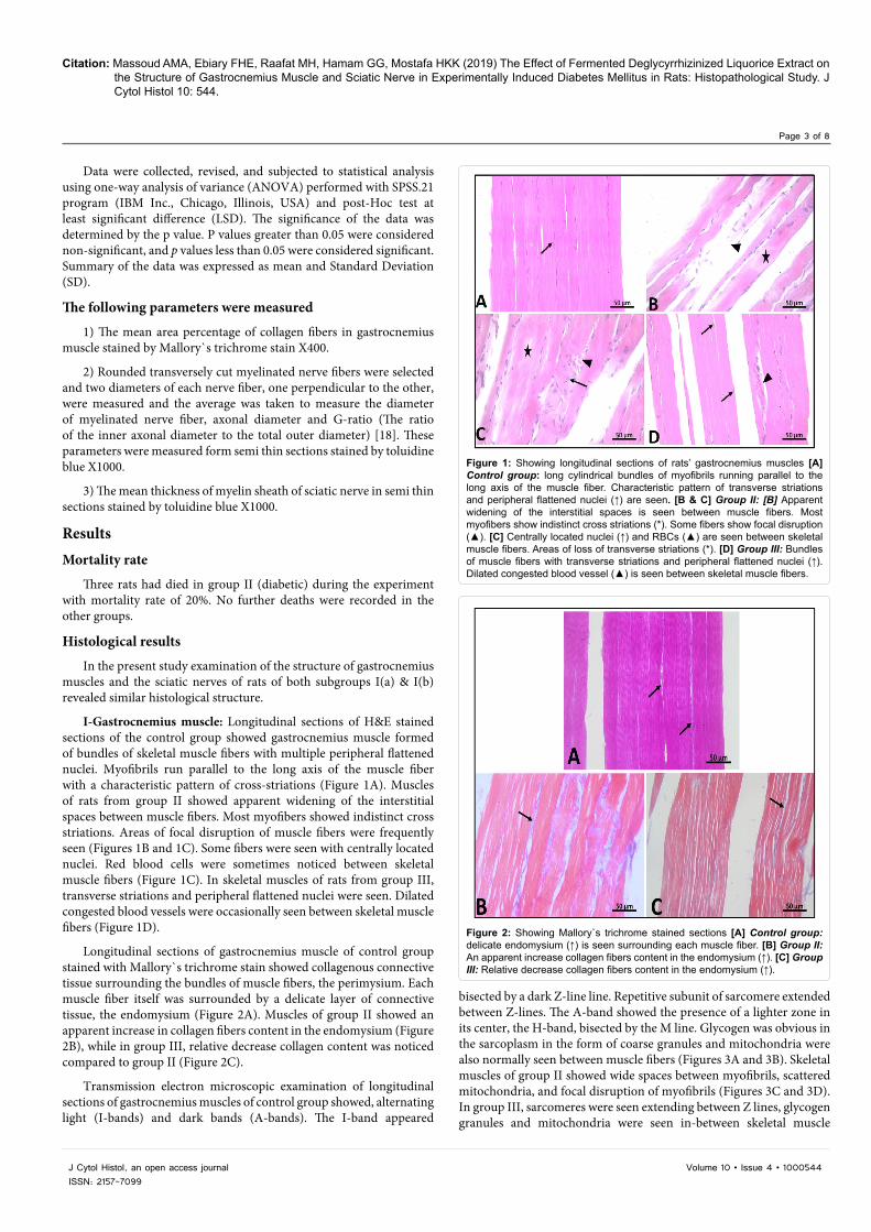
I-Gastrocnemius muscle: Longitudinal sections of H&E stained sections of the control group showed gastrocnemius muscle formed of bundles of skeletal muscle fibers with multiple peripheral flattened nuclei. Myofibrils run parallel to the long axis of the muscle fiber with a characteristic pattern of cross-striations (Figure 1A). Muscles of rats from group II showed apparent widening of the interstitial spaces between muscle fibers. Most myofibers showed indistinct cross striations. Areas of focal disruption of muscle fibers were frequently seen (Figures 1B and 1C). Some fibers were seen with centrally located nuclei. Red blood cells were sometimes noticed between skeletal muscle fibers (Figure 1C). In skeletal muscles of rats from group III, transverse striations and peripheral flattened nuclei were seen. Dilated congested blood vessels were occasionally seen between skeletal muscle fibers (Figure 1D).
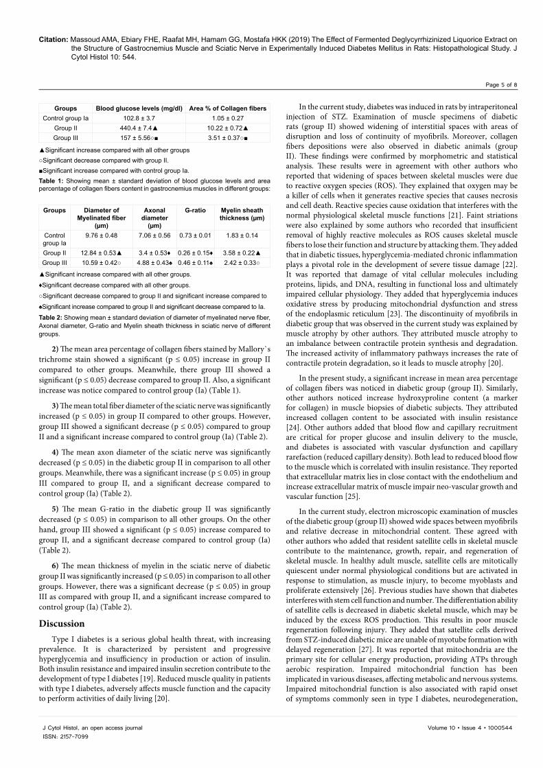
Longitudinal sections of gastrocnemius muscle of control group stained with Mallory`s trichrome stain showed collagenous connective tissue surrounding the bundles of muscle fibers, the perimysium. Each muscle fiber itself was surrounded by a delicate layer of connective tissue, the endomysium (Figure 2A). Muscles of group II showed an apparent increase in collagen fibers content in the endomysium (Figure 2B), while in group III, relative decrease collagen content was noticed compared to group II (Figure 2C).
Transmission electron microscopic examination of longitudinal sections of gastrocnemius muscles of control group showed, alternating light (I-bands) and dark bands (A-bands). The I-band appeared
bisected by a dark Z-line line. Repetitive subunit of sarcomere extended between Z-lines. The A-band showed the presence of a lighter zone in its center, the H-band, bisected by the M line. Glycogen was obvious in the sarcoplasm in the form of coarse granules and mitochondria were also normally seen between muscle fibers (Figures 3A and 3B). Skeletal muscles of group II showed wide spaces between myofibrils, scattered mitochondria, and focal disruption of myofibrils (Figures 3C and 3D). In group III, sarcomeres were seen extending between Z lines, glycogen granules and mitochondria were seen in-between skeletal muscle
Figure 1: Showing longitudinal sections of rats’ gastrocnemius muscles [A] Control group: long cylindrical bundles of myofibrils running parallel to the long axis of the muscle fiber. Characteristic pattern of transverse striations and peripheral flattened nuclei (↑) are seen. [B & C] Group II: [B] Apparent widening of the interstitial spaces is seen between muscle fibers. Most myofibers show indistinct cross striations (*). Some fibers show focal disruption (▲). [C] Centrally located nuclei (↑) and RBCs (▲) are seen between skeletal muscle fibers. Areas of loss of transverse striations (*). [D] Group III: Bundles of muscle fibers with transverse striations and peripheral flattened nuclei (↑). Dilated congested blood vessel (▲) is seen between skeletal muscle fibers.
Figure 2: Showing Mallory`s trichrome stained sections [A] Control group: delicate endomysium (↑) is seen surrounding each muscle fiber. [B] Group II: An apparent increase collagen fibers content in the endomysium (↑). [C] Group III: Relative decrease collagen fibers content in the endomysium (↑).

Page 4 of 8
Citation: Massoud AMA, Ebiary FHE, Raafat MH, Hamam GG, Mostafa HKK (2019) The Effect of Fermented Deglycyrrhizinized Liquorice Extract on the Structure of Gastrocnemius Muscle and Sciatic Nerve in Experimentally Induced Diabetes Mellitus in Rats: Histopathological Study. J Cytol Histol 10: 544.
Volume 10 • Issue 4 • 1000544J Cytol Histol, an open access journalISSN: 2157-7099
fibrils. Scattered areas of disturbed sarcomere and loss of continuity of myofibrils were occasionally noticed (Figures 3E and 3F).
II- Sciatic nerve: Transverse semi thin sections of Toluidine blue of the control group showed that the sciatic nerve was formed of densely packed nerve fibers with variable diameter which were impeded in scarce endoneurium and surrounded by connective tissue perineurium. Most of the myelinated nerve fibers show regular myelin (Figure 4A). Some nerve fibers of group II showed irregularities of myelin sheath, others showed marked thickening of the myelin sheath (Figure 4B). In addition, collapsed endoneural blood vessel lumen with thickened wall was also noticed (Figure 4C). Nerve fibers in group III showed regular myelin sheath which was nearly comparable to control group. Focal areas of myelin sheath irregularities were seen in scattered nerve fibers. Patent relatively wide lumen of an endoneural blood vessels were also detected (Figure 4D).
Transmission electron microscopic examination of control group showed that sciatic nerve with myelinated axons were having compact, regular and thick myelin sheath. The myelinated axons were enveloped by Schwann cells which had large nuclei and attenuated cytoplasm. The axoplasm contained mitochondria, microtubules and microfilament (Figure 5A). Some unmyelinated axons were also noticed (Figure 5B). Sciatic nerve fibers of group II showed invaginations of the myelin sheath towards the axoplasm with irregular outline of the axons. Focal splitting of the myelin sheath layers of some fibers was noticed (Figure 5C). Some myelinated axons were seen with disrupted vacuolated mitochondria. Unmyelinated nerve fibers and collagen fibers were also seen (Figure 5D). Myelinated axons of group III appeared with compact myelin sheath. Focal irregular thickening was seen in scattered nerve fibers. Some unmyelinated axons were seen comparable to the control group (Figure 5E).
Morphometric and statistical results
The morphometric and statistical results between the two subgroups (Ia and Ib) of the control group showed non-significant changes (p˃0.05).
1) The mean of blood glucose levels showed significant (p ≤ 0.05) increase in group II (diabetic) compared to the other groups. Meanwhile, group III (diabetic and FDGL treated) showed a significant (p ≤ 0.05) decrease compared to group II, and a significant increase compared to control group (Ia) (Table 1).
Figure 3: Showing transmission electron micrographs of rats’ gastrocnemius muscles. [A & B] Control group: [A] Regular sarcomeres extending between Z-lines (↑). Glycogen (Δ) granules and mitochondria (m) are seen between muscle fibers. [B] Dark (A) bands with a lighter zone in their centers, the H bands, are bisected by the M lines. Lighter I bands (I) are seen on either side of Z line. [C & D] Group II: [C] Wide spaces between myofibrils (*) and areas of focal disruption (↑). [D] Disruption of myofibrils continuity (*) and scattered mitochondria (↑). [E & F] Group III: [E] Sarcomeres extending between Z lines (↑), continuous myofibrils, mitochondria (Δ) and glycogen granules are seen in-between muscle fibrils. [F] Area of disturbed sarcomere (*) and loss of continuity of myofibrils (↑).
Figure 4: Showing semithin section of transversely cut sciatic nerves of rats. [A] Control group: densely packed nerve fibers with variable diameter (↑) are impeded in scarce endoneurium (◊) and surrounded by connective tissue perineurium (▲). Most of the myelinated nerve fibers show regular myelin (↑↑). [B & C] Group II: [B] Irregularities of the myelin of some nerve fibers (↑↑) are noticed. Marked thickening of the myelin sheath (▲) are also seen. [C] Collapsed endoneural blood vessel lumen with thickened wall (↑) is detected. [D] Group III: Most nerve fibers are surrounded by regular myelin sheath (↑) with scattered focal areas of myelin sheath irregularities (▲). Notice patent relatively wide lumen of an endoneural blood vessel (↑).
Figure 5: Showing transmission electron micrographs of sciatic nerves of rats. [A & B] Control group: [A] Myelinated axons are having compact, regular and thick myelin sheath (↑). The myelinated axon is enveloped by Schwann cell which has large nucleus (▲) and attenuated cytoplasm (*). The axoplasm (◊) contains mitochondria, microtubules and microfilament. [B] Some unmyelinated axons are noticed (↑↑). [C & D] Group II: [C] Irregularity of myelin sheath (↑) with irregular outline of the axons. Notice focal splitting of the myelin sheath layers of some fibers (↑↑). [D] Myelinated axon with disrupted vacuolated mitochondria (▲) is seen. Unmyelinated nerve fibers and collagen fibers are seen [E] Group III: The myelinated axons are having compact myelin sheath with focal irregular thickness (↑). Notice some unmyelinated axons (↑↑).
Page 5 of 8
Citation: Massoud AMA, Ebiary FHE, Raafat MH, Hamam GG, Mostafa HKK (2019) The Effect of Fermented Deglycyrrhizinized Liquorice Extract on the Structure of Gastrocnemius Muscle and Sciatic Nerve in Experimentally Induced Diabetes Mellitus in Rats: Histopathological Study. J Cytol Histol 10: 544.
Volume 10 • Issue 4 • 1000544J Cytol Histol, an open access journalISSN: 2157-7099
2) The mean area percentage of collagen fibers stained by Mallory`s trichrome stain showed a significant (p ≤ 0.05) increase in group II compared to other groups. Meanwhile, there group III showed a significant (p ≤ 0.05) decrease compared to group II. Also, a significant increase was notice compared to control group (Ia) (Table 1).
3) The mean total fiber diameter of the sciatic nerve was significantly increased (p ≤ 0.05) in group II compared to other groups. However, group III showed a significant decrease (p ≤ 0.05) compared to group II and a significant increase compared to control group (Ia) (Table 2).
4) The mean axon diameter of the sciatic nerve was significantly decreased (p ≤ 0.05) in the diabetic group II in comparison to all other groups. Meanwhile, there was a significant increase (p ≤ 0.05) in group III compared to group II, and a significant decrease compared to control group (Ia) (Table 2).
5) The mean G-ratio in the diabetic group II was significantly decreased (p ≤ 0.05) in comparison to all other groups. On the other hand, group III showed a significant (p ≤ 0.05) increase compared to group II, and a significant decrease compared to control group (Ia) (Table 2).
6) The mean thickness of myelin in the sciatic nerve of diabetic group II was significantly increased (p ≤ 0.05) in comparison to all other groups. However, there was a significant decrease (p ≤ 0.05) in group III as compared with group II, and a significant increase compared to control group (Ia) (Table 2).
DiscussionType I diabetes is a serious global health threat, with increasing
prevalence. It is characterized by persistent and progressive hyperglycemia and insufficiency in production or action of insulin. Both insulin resistance and impaired insulin secretion contribute to the development of type I diabetes [19]. Reduced muscle quality in patients with type I diabetes, adversely affects muscle function and the capacity to perform activities of daily living [20].
In the current study, diabetes was induced in rats by intraperitoneal injection of STZ. Examination of muscle specimens of diabetic rats (group II) showed widening of interstitial spaces with areas of disruption and loss of continuity of myofibrils. Moreover, collagen fibers depositions were also observed in diabetic animals (group II). These findings were confirmed by morphometric and statistical analysis. These results were in agreement with other authors who reported that widening of spaces between skeletal muscles were due to reactive oxygen species (ROS). They explained that oxygen may be a killer of cells when it generates reactive species that causes necrosis and cell death. Reactive species cause oxidation that interferes with the normal physiological skeletal muscle functions [21]. Faint striations were also explained by some authors who recorded that insufficient removal of highly reactive molecules as ROS causes skeletal muscle fibers to lose their function and structure by attacking them. They added that in diabetic tissues, hyperglycemia-mediated chronic inflammation plays a pivotal role in the development of severe tissue damage [22]. It was reported that damage of vital cellular molecules including proteins, lipids, and DNA, resulting in functional loss and ultimately impaired cellular physiology. They added that hyperglycemia induces oxidative stress by producing mitochondrial dysfunction and stress of the endoplasmic reticulum [23]. The discontinuity of myofibrils in diabetic group that was observed in the current study was explained by muscle atrophy by other authors. They attributed muscle atrophy to an imbalance between contractile protein synthesis and degradation. The increased activity of inflammatory pathways increases the rate of contractile protein degradation, so it leads to muscle atrophy [20].
In the present study, a significant increase in mean area percentage of collagen fibers was noticed in diabetic group (group II). Similarly, other authors noticed increase hydroxyproline content (a marker for collagen) in muscle biopsies of diabetic subjects. They attributed increased collagen content to be associated with insulin resistance [24]. Other authors added that blood flow and capillary recruitment are critical for proper glucose and insulin delivery to the muscle, and diabetes is associated with vascular dysfunction and capillary rarefaction (reduced capillary density). Both lead to reduced blood flow to the muscle which is correlated with insulin resistance. They reported that extracellular matrix lies in close contact with the endothelium and increase extracellular matrix of muscle impair neo-vascular growth and vascular function [25].
In the current study, electron microscopic examination of muscles of the diabetic group (group II) showed wide spaces between myofibrils and relative decrease in mitochondrial content. These agreed with other authors who added that resident satellite cells in skeletal muscle contribute to the maintenance, growth, repair, and regeneration of skeletal muscle. In healthy adult muscle, satellite cells are mitotically quiescent under normal physiological conditions but are activated in response to stimulation, as muscle injury, to become myoblasts and proliferate extensively [26]. Previous studies have shown that diabetes interferes with stem cell function and number. The differentiation ability of satellite cells is decreased in diabetic skeletal muscle, which may be induced by the excess ROS production. This results in poor muscle regeneration following injury. They added that satellite cells derived from STZ-induced diabetic mice are unable of myotube formation with delayed regeneration [27]. It was reported that mitochondria are the primary site for cellular energy production, providing ATPs through aerobic respiration. Impaired mitochondrial function has been implicated in various diseases, affecting metabolic and nervous systems. Impaired mitochondrial function is also associated with rapid onset of symptoms commonly seen in type I diabetes, neurodegeneration,
Groups Blood glucose levels (mg/dl) Area % of Collagen fibersControl group Ia 102.8 ± 3.7 1.05 ± 0.27
Group II 440.4 ± 7.4▲ 10.22 ± 0.72▲Group III 157 ± 5.56○■ 3.51 ± 0.37○■
▲Significant increase compared with all other groups○Significant decrease compared with group II.■Significant increase compared with control group Ia.Table 1: Showing mean ± standard deviation of blood glucose levels and area percentage of collagen fibers content in gastrocnemius muscles in different groups:
Groups Diameter of Myelinated fiber
(µm)
Axonal diameter
(µm)
G-ratio Myelin sheath thickness (µm)
Control group Ia
9.76 ± 0.48 7.06 ± 0.56 0.73 ± 0.01 1.83 ± 0.14
Group II 12.84 ± 0.53▲ 3.4 ± 0.53♦ 0.26 ± 0.15♦ 3.58 ± 0.22▲Group III 10.59 ± 0.42○ 4.88 ± 0.43♠ 0.46 ± 0.11♠ 2.42 ± 0.33○
▲Significant increase compared with all other groups.
♦Significant decrease compared with all other groups.
○Significant decrease compared to group II and significant increase compared to
♠Significant increase compared to group II and significant decrease compared to Ia.
Table 2: Showing mean ± standard deviation of diameter of myelinated nerve fiber, Axonal diameter, G-ratio and Myelin sheath thickness in sciatic nerve of different groups.
Page 6 of 8
Citation: Massoud AMA, Ebiary FHE, Raafat MH, Hamam GG, Mostafa HKK (2019) The Effect of Fermented Deglycyrrhizinized Liquorice Extract on the Structure of Gastrocnemius Muscle and Sciatic Nerve in Experimentally Induced Diabetes Mellitus in Rats: Histopathological Study. J Cytol Histol 10: 544.
Volume 10 • Issue 4 • 1000544J Cytol Histol, an open access journalISSN: 2157-7099
and muscle loss. Treatments that enhance mitochondrial function can delay the progression of these diseases [28].
Animal models of diabetes do not reach the severity of human diabetic neuropathy but relatively mild neurophysiological deficits and minor morphometric changes. The lack of degenerative neuropathy in diabetic rodent models seems to be a consequence of the shorter length of the axons or the shorter animal life span. Diabetes-induced demyelination needs many weeks or even months before it can be evident by morphometrical analysis [29].
In the current work, the sciatic nerve in diabetic animals (group II) showed irregularity and invaginations of the myelin sheath. Focal splitting of the myelin sheath layers and vacuolated mitochondria within the axoplasm were also noticed. These results were agreed with some authors who reported that peripheral neuropathy is a very common disease complication in diabetic patients, about 30-90% of patients with diabetes has peripheral neuropathy [5]. Furthermore, increased vessel wall thickness was noticed in diabetic group of the current study. It was explained that diabetic-induced sustained hyperglycemia is responsible for nerve injury in those patients. They found an increase in the vessel wall thickness eventually could led to nerve ischemia resulting in anaerobic metabolism and decrease in ATP production. This lack of energy hinders ATPase ion pump in nerve fibers and causes sodium, calcium and water accumulation and consequently degeneration of fibers and endoneural edema [30]. Other authors added that ROS and reactive nitrogen species resulting from chronic hyperglycemia are important cause of diabetic peripheral neuropathy. These free radicals and some unidentified metabolic factors activate the nuclear enzyme poly ADP-ribose polymerase, which is a fundamental mechanism for complications observed in diabetic neuropathy [31]. Diabetic peripheral neuropathy is characterized by pain, paraesthesia and sensory loss [6]. Moreover, it was postulated that accumulation of ROS affects Schwan cells [32]. These cells might be responsible for regulating the caliber of the axon, matching it to the thickness of the myelin sheath through a mechanism that involves myelin-associated glycoprotein [33]. These findings could explain the results of our study that revealed a significant increase in the myelin sheath thickness, its focal lamellar splitting, and decrease in the axonal diameter as well as in the G-ratio. It was reported that G-ratio is widely utilized as a functional and structural index of optimal axonal myelination [18].
Many conventional drugs used to manage hyperglycemia; fail to provide long-term control. Most anti-diabetic agents have serious adverse side effects. So, there is a rising trend all over the world to use supplementary/complimentary treatments to improve diabetic management. Among the known bioactive compounds, plant polyphenols have gained much attention and popularity because of their antihyperglycemic effects and minimal side effects [34]. Liquorice is one of the oldest herbs used in traditional medicine due to its wide pharmacological features [35]. Meanwhile, one of the most commonly reported side effects of liquorice supplementation is elevated blood pressure, hypokalemia and sodium retention [36]. This is thought to be due to pseudo-aldosterone-like effects of liquorice. The glycyrrhetenic acid; primary active component of liquorice; inhibits peripheral metabolism of cortisol, which binds to mineralocorticoid receptors as aldosterone. This leads to water retention and hypertension. So, a DGL preparation was developed to reduce its adverse effects [37]. Additionally, fermentation of liquorice produces large amounts of essential enzymes as amylase and lipase which proved to be deficient in diabetic patients [38]. Previously, glycyrrhizin was used to be removed form liquorice by boiling forming DGL. Unfortunately, boiling will
prevent fermentation process (which is essential for production of amylase and lipase) by destruction of microorganisms needed for fermentation process. Hence FDGL was prepared without boiling by Massoud, 2011 according to the European Patent Specification Ep 1 925 312 B1 [12].
In the present study FDGL treatment of diabetic rats (group III) showed amelioration of both muscle and nerve complications associated with diabetes which showed pictures comparable to the control.
The gastrocnemius muscle of rats treated with FDGL in the group III showed distinct transverse striations, peripheral nuclei; sarcomeres extended between two Z lines and mitochondria in-between myofibrils. Minimal amounts of collagen fibers were also seen in the endomysium. Regarding the sciatic nerve, it showed regular myelin sheath and patent thin walled relatively wide lumen of endoneural blood vessels. The myelinated axons appeared with compact myelin sheath while, some unmyelinated axons were also noticed nearly comparable to that of the control. Morphometric and statistical analysis confirmed these results.
The body’s antioxidant defenses can respond to conditions of increased oxidative stress with a compensatory mechanism for the chronic overproduction of free radicals. This might explain the elevation in catalase and superoxide dismutase activities as well as reduced glutathione GSH content in diabetic muscles. Administration of FDGL could activate these antioxidant mechanisms [39]. These agreed with some authors who reported that Liquorice extract ameliorated diabetic myopathy and neuropathy by activating the endogenous antioxidant effects [40]. Other authors confirmed that antioxidative effects of DGL itself could be attributed to therapeutic effects [41]. In the present study, liquorice ethanolic extract showed significant antihyperglycemic activity. This antihyperglycemic effect is consistent with a previous relatively old but important study, in which feeding the liquorice ethanolic extract for four weeks decreased blood glucose levels in genetically diabetic mice. They reported that the protective effect of this plant might be through modulating the oxidative stress caused by the hyperglycemia-induced generation of free radicals. They suggested that DGL ameliorate the diabetic myopathy and neuropathy through Peroxisome proliferator-activated receptor γ (PPAR) ligands [42]. Other authors added that PPAR ligands such as thiazolidinediones are effective against diabetic myopathy and neuropathy. They showed that non-aqueous fractions of liquorice extracted with ethanol, ethyl acetate and acetone, but not an aqueous extract, had PPAR ligand-binding activity. They found prenylflavonoids including glycycoumarin, glycyrin, dehydroglyasperin C and dehydroglyasperin D. These four substances were identified as active compounds with PPAR ligand-binding activity [43]. It was also confirmed that DGL ethanolic extract contained these four active compounds at a total concentration of 16.7/100 g extract. They confirmed the anti-hyperglycemic, anti-inflammatory and free radical scavenging effects against diabetic complications [44].
Rui et al. summarized the current knowledge on anti-inflammatory properties and mechanisms of compounds isolated from liquorice mainly from year 2010 to 2016 without language restriction. Fortunately, they reported the anti-inflammatory properties and mechanisms of liquorice and its natural compounds; they introduced the related clinical drugs, evaluated the safety and obtained new insights for further research of liquorice [34].
ConclusionDepending on our findings we could conclude that fermented
deglycyrrhizinated liquorice extract ameliorated the hazardous effect
Page 7 of 8
Citation: Massoud AMA, Ebiary FHE, Raafat MH, Hamam GG, Mostafa HKK (2019) The Effect of Fermented Deglycyrrhizinized Liquorice Extract on the Structure of Gastrocnemius Muscle and Sciatic Nerve in Experimentally Induced Diabetes Mellitus in Rats: Histopathological Study. J Cytol Histol 10: 544.
Volume 10 • Issue 4 • 1000544J Cytol Histol, an open access journalISSN: 2157-7099
of diabetes mellitus on the structure of both gastrocnemius muscle and sciatic nerve of rats.
RecommendationFurther investigation is highly recommended to detect the effect
for FDGL extract administration on different time intervals for further evaluating its therapeutic potentials.
Conflicts of Interest
The authors declare no conflicts of interest.
References
1. Whiting DR, Guariguata L, Weil C, Shaw J (2011) IDF diabetes atlas: global estimates of the prevalence of diabetes for 2011 and 2030. Diabetes Res Clin Pract 94: 311-321.
2. Nathan D (2015) Diabetes: advances in diagnosis and treatment. JAMA 314: 1052-1062.
3. Kamei Y, Miura S, Suzuki M, Kai Y, Mizukami J, et al. (2004) Skeletal muscle FOXO1 (FKHR) transgenic mice have less skeletal muscle mass, down-regulated Type I (slow twitch/red muscle) fiber genes, and impaired glycemic control. J Biol Chem 279: 41114-41123.
4. D’Souza DM, Al-Sajee D, Hawke TJ (2013) Diabetic myopathy: Impact of diabetes mellitus on skeletal muscle progenitor cells. Front Physiol 4: 379.
5. Callaghan B, Cheng H, Stables C, Smith A, Felman F (2012) Diabetic neuropathy: manifestations and current treatments. Lancet Neurol 11: 521-534.
6. Singh R, Kishore L, Kaur N (2014) Diabetic peripheral neuropathy: current perspectives and future directions. Pharmacol Res 80: 21-35.
7. Asl MN, Hosseinzadeh H (2008) Review of pharmacological effects of Glycyrrhiza sp. and its bioactive compounds. Phytother Res 22: 709-724.
8. Rizzato G, Scalabrin E, Radaelli M, Capodaglio G, Piccolo O (2017) A new exploration of licorice metabolome. Food Chem 221: 959-968.
9. Rajandeep K, Harpreet K, Ajaib D (2013) Glycyrrhiza Glabra: A phytopharmacological Review. IJPSR 4: 2470-2477.
10. Giulia P, Laura C, Sónia S, Francisca R, Beatriz P (2018) Liquorice (Glycyrrhiza glabra): A phytochemical and pharmacological review. Phytother Res 32: 2323-2339.
11. Aughsteen A, Abu Umair M, Mahmoud S (2005) Biochemical analysis of serum pancreatic amylase and lipase enzymes in patients with type 1 and 2 diabetes mellitus. Saudi Med J 26: 73-77.
12. Massoud AMA (2011) Global Patent Index - EP 1925312 B1.
13. Zangiabadi N, Sheibani V, Asadi-Shekaari M, Shabani M, Jafari M, et al. (2011) Effects of Melatonin in Prevention of Neuropathy in STZ-Induced Diabetic Rats. American Journal of Pharmacology and Toxicology 6: 59-67.
14. Amaral S, Santos MS, Seica R, Ramalho SJ, Moreno AJ (2006) Effects of hyperglycemia on sperm and testicular cells of Goto-Kakizaki and streptozotocin-treated rat models for diabetes. Theriogenology 66: 2056-2067.
15. Buck WB, Osweiter GD, Van GA (1976) Clinical and diagnostic veterinary toxicology. Kendall/Hunt Publishing Company 52011.
16. Afifi N, Ramadan A, El-Aziz MI, Saki E (1991) Influence of dimethoate on testicular and epididymal organs, testosterone plasma level and their tissue residues in rats. Dtsch Tierarztl Wochenschr 98: 419-423.
17. Gamble M, Bancroft D, John D (2013) Theory and practice of histological techniques. (7th edtn), USA: Churchill Livingston.
18. Abd E Samad AA, Raafat MH, Shokry Y, Abu Zahra FA, Abdellah AM (2015) Histological study on the role of bone marrow-derived mesenchymal stem cells on the sciatic nerve and the gastrocnemius muscle in a model of sciatic nerve crush injury in albino rats. Egypt J Histol 38: 438-451
19. Li Y, Yi X, Liu C, Kong D, Zhang J, et al. (2017) Myricetin: a potent approach for the treatment of type 2 diabetes as a natural class B GPCR agonist. FASEB J 31: 2603-2611.
20. Perry BD, Caldow MK, Brennan-Speranza TC, Sbaraglia M, Jerums G, et al. (2016) Muscle atrophy in patients with Type 2 Diabetes Mellitus: roles of
inflammatory pathways, physical activity and exercise.. Exerc Immunol Rev 22: 94-109.
21. Pandey KB, Rizvi SI (2014) Role of red grape polyphenols as antidiabetic agents. Integr Med Res 3: 119-125.
22. Ullah A, Khan A, Khan I (2016) Diabetes mellitus and oxidative stress- A concise Review. Saudi Pharm J 24: 547-553.
23. Chagas VT, Coelho RM, Gaspar RS, da Silva SA, Mastrogiovanni M, et al. (2018) Protective Effects of a Polyphenol-Rich Extract from Syzygium cumini (L.) Skeels Leaf on Oxidative Stress-Induced Diabetic Rats. Oxid Med Cell Longev 18.
24. Berria R, Wang L, Richardson DK, Finlayson J, Belfort R, et al. (2006) Increased collagen content in insulin-resistant skeletal muscle. Am J Physiol Endocrinol Metab 290: E560-565.
25. Williams AS, Kang L, Wasserman DH (2015) The Extracellular Matrix and Insulin Resistance. Trends Endocrinol Metab 26: 357-366.
26. Fujimaki S, Kuwabara T (2017) Diabetes-Induced Dysfunction of Mitochondria and Stem Cells in Skeletal Muscle and the Nervous System. Int J Mol Sci 18: E2147.
27. Kurosaka M, Naito H, Ogura Y, Machida S, Katamoto S (2012) Satellite cell pool enhancement in rat plantaris muscle by endurance training depends on intensity rather than duration. Acta Physiol 205: 159-166.
28. Jung H, Lee D, Ryu HG, Choi B, Go Y, et al. (2017) Myricetin improves endurance capacity and mitochondrial density by activating SIRT1 and PGC-1α. Sci Rep 7: 6237.
29. Ariza L, Pagès G, García-Lareu B, Cobianchi S, Otaegui PJ, et al. (2014) Expérimental diabètes in néonatal mice induces early peripheral sensorimotor neuropathy. Neuroscience 274: 250-9.
30. Zychowska M, Rojewska E, Przewlocka B, Mika J (2013) Mechanisms and pharmacology of diabetic neuropathy-experimental and clinical studies. Pharmacol Rep 65: 1601-10.
31. Premkumar LS, Pabiddi RM (2013) Diabetic peripheral neuropathy: role of reactive oxygen and nitrogen species. Cell Biochem Biophys 67: 373-383.
32. Yang X, Yao W, Shi H, Liu H, Li Y, et al. (2016) Paeoniflorin protects Schwann cells against high glucose induced oxidative injury by activating Nrf2/ARE pathway and inhibiting apoptosis. J Ethnopharmacol 185: 361-9.
33. Nguyen T, Mehta NR, Conant K, Kim KJ, Jones M, et al. (2009) Axonal protective effects of the myelin-associated glycoprotein. J Neurosci 29: 630-7.
34. Rui Yang, BC Yuan, YS Ma, Shan Zhou, Ying Liu (2017) The anti-inflammatory activity of licorice, a widely used Chinese herb. Pharm Biol 55: 5-18.
35. Mehmet A, Nevin S (2017) A Review: Pharmacological Effects of Licorice (Glycyrrhiza glabra) on Human Health. IJBCS 6: 12-26.
36. Jalal Z, Zahra K, Masoud G (2013) Licorice (Glycyrrhiza glabra Linn) as a valuable medicinal plant. Int J Adv Biol Biom Res 1: 1281-1288.
37. Ghulam D, Muhammad R. (2016) Review - Glycyrrhiza glabra L. (Liquorice). Pak J Pharm Sci 29:1727-1733.
38. Massoud AMA, El Ebiary FH, Al- khalek HAA, Gawad SA (2019) Possible therapeutic role of fermented deglycyrrhizinized liquorice extract on experimentally induced diabetic keratopathy in Rats. Histological Study. Cytol Histol Rep 2: 107.
39. Lee HN, Cho HJ, Lim DY, Kang YH, Lee KW, et al. (2013) Mechanisms by which licochalcone E exhibits potent anti-inflammatory properties: studies with phorbol ester-treated mouse skin and lipopolysaccharide stimulated murine macrophages. Int J Mol Sci 14: 10926-10943.
40. Yehuda I, Madar Z, Leikin-Frenkel A, Tamir S (2015) Glabridin, an isoflavan from licorice root, downregulates iNOS expression and activity under high-glucose stress and inflammation. Mol Nutr Food Res 59: 1041-1052.
41. Xiao ZW, Zhang W, Ma L, Qiu ZW (2014) Therapeutic effect of magnesium isoglycyrrhizinate in rats on lung injury induced by paraquat poisoning. Eur Rev Med Pharmacol Sci 18: 311-320.
42. Mae T, Kishida H, Nishiyama T, Tsukagawa M, Konishi E, et al. (2003) A licorice ethanolic extract with peroxisome proliferators activated receptor gamma ligand binding activity affects diabetes in KKAy mice, abdominal obesity in diet induced obese C57BL mice and hypertension in spontaneously hypertensive rats. J Nutr 133: 3369-3377.

Page 8 of 8
Citation: Massoud AMA, Ebiary FHE, Raafat MH, Hamam GG, Mostafa HKK (2019) The Effect of Fermented Deglycyrrhizinized Liquorice Extract on the Structure of Gastrocnemius Muscle and Sciatic Nerve in Experimentally Induced Diabetes Mellitus in Rats: Histopathological Study. J Cytol Histol 10: 544.
Volume 10 • Issue 4 • 1000544J Cytol Histol, an open access journalISSN: 2157-7099
43. Fu Y, Chen J, Li YJ, Zheng YF, Li P (2013) Antioxidant and anti-inflammatory activities of six flavonoids separated from licorice. Food Chem 141: 1063-1071.
44. Imai K, Takagi Y, Iwazaki A, Nakanishi K (2013) Radical scavenging ability of glycyrrhizin. Free Radic Antioxid 3: 40-42.