Embed Size (px)
Citation preview

EZH2 expands breast stem cells through activationof NOTCH1 signalingMaria E. Gonzaleza, Heather M. Moorea,b, Xin Lic, Kathy A. Toya, Wei Huanga, Michael S. Sabeld, Kelley M. Kidwelle,and Celina G. Kleera,b,1
aDepartment of Pathology and Comprehensive Cancer Center, University of Michigan, Ann Arbor, MI 48109; bCellular and Molecular Biology Program,University of Michigan, Ann Arbor, MI 48109; cDepartment of Pathology, East Carolina University, Greenville, NC 27858; dDepartment of Surgery,University of Michigan, Ann Arbor, MI 48109; and eDepartment of Biostatistics, University of Michigan, Ann Arbor, MI 48109
Edited by Carlos L. Arteaga, Vanderbilt University School of Medicine, Nashville, TN, and accepted by the Editorial Board January 7, 2014 (received for reviewMay 13, 2013)
Breast cancer is the second-leading cause of cancer-related deathsin women, but the details of how it begins remain elusive. Increasingevidence supports the association of aggressive triple-negative (TN)breast cancer with heightened expression of the Polycomb groupprotein Enhancer of Zeste Homolog 2 (EZH2) and increased tumor-initiating cells (TICs). However, mechanistic links between EZH2and TICs are unclear, and direct demonstration of a tumorigenicfunction of EZH2 in vivo is lacking. Here, we identify an unrecognizedEZH2/NOTCH1 axis that controls breast TICs in TN breast carcino-mas. EZH2 overexpression increases NOTCH1 expression and signal-ing, and inhibition of NOTCH1 activity prevents EZH2-mediatedstem cell expansion in nontumorigenic breast cells. We uncover aunique role of EZH2 in activating, rather than repressing, NOTCH1signaling through binding to the NOTCH1 promoter in TN breastcancer cells. EZH2 binding is independent of its catalytic histone H3lysine 27 methyltransferase activity and of the Polycomb RepressiveComplex 2 but corresponds instead to transcriptional activationmarks. In vivo, EZH2 knockdown decreases the onset and volumeof xenografts derived from TN breast TICs. Conversely, transgenicEZH2 overexpression accelerates mammary tumor initiation andincreases NOTCH1 activation in mouse mammary tumor virus-neumice. Consonant with these findings, in clinical samples, high levelsof EZH2 are significantly associated with activated NOTCH1 proteinand increased TICs in TN invasive carcinomas. These data reveala functional and mechanistic link between EZH2 levels, NOTCH1signaling activation, and TICs, and provide previously unidentifiedevidence that EZH2 enhances breast cancer initiation.
Invasive breast carcinoma arises in the terminal-duct-lobularunit and progresses through phases of increasing proliferation
and altered differentiation to atypical ductal hyperplasia and car-cinoma in situ. Anaplasia, which describes cells lacking differenti-ation, is a hallmark of cancer. Dysregulation of genes governingcell type identity may lead to malignant transformation (1). Thetranscriptional memory of cells is tightly regulated through epige-netic mechanisms largely by Polycomb and Trithorax group pro-teins (2). Enhancer of Zeste Homolog 2 (EZH2) is the catalyticsubunit of Polycomb Repressive Complex 2 (PRC2), which silencesgene transcription through trimethylation of histone H3 on lysine27 (H3K27me3) (3). EZH2 protein is up-regulated in multiplemalignancies (4, 5), where its oncogenic activity is thought to beprimarily mediated by silencing tumor suppressor genes (6). Re-cent evidence implicates EZH2 in transcriptional activation (7–10),but the mechanisms are not well-defined.EZH2 is up-regulated in clinically aggressive breast carcino-
mas, where it independently predicts survival (11). EZH2 over-expression is significantly associated with triple-negative (TN)carcinomas, a biologically aggressive group of breast cancer char-acterized by lack of estrogen and progesterone receptor expressionand absence of HER-2/neu overexpression (11). In benign breasttissues, elevated levels of EZH2 protein signal future develop-ment of breast cancer up to 12 y before diagnosis, indicating thatEZH2 up-regulation precedes morphological atypia or carcinoma
(12). Recently, EZH2 has been shown to play a role in self-renewalof breast tumor-initiating cells (TICs) (13). However, direct dem-onstration that EZH2 promotes breast cancer initiation is lack-ing, and the responsible mechanisms need further investigation.Our data identify a unique molecular mechanism by which EZH2promotes breast cancer development and provide support fortargeting the gene activating function of EZH2 in TN invasivebreast cancer.
ResultsEZH2 Knockdown Reduces TICs and Inhibits the NOTCH1 Pathway inBreast Cancer. To examine the effect of EZH2 on the TIC pop-ulations of TN breast cancer, we used primary human breastcancer cells and SUM149 and MDA-MB-231 cell lines. As is thecase for clinical samples of TN breast cancer, these cells exhibithigh endogenous levels of EZH2 in comparison with benignbreast cells (14). EZH2 knockdown (KD) was achieved throughstable lentiviral-mediated short hairpin RNA interference (shRNA)previously developed in our laboratory (14). To reconstitute EZH2expression in KD cells, we used a wild-type EZH2-encoding, myc-tagged adenovirus (11) (Fig. 1A).To understand the role of EZH2 in the regulation of breast
TICs, we performed mammosphere assays, based on the prop-erty of TICs to survive in nonadherent, serum-free culture con-ditions (15). EZH2 KD in SUM149 and MDA-MB-231 cellsreduced sphere numbers compared with controls (Fig. 1A and
Significance
Triple-negative breast cancers comprise 10% of invasive breastcarcinomas but are responsible for a disproportionate numberof deaths and remain poorly understood. Unfortunately, cur-rent therapies are only weakly effective, and the median dis-ease-free survival is 4 y among young women. Clinical studiessupport the relevance of Enhancer of Zeste Homolog 2 (EZH2)overexpression to the progression of triple-negative breastcarcinomas. Our study shows that EZH2 acts as an activator ofthe NOTCH1 promoter and signaling to expand the stem cellpool, leading to accelerated breast cancer initiation and growth.We discovered that this function is independent of EZH2 histonemethyltransferase activity and of its Polycomb Repressive Com-plex 2-binding partners, paving the way for novel therapeuticstrategies.
Author contributions: M.E.G., X.L., and C.G.K. designed research; M.E.G., H.M.M., X.L.,K.A.T., and W.H. performed research; M.S.S. provided clinical tissue microarray infor-mation; M.E.G., H.M.M., X.L., W.H., K.M.K., and C.G.K. analyzed data; and M.E.G.,H.M.M., and C.G.K. wrote the paper.
The authors declare no conflict of interest.
This article is a PNAS Direct Submission. C.L.A. is a guest editor invited by theEditorial Board.1To whom correspondence should be addressed. E-mail: [email protected].
This article contains supporting information online at www.pnas.org/lookup/suppl/doi:10.1073/pnas.1308953111/-/DCSupplemental.
3098–3103 | PNAS | February 25, 2014 | vol. 111 | no. 8 www.pnas.org/cgi/doi/10.1073/pnas.1308953111

Fig. S1A), which was effectively rescued by EZH2 reexpressionin SUM149 EZH2 KD cells (Fig. 1A). To identify TICs, we alsoused the positive activity of aldehyde dehydrogenase 1 (ALDH1)measured by the ALDEFLUOR assay (16) and the cell surfacemarkers CD44+/CD24− (17). In SUM149 cells, EZH2 mRNAexpression was higher in the sorted ALDH1+ population vs. theALDH1− population, indicating that EZH2 is preferentiallyexpressed at higher levels in TICs vs. non-TICs (Fig. 1B). EZH2KD in SUM149 and MDA-MB-231 cells significantly reducedthe percentage of ALDH1+ and CD44+/CD24− cells comparedwith controls (Fig. S1 A and B). Extending these observationsto human breast cancer, EZH2 KD decreased sphere numbersand reduced the ALDH1+ and CD44+/CD24− populations inprimary cancer cells derived from two patients with TN invasivecarcinomas (Fig. S1 F and G).
The in vivo consequences of decreased TICs attributable toEZH2 KD were investigated by injecting ALDH1+ and ALDH1−
populations of SUM149 EZH2 KD and controls into the clearedmammary fat pads of NOD/SCID mice. EZH2 KD in SUM149cells significantly delayed tumor onset and decreased the tumorvolume of ALDH1+ cells compared with controls, whereas it hadno significant effect on the ALDH1− populations (Kaplan–Meier, log-rank P = 0.0019; and mixed-regression model, P <0.05; Fig. 1C, Fig. S1C, and Table S1).To search for critical genes and pathways mediating the effect
of EZH2 on TICs, we used a stem cell signaling focused PCRarray comparing the ALDH1+ and ALDH1− populations ofSUM149 EZH2 KD and control cells. NOTCH1 was one of themost significantly down-regulated genes by EZH2 KD in theALDH1+ population compared with the ALDH1− cells (9,687fold vs. 18 fold, respectively; Student t test P < 0.00001; Fig. S2 Aand B). Real-time RT-PCR of NOTCH signaling pathway genesvalidated these results and showed that EZH2 KD most signif-icantly reduces the mRNA levels of NOTCH1 compared with theother NOTCH receptors and deregulates NOTCH signalingpathway components in the ALDH1+ population (Fig. 1D). Thiseffect was also observed in vivo because xenografts formed byEZH2 KD ALDH1+ cells exhibited decreased NOTCH1 sig-naling proteins compared with controls (Fig. S2C). Interrogationof publicly available human breast cancer cDNA array datasetsusing Oncomine confirmed the significant and hitherto unknownassociation between EZH2 and NOTCH1 mRNA expression inbreast carcinomas from six independent datasets (Fig. S3).EZH2 KD in MDA-MB-231, SUM149, and patient-derived
breast cancer cells down-regulated NOTCH1 intracellular domain(NICD1) protein, the activated intracellular form of NOTCH1,and reduced the expression of NOTCH pathway proteins, consis-tent with the mRNA data (Fig. S1 D, F, and G). Ectopic expres-sion of EZH2 was sufficient to rescue NICD1 protein expressionin SUM149 and MDA-MB-231 EZH2 KD cells (Fig. S1E). Col-lectively, we provide evidence that the TIC-enriched populationin TN breast cancer manifests increased NOTCH1 signaling inan EZH2-dependent manner and exhibits gene expression sig-natures of stemness.
An EZH2/NOTCH1 Axis Regulates Stem Cells. Endogenous EZH2mRNA levels in primary epithelial cells derived from mammo-plasties are higher in the mammosphere forming populationcompared with adherent cell cultures (Fig. S4A). We used aconditional doxycycline (DOX)-mediated system to overexpressEZH2 in MCF10A cells (18). Whereas DOX treatment ofMCF10A-pLVX-EZH2 cells significantly induced EZH2 over-expression in ALDH1+ and ALDH1− cells, EZH2 mRNA levelswere significantly higher in ALDH1+ cells (Fig. S4B). Consistently,DOX-induced EZH2 overexpression increased NOTCH1 mRNAlevels in ALDH1+ MCF10A cells (Fig. S4C). DOX-mediatedEZH2 overexpression in MCF10A-pLVX-EZH2 cells in-creased NICD1 protein, mammospheres, and the percentage ofCD44+/CD24− and ALDH1+ cells compared with untreated cells(Fig. 2 A–C and Fig. S4D). Reduction of NOTCH activationby pretreatment with a γ-secretase inhibitor (GSI) or NOTCH1siRNA was sufficient to prevent these effects (Fig. 2 A–C andFig. S4D). Demonstrating the importance of this pathway to hu-man breast cancer, expression of constitutively active NICD1 (19)in patient-derived breast cancer cells effectively rescued the de-creased sphere formation due to EZH2 KD (Fig. S1H).
EZH2 Regulates NOTCH Transcriptional Activity and Expands StemCells in a Histone Methyltransferase Activity-Independent Manner.To investigate the effect of EZH2 on NOTCH signaling, weused a lentiviral NOTCH reporter vector that drives the ex-pression of GFP under the minimal essential CMV promoterdownstream of NOTCH transcriptional response elements (20).
D
BA
Ad-Ctrl
EZH2
β-actin
shEZH2 +- + +- - -+
Ad-EZH2 - - - +
SUM149
Rel
ativ
e EZ
H2
mR
NA
C
Fig. 1. EZH2 KD reduces TICs in TN breast cancer and leads to NOTCH1pathway inhibition. (A) Immunoblots of SUM149 cells with EZH2-targetedshRNA (shEZH2) compared with scrambled shRNA. EZH2 was transientlyrescued using a myc-tagged, EZH2-encoding adenovirus. Ad-Ctrl, adenoviruscontrol. Bars show the average sphere numbers ± SD per 5 × 10⁴ plated cells(*P = 0.0001; **P ≤ 0.0004). Representative images of spheres after 7 d inculture. (Magnification: 200×.) (B) EZH2 quantitative RT-PCR in SUM149ALDH1+ and ALDH1− cells (*P = 0.0001). (C) ALDH1+ and ALDH1− pop-ulations of SUM149 EZH2 KD or control cells were isolated by flow cytom-etry, and 1 × 10⁴ cells were injected into the cleared mammary fat pads ofNOD/SCID mice (n = 10 mice per condition). EZH2 KD significantly decreasesthe volume of tumors formed by SUM149 ALDH1+ cells compared withcontrols. Average tumor volume ± SEM for weeks 5–10 postinjection for allconditions (mixed regression model, *P = 0.04 and **P = 0.004). (D) Cellsdescribed in C were subjected to qRT-PCR for NOTCH family genes.
Gonzalez et al. PNAS | February 25, 2014 | vol. 111 | no. 8 | 3099
MED
ICALSC
IENCE
S

We generated myc-tagged EZH2 deletion mutants involving theamino-terminal homology domains I and II (ΔHI and ΔHII), thecarboxyl-terminal SET domain (ΔSET), and the nuclear local-ization signal (ΔNLS) in adenoviral vectors and expressed themin MCF10A cells (Fig. 3A and Fig. S4E). NICD1 protein wasup-regulated by ectopic expression of wild-type EZH2, but notof ΔSET, ΔNLS, ΔHI, or ΔHII (Fig. S4E). Functionally, alldeletion mutants blocked the ability of EZH2 to enhanceNOTCH transcriptional activity and to increase spheres and thepercentage of CD44+/CD24− cells (Fig. 3B and Fig. S4 F andG). To elucidate whether the observed effects of ΔSET areattributable to its enzymatic function, we overexpressed theEZH2-H689A mutant, which has reduced histone methyl-transferase (HMT) activity, in MCF10A cells (21). Our datashow that EZH2-H689A increased NOTCH1 signaling andsphere numbers to levels similar to wild-type EZH2, suggesting
that the HMT activity may not be required for these functions(Fig. 3C).
EZH2 Binds to the Proximal NOTCH1 Promoter to Activate Transcription.EZH2 has been reported to function as a transcriptional re-pressor, but there is recent evidence supporting an activating roleby yet unclear mechanisms (7–10). In TN breast cancer, EZH2 hasbeen shown to form a complex with RelA and RelB to activatetranscription (9). We hypothesized that EZH2-induced NOTCH1up-regulation may be linked directly to an ability to bind to theNOTCH1 promoter. We performed chromatin immunoprecipi-tation (ChIP) assays on primary nontumorigenic breast epithelialcells transduced with adenoviral vectors containing wild-typeEZH2 or the ΔSET and ΔHII mutants. These mutants were se-lected because the SET domain is required for HMT activity andthe HII domain has been reported to promote gene activation(5, 7). We used primers targeting the NOTCH1 promoter regionfrom −532 to −4510 base pairs upstream of the transcription startsite (Fig. 4A). Primers flanking the GAPDH promoter and theMYT1 promoter, a known direct transcriptional repression targetof EZH2 through H3K27me3 (22), were used as negative andpositive binding controls, respectively. Upon overexpression ofwild-type EZH2, we observed a significant increase in EZH2
C
DOX GSI
NICD1
EZH2
α-tubulin
++
pLVX-EZH2
--- +
B
AC
D44
CD24
1% 9% 0.4%
pLVX-EZH2DOX GSI
+-
--
++
NICD1
EZH2
β-actin
- + +-DOX + + + +
NOTCH1-siRNA
MCF10A
Fig. 2. NOTCH1 pathway activation is required for EZH2-mediated breaststem cell expansion. (A, Left) Immunoblots of MCF10A pLVX-EZH2 cellsDOX-induced and controls probed with anti-EZH2 and anti-NICD1. GSI(17 nM for 3 d) was added 24 h before DOX. Representative images ofmammospheres after 7 d in culture. (Magnification: 200×.) (A, Right)mammosphere assay of MCF10A pLVX and pLVX-EZH2 cells DOX-inducedand controls, with or without GSI Average sphere number ± SD per 5 × 10⁴
plated cells in the secondary generation (*P < 0.0001). (B) Flow cytometricassays to detect CD44+/CD24− populations in MCF10A pLVX-EZH2 Dox-induced and controls. GSI was added (1.7 nM for 7 d) 24 h before DOX.Percentages are expressed ± SD (*P ≤ 0.005). (C) Immunoblots and mam-mosphere assays of MCF10A pLVX and pLVX-EZH2 cells DOX-induced andcontrols, treated with NOTCH1 siRNA or scrambled controls 24 h beforeDOX. Average sphere number ± SD per 5 × 10⁴ plated cells in the secondarygeneration (*P < 0.0005).
CA
β-actin
Histone H3
EZH2
H3K27me3
NICD1
HES1
MCF10A
NOTCH Transcriptional ActivityB
22501850
1600
2000
2050
EZH2ΔSET
ΔHIΔHII
ΔNLS
MCF10A
Fig. 3. EZH2 regulates NOTCH transcriptional activity and expands stemcells in an HMT activity-independent manner. (A) Schematic diagram of myc-tagged, EZH2 deletion mutants: ΔSET, ΔHI (homology domain I), ΔHII(homology domain II), and ΔNLS (nuclear localization signal). (B, Top)GFP-NOTCH promoter reporter assay of MCF10A cells overexpressingfull-length EZH2, EZH2 deletion mutants, or controls. Percentages ofGFP-expressing cells ± SD (*P = 0.0004). (B, Middle and Bottom) Mammo-sphere assays and representative images after 7 d. (Magnification: 200×.) Av-erage number of mammospheres ± SD (*P < 0.0001). (C, Upper) Immunoblotof MCF10A cells transduced with EZH2 and EZH2-H689A mutant probed withanti-NICD1, anti-HES1, anti-H3K27me3, and anti-histone H3. (C, Lower) Mam-mosphere assay. Average sphere numbers per 5 × 10⁴ plated cells in the secondgeneration ± SD (*P ≤ 0.0001).
3100 | www.pnas.org/cgi/doi/10.1073/pnas.1308953111 Gonzalez et al.
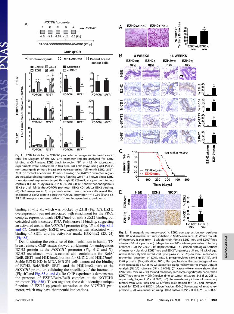
binding at −1.2 kb, which was blocked by ΔHII (Fig. 4B). EZH2overexpression was not associated with enrichment for the PRC2complex repression mark H3K27me3 or with SUZ12 binding butcoincided with increased RNA Polymerase II binding, suggestingan activated area in the NOTCH1 promoter (Fig. 4B and Fig. S5 Aand C). Consistently, EZH2 overexpression was associated withbinding of SET1 and its activation mark, H3K4me2 (23, 24)(Fig. S5).Demonstrating the existence of this mechanism in human TN
breast cancer, ChIP assays showed enrichment for endogenousEZH2 protein at the NOTCH1 promoter (Fig. 4 C and D).EZH2 recruitment was associated with enrichment for RelA/RelB, SET1, and H3K4me2, but not for SUZ12 and H3K27me3.Stable EZH2 KD in MDA-MB-231 cells decreased the bindingof EZH2, RelA/RelB, SET1, and the H3K4me2 mark at theNOTCH1 promoter, validating the specificity of the interaction(Fig. 4C and Fig. S5 A and B). Re-ChIP experiments demonstratethe presence of EZH2/RelA/RelB complex at the NOTCH1promoter (Fig. S5B). Taken together, these data identify a uniquefunction of EZH2 epigenetic activation at the NOTCH1 pro-moter, which may have therapeutic implications.
Fold
Enr
ichm
ent R
elat
ive
to GAPDH
A
Nontumorigenic
EZH
2H
3K27
me3
SUZ1
2
B C MDA-MB-231 Patient breastcancer cells
D
ChIP qPCR
CAGGAGGGGCGCCGGGACACGC (22bp)
NOTCH1BCDE
NOTCH1 promoterA
-4.5 -3.2 -2.68 -1.2 -0.5 (kb)
Fig. 4. EZH2 binds to the NOTCH1 promoter in benign and in breast cancercells. (A) Diagram of the NOTCH1 promoter regions analyzed for EZH2binding in ChIP assays. EZH2 binds to region “B” at −1.2 kb; subsequentexperiments were performed in this area. (B) ChIP assays using qRT-PCR innontumorigenic primary breast cells overexpressing full-length EZH2, ΔSET,ΔHII, or control adenovirus. Primers flanking the GAPDH promoter regionare negative binding controls. Primers flanking MYT1, a known direct EZH2transcriptional repression target through H3K27me3, are positive bindingcontrols. (C) ChIP assays (as in B) in MDA-MB-231 cells show that endogenousEZH2 protein binds the NOTCH1 promoter. EZH2 KD reduces EZH2 binding.(D) ChIP assays (as in B) in patient-derived breast cancer cells reveal thatendogenous EZH2 protein binds the NOTCH1 promoter. *P < 0.05 (B and C).All ChIP assays are representative of three independent experiments.
C
EZH2wt;neu
EZH2+;neu
8 WEEKSB
EZH2+;neuEZH2wt;neuA
EZH2 NICD1H&E
EZH
2wt;
neu
EZH
2+;
neu
D
EZH2wt;neu
EZH2+;neu
16 WEEKS
EZH
2N
ICD
1H
&E
p-ST
AT3
Ki-6
7
EZH
2wt;n
eu
EZH
2+;n
eu
Fig. 5. Transgenic mammary-specific EZH2 overexpression up-regulatesNOTCH1 and accelerates tumor initiation in MMTV-neu mice. (A) Whole mountsof mammary glands from 16-wk-old virgin female EZH2+;neu and EZH2wt;neumice (n = 10 mice per group). (Magnification: 200×.) Average number of tertiarybranches ± SD (*P = 0.01). (B) Representative H&E-stained histological sectionsof mammary glands of EZH2+;neu and EZH2wt;neu mice at 8 and 16 wk of age.Arrow shows atypical intraductal hyperplasia in EZH2+;neu mice. Immunohis-tochemical detection of EZH2, NICD1, phosphorylated-STAT3 (p-STAT3), andKi-67 proteins. (Magnification: 400×.) Bar graphs show the percentages of rel-ative expression ± SD at 16 wk quantified using Framework for Image DatasetAnalysis (FRIDA) software (*P < 0.0004). (C) Kaplan–Meier curve shows thatEZH2+;neu mice (n = 30) formed mammary carcinomas significantly earlier thanEZH2wt;neu mice (n = 25) (median time to tumor initiation: 243 d vs. 295 d,respectively; log-rank P < 0.0001). (D) Representative pictures of mammarytumors from EZH2+;neu and EZH2wt;neu mice stained for H&E and immunos-tained for EZH2 and NICD1. (Magnification: 400×.) Percentage of relative ex-pression ± SD was quantified using FRIDA software (*P < 0.003, **P = 0.009).
Gonzalez et al. PNAS | February 25, 2014 | vol. 111 | no. 8 | 3101
MED
ICALSC
IENCE
S

Transgenic EZH2 Overexpression Up-Regulates NOTCH1, IncreasesStem Cells, and Accelerates Tumor Initiation. Because of tumor la-tency, the mouse mammary tumor virus (MMTV)-neu mousemodel is well-suited to test the effect of EZH2 overexpression onaccelerating tumor initiation (25). Mammary-specific EZH2transgenic mice developed in our laboratory (26) were crossedwith MMTV-neu mice (25). Female 8-wk-old virgin EZH2+;neumice exhibited ductal hyperbranching compared with EZH2wt;neumice (Fig. 5A). Virgin EZH2+;neu mice developed atypicalintraductal hyperplasia similar to human disease, had up-regu-lation of NOTCH1 signaling proteins, and increased cell pro-liferation, compared with EZH2wt;neu mice (Fig. 5B). EZH2+;neu mice formed invasive mammary carcinomas significantlyearlier than EZH2wt;neu mice (Kaplan–Meier, log-rank P <0.0001; Fig. 5C). Although no notable histopathological differ-ences were apparent, EZH2+;neu tumors exhibited increasedEZH2 and NICD1 (Fig. 5D). Collectively, these data providedirect in vivo evidence that precancerous EZH2 up-regulationpromotes atypical epithelial hyperplasia with heightened NOTCH1pathway activation and that EZH2 overexpression is sufficientto accelerate tumor initiation in MMTV-neu mice.Flow cytometric analyses in the lineage negative (Lin‒) pop-
ulation using ESA and CD49f, shown to delineate cellular sub-sets in MMTV-neu mice and in the human breast (27, 28),demonstrated that EZH2+;neu glands had increased stem cells(Lin− ESAmed CD49f high) and progenitors (Lin− ESAhigh CD49fmed) compared with EZH2wt,neu glands (Fig. S6A). The transi-tion from preneoplasia to tumor formation in MMTV-neu miceis characterized by an increase in ESA-, CD49f-, and CD61-expressing cells detected by flow cytometry (28). Using dualimmunohistochemistry, we detected areas of increased numbersof tumor cells expressing these markers in EZH2+;neu tumorscompared with EZH2wt;neu tumors (Fig. S6D).Transplantation experiments (29) showed that stem cells, but
not progenitor cells, isolated from preneoplastic EZH2+;neuand EZH2wt;neu mammary glands exhibited in vivo gland-reconstituting activity. Of note, stem cells from EZH2+;neuglands formed hyperplastic outgrowths with increased numberand size of terminal end buds, higher NICD1 and Ki-67 expres-sion, and elevated percentages of stem cells compared withEZH2wt;neu outgrowths (Fig. S6 B and C).
EZH2/NICD1 Axis in TN Breast Cancer Tissues. To examine whetherEZH2-mediated regulation of NOTCH1 and TICs exists in tu-mor tissues, tissue microarrays from 143 primary invasive breastcarcinoma patients (67 luminal, 9 HER-2/neu over-expressing, 58TN, and 9 unknown subtype) were interrogated for EZH2 intandem with NICD1 and the presence of CD44+/CD24− pop-ulations by immunohistochemistry. Segregating the patient cohortinto EZH2low vs. EZH2high and NICD1+ vs. NICD1− groups, astatistically significant association was identified wherein 77%of the EZH2high scoring patients were also NICD1+ (χ2 test,P < 0.0001; Fig. 6 A and B). Concordant EZH2high and NICD1+
expression was significantly associated with higher histologicalgrade, a measure of poor tumor differentiation, and with thepresence of CD44+/CD24− cancer cells (χ2 test, P = 0.006 andP = 0.0008, respectively; Fig. 6 B and C and Table S2). Furthercharacterization of the dataset indicates that of the 58 tumorscategorized as TN breast cancers, 83% (48 patients) fall intothe EZH2high subgroup. Tumors with concordant EZH2high andNICD1+ are more likely to be TN, whereas those tumorswithout EZH2high and NICD1+ expression are more likely to beluminal (χ2 test, P = 0.029; Fig. 6C and Table S2).
DiscussionEZH2 is an independent marker of recurrence and metastasis inwomen with breast cancer, where EZH2 overexpression occursmainly in TN compared with luminal tumors (11). Despite major
advances in diagnosis and treatment, there is a considerable gapin our understanding of the mechanisms that induce breastcancer development and progression. In this study, we identifya previously undescribed role for EZH2 in regulating NOTCH1-dependent breast TIC expansion and show that EZH2 has a di-rect role in breast cancer progression.We found that EZH2 expression regulates the abundance of
TICs in vitro and in vivo. Ectopic EZH2 expression increased thestem cell pool in nontumorigenic breast cells, whereas EZH2down-regulation reduced the breast TIC population in vitro andin xenograft studies. Our data strengthen those from an earlierstudy showing that EZH2 can promote breast TIC expansion(13) and further demonstrate the consequences of EZH2 levelson breast cancer initiation. EZH2 down-regulation in TN breastcancer cells retarded breast cancer initiation. Providing pre-viously unidentified in vivo evidence for a role of EZH2 in breast
EZH2 NICD1
Cas
e 1
Cas
e 2
Cas
e 3
CD44: RedCD24: BrownH&E
EZH2
Low High Total p-value
NICD1
Negative 39 (70.9%) 20 (22.7%) 59 <0.0001
Positive 16 (29.1%) 68 (77.3%) 84
Total 55 88 143
Association between EZH2 and NICD1 Levels in Breast CancerA
B
C
Fig. 6. EZH2 overexpression is associated with high NICD1 and increased TICsin human breast cancer tissues. (A) Distribution of EZH2 and NICD1 expressionin the patient cohort (n = 143) (χ2 test, P < 0.0001). (B) Human breast cancertissue samples immunostained for EZH2 and NICD1 and coimmunostainedfor CD44 (red) and CD24 (brown). Case 1 is a luminal-type invasive carcinomawith low EZH2 expression, negative NICD1, and CD44−/CD24+. Case 2 is a TNinvasive carcinoma with high EZH2 expression and positive NICD1 and containsCD44+/CD24− cancer cells. Case 3 is a representative picture of a tumor em-bolus in a breast lymphatic with high EZH2 and high NICD1 and containsCD44+/CD24− cancer cells. (Magnification: 400×.) (C) Percentage of invasivecarcinomas with concordant EZH2high and NICD1+ expression according to thepresence of CD44+/CD24− cancer cells (Left) and according to breast cancersubtypes (Right). We found a significant association between the EZH2high/NICD1+ phenotype and the presence of CD44+/CD24− cells and the TN breastcancer subtype (χ2 test: P = 0.0008 and P = 0.029, respectively).
3102 | www.pnas.org/cgi/doi/10.1073/pnas.1308953111 Gonzalez et al.

cancer initiation in transgenic models, overexpression of EZH2in MMTV-neu mice decreased the latency to breast cancer on-set. The role of EZH2 in promoting breast stem cell expansionand cancer initiation sheds light on our previous study showingthat EZH2 is increased in histologically normal breast tissuesfrom women up to 12 y before they develop breast cancer (12).From a clinical perspective, blocking EZH2 may prevent orameliorate breast cancer initiation in women with high EZH2protein expression levels in their breast epithelium.Despite interest in the association between EZH2 functions,
breast TICs, and TN breast cancer (13), the molecular mechanismsunderlying the tumorigenic function of EZH2 in this cancer subtypeand the relationship to NOTCH1 signaling have not yet been con-sidered. Furthermore, whereas a role for NOTCH1 in breast tu-morigenesis has been established in vivo (30), the factors regulatingincreased NOTCH1 expression and signaling in breast cancer cellsare largely unknown (31, 32). We show that EZH2 is a regulator ofNOTCH1 expression and pathway activation in TN breast cancerand that NOTCH signaling activation is required for EZH2-de-pendent stem cell expansion. The association and mechanistic linkbetween EZH2 and NOTCH1 was validated in vitro, in vivo, and inhuman breast cancer samples. The relevance of our findings isfurther supported by a research study, which, by using the NOTCH-GFP reporter assay used here, demonstrated that NOTCH activityidentifies the cancer stem cell population in lung carcinomas (20).Substantial studies show that the canonical function of EZH2
is exerted via transcriptional repression through its HMT activityon H3K27 (4, 11, 13, 22). Most studies have focused on PRC2-mediated repression of tumor suppressor genes as the mainoncogenic mechanism of EZH2 (6). More recently, EZH2 wasshown to activate transcription via non–PRC2-mediated mech-anisms, including interaction with RelA/RelB to activate NF-κBtargets (9). Our study defines a unique role and mechanism forEZH2 in TN breast cancer, whereby EZH2 binds to the NOTCH1promoter and induces epigenetic activation. We demonstratethat the amino-terminal HII domain of EZH2 mediates binding
to the NOTCH1 promoter and that the HMT activity of EZH2 isnot required for NOTCH1 transcriptional activation. Our resultsare consistent with a recent study documenting the importanceof the amino-terminal domain of EZH2 in enhancing genetransactivation through an HMT-independent mechanism inbreast cancer (7). Together, our data strengthen the emergingnotion that overexpressed EZH2 may function through non-canonical mechanisms leading to activation of target genes in anHMT-independent manner.In clinical invasive breast cancer samples, high EZH2 and
NICD1 are significantly coexpressed in TN compared with luminaltumors, supporting the contention that the EZH2/NICD1 axis isoperative in vivo and in humans. Furthermore, EZH2high/NICD1+
tumors are more frequently poorly differentiated and exhibit highnumbers of TICs compared with tumors without this phenotype. Inconclusion, our findings establish a previously unrecognized linkbetween EZH2, NOTCH1 signaling activation, and TICs in TNinvasive carcinomas. These data advance the current understandingof the mechanisms of EZH2 in breast cancer and lend support tothe emerging transcriptional activating role of EZH2. By providingpreviously unidentified direct evidence that EZH2 overexpressionaccelerates breast cancer initiation in vivo our work paves the way totargeting EZH2 to halt breast cancer progression.
Materials and MethodsDetailed protocols regarding cell culture, vectors, pharmacologic treatments,Western blot analyses, antibodies, microarrays, mammosphere assays, flowcytometry, ChIP analyses, immunohistochemistry, and animal studies aredescribed in SI Materials and Methods. The breast cancer patient cohort hasbeen described previously (see SI Materials and Methods). Original Westernblots are shown in Fig. S7.
ACKNOWLEDGMENTS. This work was supported by National Institutes ofHealth Grants CA107469, CA125577, and CA154224 (to C.G.K.), Departmentof Defense Breast Cancer Research Program Predoctoral Traineeship AwardBC093828 (to H.M.M.), and in part through the University of Michigan’sCancer Center Support Grant P30 CA046592.
1. Jacobs JJ, Kieboom K, Marino S, DePinho RA, van Lohuizen M (1999) The oncogeneand Polycomb-group gene bmi-1 regulates cell proliferation and senescence throughthe ink4a locus. Nature 397(6715):164–168.
2. Francis NJ, Kingston RE (2001) Mechanisms of transcriptional memory. Nat Rev MolCell Biol 2(6):409–421.
3. Cao R, et al. (2002) Role of histone H3 lysine 27 methylation in Polycomb-group si-lencing. Science 298(5595):1039–1043.
4. Chang CJ, HungMC (2012) The role of EZH2 in tumour progression. Br J Cancer 106(2):243–247.
5. Min J, et al. (2010) An oncogene-tumor suppressor cascade drives metastatic prostatecancer by coordinately activating Ras and nuclear factor-kappaB. Nat Med 16(3):286–294.
6. Bracken AP, et al. (2003) EZH2 is downstream of the pRB-E2F pathway, essential forproliferation and amplified in cancer. EMBO J 22(20):5323–5335.
7. Shi B, et al. (2007) Integration of estrogen and Wnt signaling circuits by the polycombgroup protein EZH2 in breast cancer cells. Mol Cell Biol 27(14):5105–5119.
8. Xu K, et al. (2012) EZH2 oncogenic activity in castration-resistant prostate cancer cellsis Polycomb-independent. Science 338(6113):1465–1469.
9. Lee ST, et al. (2011) Context-specific regulation of NF-κB target gene expression byEZH2 in breast cancers. Mol Cell 43(5):798–810.
10. Asangani IA, et al. (2013) Characterization of the EZH2-MMSET histone methyl-transferase regulatory axis in cancer. Mol Cell 49(1):80–93.
11. Kleer CG, et al. (2003) EZH2 is amarker of aggressive breast cancer and promotes neoplastictransformation of breast epithelial cells. Proc Natl Acad Sci USA 100(20):11606–11611.
12. Ding L, Erdmann C, Chinnaiyan AM, Merajver SD, Kleer CG (2006) Identification ofEZH2 as a molecular marker for a precancerous state in morphologically normalbreast tissues. Cancer Res 66(8):4095–4099.
13. Chang CJ, et al. (2011) EZH2 promotes expansion of breast tumor initiating cellsthrough activation of RAF1-β-catenin signaling. Cancer Cell 19(1):86–100.
14. Gonzalez ME, et al. (2009) Downregulation of EZH2 decreases growth of estrogen re-ceptor-negative invasive breast carcinoma and requires BRCA1. Oncogene 28(6):843–853.
15. Dontu G, et al. (2003) In vitro propagation and transcriptional profiling of humanmammary stem/progenitor cells. Genes Dev 17(10):1253–1270.
16. Ginestier C, et al. (2007) ALDH1 is a marker of normal and malignant human mam-mary stem cells and a predictor of poor clinical outcome. Cell Stem Cell 1(5):555–567.
17. Al-Hajj M, Wicha MS, Benito-Hernandez A, Morrison SJ, Clarke MF (2003) Prospectiveidentification of tumorigenic breast cancer cells. Proc Natl Acad Sci USA 100(7):3983–3988.
18. Gonzalez ME, et al. (2011) Histone methyltransferase EZH2 induces Akt-dependentgenomic instability and BRCA1 inhibition in breast cancer. Cancer Res 71(6):2360–2370.
19. Pui JC, et al. (1999) Notch1 expression in early lymphopoiesis influences B versus Tlineage determination. Immunity 11(3):299–308.
20. Hassan KA, et al. (2013) Notch pathway activity identifies cells with cancer stem cell-like properties and correlates with worse survival in lung adenocarcinoma. ClinCancer Res 19(8):1972–1980.
21. Kim E, et al. (2013) Phosphorylation of EZH2 activates STAT3 signaling via STAT3methylation and promotes tumorigenicity of glioblastoma stem-like cells. Cancer Cell23(6):839–852.
22. Cao Q, et al. (2008) Repression of E-cadherin by the polycomb group protein EZH2 incancer. Oncogene 27(58):7274–7284.
23. Liang G, et al. (2004) Distinct localization of histone H3 acetylation and H3-K4methylation to the transcription start sites in the human genome. Proc Natl Acad SciUSA 101(19):7357–7362.
24. South PF, Harmeyer KM, Serratore ND, Briggs SD (2013) H3K4 methyltransferase Set1is involved in maintenance of ergosterol homeostasis and resistance to Brefeldin A.Proc Natl Acad Sci USA 110(11):E1016–E1025.
25. Guy CT, et al. (1992) Expression of the neu protooncogene in themammary epithelium oftransgenic mice induces metastatic disease. Proc Natl Acad Sci USA 89(22):10578–10582.
26. Li X, et al. (2009) Targeted overexpression of EZH2 in the mammary gland disrupts ductalmorphogenesis and causes epithelial hyperplasia. Am J Pathol 175(3):1246–1254.
27. Lim E, et al.; kConFab (2009) Aberrant luminal progenitors as the candidate target pop-ulation for basal tumor development in BRCA1 mutation carriers. Nat Med 15(8):907–913.
28. Lo PK, et al. (2012) CD49f and CD61 identify Her2/neu-induced mammary tumor-initiating cells that are potentially derived from luminal progenitors and maintainedby the integrin-TGFβ signaling. Oncogene 31(21):2614–2626.
29. Guo W, et al. (2012) Slug and Sox9 cooperatively determine the mammary stem cellstate. Cell 148(5):1015–1028.
30. Hu C, et al. (2006) Overexpression of activated murine Notch1 and Notch3 in trans-genic mice blocks mammary gland development and induces mammary tumors. Am JPathol 168(3):973–990.
31. Haughian JM, et al. (2012) Maintenance of hormone responsiveness in luminal breastcancers by suppression of Notch. Proc Natl Acad Sci USA 109(8):2742–2747.
32. Clementz AG, Rogowski A, Pandya K, Miele L, Osipo C (2011) NOTCH-1 and NOTCH-4are novel gene targets of PEA3 in breast cancer: Novel therapeutic implications.Breast Cancer Res 13(3):R63.
Gonzalez et al. PNAS | February 25, 2014 | vol. 111 | no. 8 | 3103
MED
ICALSC
IENCE
S