Embed Size (px)
Citation preview
Eye Trauma
Eye Trauma
• Ocular injuries may be from blunt, penetrating or perforating injuries
• Intervene before obtaining vision• Thorough ocular examination for soft tissue • Check for canalicular integrity• Always rule out globe rupture• May be anterior or posterior– High index of suspicion for ruptured globe, foreign
body
General Guidelines
• Complete history/nature of injury• Thorough and methodical ocular examination• “First, do no harm”
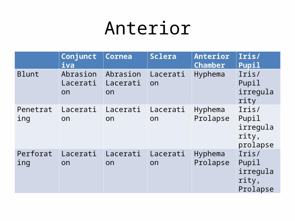
AnteriorConjunctiva Cornea Sclera Anterior
ChamberIris/Pupil
Blunt AbrasionLaceration
AbrasionLaceration
Laceration Hyphema Iris/Pupil irregularity
Penetrating Laceration Laceration Laceration HyphemaProlapse
Iris/Pupil irregularity, prolapse
Perforating Laceration Laceration Laceration HyphemaProlapse
Iris/Pupil irregularity, Prolapse
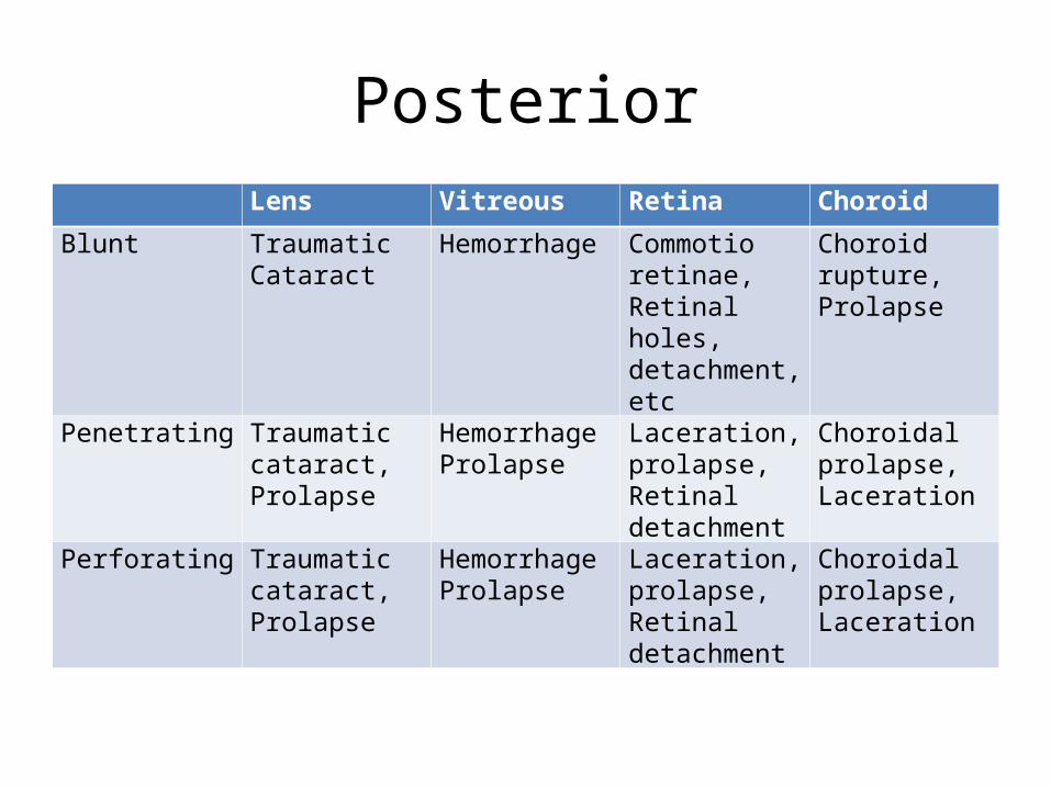
PosteriorLens Vitreous Retina Choroid
Blunt Traumatic Cataract
Hemorrhage Commotio retinae, Retinal holes, detachment, etc
Choroid rupture, Prolapse
Penetrating Traumatic cataract, Prolapse
HemorrhageProlapse
Laceration, prolapse, Retinal detachment
Choroidal prolapse, Laceration
Perforating Traumatic cataract, Prolapse
HemorrhageProlapse
Laceration, prolapse, Retinal detachment
Choroidal prolapse, Laceration

CORNEAL ABRASION SECONDARY TO THERMAL BURN
• History:– Exposure to welding or sun lamps without protective
eyewear, UV exposure – snow blindness• Symptoms– Moderate to severe ocular pain– Foreign body sensation– Red eye– Tearing– Symptoms worst within 6-12 hours of exposure
• Critical signs– Confluent epithelial defects in interpalpebral distribution
seen by fluorescein staining
• Work up– History of exposure– Slit lamp exam– Rule out possibility of chemical burns
• Treatment– Cycloplegics
• They help with ciliary spasm– Antibiotics– Analgesics– Optional pressure patch (for faster corneal healing)

CORNEAL ABRASION SECONDARY TO CHEMICAL BURNS
• One of the true emergencies in ophthalmology• Emergency treatment:– Copious eye irrigation with saline or ringer’s lactate
solution for at least 30 minutes– When it happens in the house, wash with water– Irrigation volume vary with chemical and duration of
exposure– Ideally, use of litmus paper to determine neutrality – Why not basic solution to counteract acid, instead of
water? Do not irrigate with opposite pH because exothermic reaction will occur and make the burn worse
• ACID vs ALKALI– Acid burns cause denaturation of tissue proteins
(serve as buffer so it does not penetrate)– Alkali saponifies fatty acids thus causing deeper
penetration– More devastating injury with alkali burn
Mild to moderate burns
– Scattered corneal epithelial defects– No significant areas of perilimbal ischemia– Chemosis - edema of the conjunctiva of the eye
Work-Up and Treatment• Work up
– Slit lamp examination with fluorescein staining• Treatment
– Copious irrigation with sweeping of fornices– Cycloplegia
• Paralysis of the ciliary muscle, resulting in a loss of accommodation. • Cycloplegic drugs, including atropine, cyclopentolate, homatropine,
scopolamine, and tropicamide, are indicated for use in cycloplegic refractions and the treatment of uveitis.
– Antibiotic– Artificial tears
• promotes healing for minor injuries– Oral analgesics
Severe burns
• Critical signs– Pronounced chemosis with conjunctival blanching– Corneal edema and opacification– Moderate to severe anterior chamber reaction– IOP increase
• Work up– Same as thermal burns– Repeat staining since defect may be slow to take
up
Treatment• Irrigation
– Admission may be necessary• Debride necrotic tissue/foreign body• Cycloplegia• Antibiotic• Steroid if significant anterior chamber or corneal inflammation
present– But in other cases, no steroids because it may retard epithelial healing
• May put on pressure patch• Anti-glaucoma meds for IOP increase• Lysis of conjunctival adhesions by using glass rod • Artificial tears
– Because most patients cannot move the eye anymore due to adhesions
Follow up
• Close monitoring– IOP
• Tapering of steroids after 7-10 days to allow for epithelial healing
• Artificial tears
PERIOCULAR TRAUMA
Types of periocular trauma
• Soft tissue injuries– Contusion– Avulsion– Puncture– Lacerations – complex or simple; deep or
superficial
• Fractures
Lid Injury
• Lids – outermost protective mechanism• Reflex closure before most injuries• Lacerations most common• Lid closure – cranial nerve VII
Periorbital contusion hematoma:
• Periocular edema and hematoma• Chemosis• Good vision• Subconjunctival Hemorrhage• Ptosis• Intact EOMs• No palpable fractures or defects• Ask for diagnostics just in case you are missing a
fracture• Cold compress• Anti-inflammatory meds
Pre-septal fat contusion / Lid laceration
• Considerations:– Lid margin vs. non lid margin– Pre-septal fat– r/o canalicular involvement– r/o globe rupture
• Non margin laceration– thorough ocular exam– Primary repair– Antibiotics– Analgesics
Eyelid Margin laceration
• align the eyelid margin• need to move tissue around• use of flaps and grafts dependent on tissue
defect
CONJUNCTIVAL LACERATION WITH CORNEAL ABRASION
• Example: 32 y/o M basketball player, accidentally poked on right eye
• Signs and Symptoms– Sharp pain, photophobia, FB sensation, tearing, red
eye– staining of conjunctiva, exposed white sclera is
appreciated– VA 20/50
• Work up– Slit lamp exam with fluorescein staining– Lid eversion (to rule out foreign body)
• Treatment– Antibiotic coverage– Artificial tears– Cycloplegic– Patching (gives a banding effect)– Repair of laceration if very large– DO NOT GIVE steroid drops• delays repair of epithelium
Case continued…
• Same patient• 2 days later, complaining of throbbing pain,
photophobia• VA 20/40• Cells and flare in the AC– aqueous humor in anterior chamber must be
pristine clean
Critical Signs
• Photophobia• Either poorly dilating pupil or large pupil• Conjunctival injection• Cells and flare
• Work up– Slit lamp exam– IOP check
• Differentials– Corneal abrasion• still considered because he may have not used his
patch delayed healing– Traumatic microhyphema– Traumatic iritis
• Treatment– Cyclopegic– Steroid if no improvement in 5-7 days
• Follow up– One week– Discontinue meds if resolved– Check in one month for post trauma sequelae

HYPHEMA
• Any gross blood in the anterior chamber is hyphema; micro means suspended amount in aqueous humor
• Signs and Symptoms– Pain and blurred vision– VA 20/80– Gross blood noted on anterior chamber
• Work up– Extensive history– Complete ocular exam
Hyphema grading
• Microhyphema• Gr I – 25%• Gr II – 50%• Gr III – 75%• Gr IV – 100%
Treatment
• Bed rest• Eye shield (but do not press the eye during PE
to avoid more bleeding)• Long acting cycloplegic• Mild analgesic• Consider steroids• Consider anti-glaucoma drugs if IOP is high• Aminocaproic acid
When to admit for hospitalization?
– Poor VA on presentation– Blood dyscrasia with increased IOP– Medically uncontrollable IOP– Large initial hyphema– Delayed presentation to MD– Large amount of recent NSAID intake
Follow up
• Close follow up especially for patients with increased risk for re-bleed
• Golden period of 3-5 days risk• Refrain from vigorous activity for about 2
weeks• Follow up in 2-4 weeks for possible sequelae– initial grading of hyphema to monitor
improvement later• Yearly check if extensive
Surgery
• Corneal stromal blood staining• Significant visual deterioration• Total blood filling in AC• Persistent clot packed in angle• IOP increase with maximal medical therapy

TRAUMATIC CATARACTS
• Secondary to blunt or penetrating ocular trauma
• Form stellate- or rosette-shaped posterior axial opacities that may be stable or progressive
• Lens dislocation and subluxation are commonly found in conjunction with traumatic cataract
Signs and Symptoms
• Mechanism of injury - Sharp versus blunt• Past ocular history - Previous eye surgery, glaucoma,
retinal detachment, diabetic eye disease• Past medical history - Diabetes, sickle cell, Marfan
syndrome, homocystinuria, hyperlysinemia, sulfate oxidase deficiency
• Visual complaints– Decreased vision– Monocular diplopia– Binocular diplopia– Pain
Complete ophthalmic examination• Vision and pupils - Presence of afferent pupillary defect (APD)
indicative of traumatic optic neuropathy• Extraocular motility - Orbital fractures or traumatic nerve palsy• Intraocular pressure - Secondary glaucoma, retrobulbar
hemorrhage• Anterior chamber - Hyphema, iritis, shallow chamber, iridodonesis,
angle recession• Lens - Subluxation, dislocation, capsular integrity (anterior and
posterior), cataract (extent and type), swelling, phacodonesis• Vitreous - Presence or absence of hemorrhage, posterior vitreous
detachment• Fundus - Retinal detachment, choroidal rupture, commotio retinae,
preretinal hemorrhage, intraretinal hemorrhage, subretinal hemorrhage, optic nerve pallor, optic nerve avulsion
Workup
• B-scan - If the posterior pole cannot be visualized
• A-scan - Prior to cataract extraction• CT scan of the orbits - Fractures and foreign
bodies
Treatment
• If glaucoma is a problem, control intraocular pressure with standard medications. Add corticosteroids if lens particles are the cause or if iritis is present.
• Focal cataract– Observation is warranted if the cataract is outside the
visual axis.– Miotic therapy may be of benefit if the cataract is close to
the visual axis.• In some cases of lens subluxation, miotics may correct
monocular diplopia. Mydriatics may allow for vision around the lens with aphakic correction.
Indications for Surgery
• Unacceptable decreased vision• Obstructed view of posterior pathology• Lens-induced inflammation or glaucoma• Capsular rupture with lens swelling• Other trauma-induced ocular pathology
necessitating surgery
Surgical Care
• Preoperative capsular integrity and zonular stability should be surmised.• In cases of posterior dislocation without glaucoma, inflammation, or visual
obstruction, surgery may be avoided.• Standard phacoemulsification may be performed
– Lens capsule intact– Sufficient zonular support
• Intracapsular cataract extraction– anterior dislocation or extreme zonular instability
• can cause pupillary block glaucoma.• Pars plana lensectomy and vitrectomy may be best in cases of posterior
capsular rupture, posterior dislocation, or extreme zonular instability.• Automated irrigation/aspiration can be used in patients younger than 35
years.• Lens implantation

TRAUMATIC VITREOUS HEMORRHAGE
• Extravasation of blood into one of the several potential spaces formed within and around the vitreous body
Signs and Symptoms
• present with a complaint of visual haze, floaters, cloudy vision or smoke signals, photophobia, and perception of shadows and cobwebs.
• Small vitreous hemorrhage often is perceived as new multiple floaters,
• Moderate vitreous hemorrhage is perceived as dark streaks, and
• Dense vitreous hemorrhage tends to significantly decrease vision even to light perception.
• Ophthalmoscopic examination reveals blood within the vitreous gel and/or the anterohyaloid or retrohyaloid spaces.
Treatment
• No treatment unless very extensive hemorrhage
• Even choroidal ruptures, if they are not prolapsed, no need to repair, just wait
• Usually clears without therapy
Surgical Care
• Indications for surgical removal of the vitreous blood include the following:– Vitreous hemorrhage associated with detached
retina– Long-standing vitreous hemorrhage with duration
greater than 2-3 months– Vitreous hemorrhage associated with rubeosis– Vitreous hemorrhage associated with hemolytic or
ghost-cell glaucoma

RUPTURED GLOBE
• Significantly decreased VA• Shallow or flat AC• Altered size, position of pupil• Visible tracks through the lens or vitreous tracing
the line of passage of FB• Marked conjunctival chemosis• Subconjunctival hemorrhage• Total hyphema with low pressure• Positive Seidel’s test
• “Do no harm”– Avoid applying pressure on the eye– Avoid straining
• May be from blunt, penetrating or perforating mechanism so know the history
• Eye shield

• NPO (in preparation for possible surgery later)• Antibiotic coverage• Tetanus prophylaxis• Consider anti-emetics (so that staining is avoided) • Ancillary test– Rule out occult rupture with scans
• Arrange for immediate surgical repair– Any delay sympathetic ophthalmia affecting the other
eye– When do you remove the eye? Consistent finding of NLP
by 3 consultants

FOREIGN BODIES
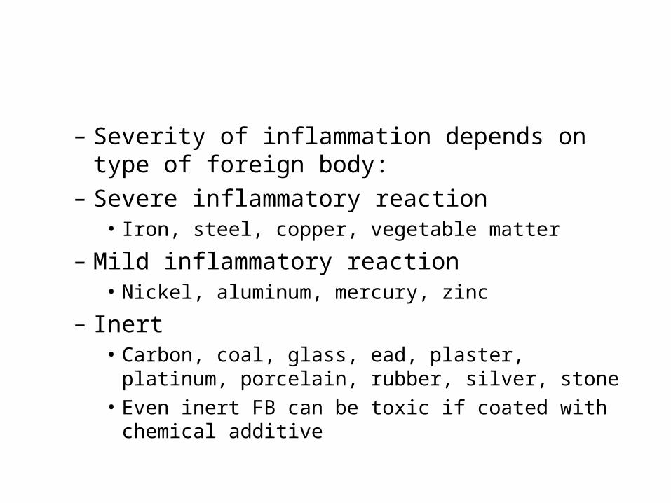
– Severity of inflammation depends on type of foreign body:
– Severe inflammatory reaction• Iron, steel, copper, vegetable matter
– Mild inflammatory reaction• Nickel, aluminum, mercury, zinc
– Inert• Carbon, coal, glass, ead, plaster, platinum, porcelain, rubber,
silver, stone• Even inert FB can be toxic if coated with chemical additive
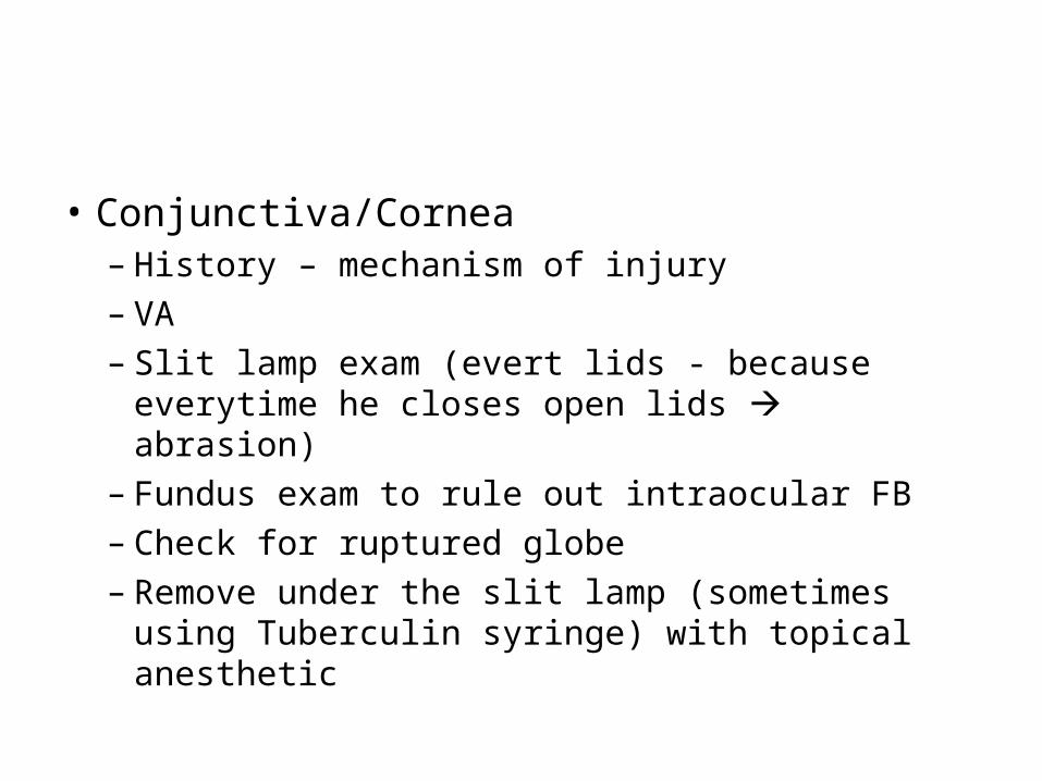
• Conjunctiva/Cornea– History – mechanism of injury– VA– Slit lamp exam (evert lids - because everytime he
closes open lids abrasion)– Fundus exam to rule out intraocular FB– Check for ruptured globe– Remove under the slit lamp (sometimes using
Tuberculin syringe) with topical anesthetic
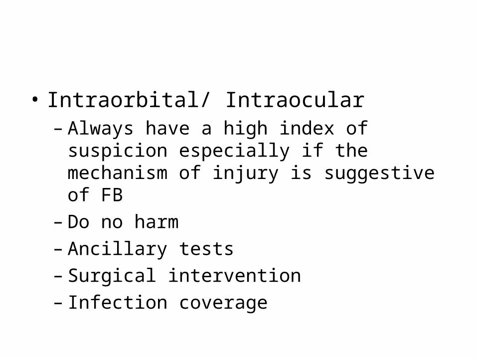
• Intraorbital/ Intraocular– Always have a high index of suspicion especially if
the mechanism of injury is suggestive of FB– Do no harm– Ancillary tests– Surgical intervention– Infection coverage