Embed Size (px)
Citation preview
Deletion of mouse MsrA results in HBO-induced cataract: MsrArepairs mitochondrial cytochrome c
LA Brennan,1 W Lee,1 T Cowell,1 F Giblin,2 M Kantorow1
1Biomedical Sciences Department, Charles E. Schmidt College of Biomedical Science, Florida Atlantic University, Boca Raton, FL;2Eye Research Institute, Oakland University, Rochester, MI
Purpose: Considerable evidence indicates a role for methionine sulfoxide reductase A (MsrA) in lens cell resistance tooxidative stress through its maintenance of mitochondrial function. Correspondingly, increased protein methioninesulfoxide (PMSO) is associated with lens aging and human cataract formation, suggesting that loss of MsrA activity isassociated with this disease. Here we tested the hypothesis that loss of MsrA protein repair is associated with cataractformation. To test this hypothesis we examined the effect of MsrA deletion on lens opacity in mice treated with hyperbaricoxygen, identified lens mitochondrial proteins oxidized upon deletion of MsrA and determined the ability of MsrA torepair the identified proteins.Methods: Wild-type and MsrA knockout mice were treated or not treated with 100 treatments of hyperbaric oxygen(HBO) over an 8 month period and lenses were examined by in vivo light scattering measurements documented by slit-lamp imaging. Co-immunoprecipitation of MsrA was conducted against five specific protein representatives of the fivecomplexes of the electron transport chain in addition to cytochrome c (cyt c). Cyt c in lens protein from the knockout andwild-type lenses was subjected to cyanogen bromide (CNBr) cleavage to identify oxidized methionines. Methionine-specific CNBr cleavage was used to differentiate oxidized and un-oxidized methionines in cyt c in vitro and the ability ofMsrA to restore the activity of oxidized cyt c was evaluated. Mass spectrometry analysis of cyt c was used to confirmoxidation and repair by MsrA in vitro.Results: HBO treatment of MsrA knockout mice led to increased light scattering in the lens relative to wild-type mice.MsrA interacted with four of the five complexes of the mitochondrial electron transport chain as well as with cyt c. Cytc was found to be aggregated and degraded in the knockout lenses consistent with its oxidation. In vitro analysis of oxidizedcyt c revealed the presence of two oxidized methionines (met 65 and met 80) that were repairable by MsrA. Repair of theoxidized methionines in cyt c restored the activity of cytochrome c oxidase and reduced cytochrome c peroxidase activity.Conclusions: These results establish that MsrA deletion causes increased light scattering in mice exposed to HBO andthey identify cyt c as oxidized in the knockout lenses. They also establish that MsrA can restore the in vitro activity ofcyt c through its repair of PMSO. These results support the hypothesis that MsrA is important for the maintenance of lenstransparency and provide evidence that repair of mitochondrial cyt c by MsrA could play an important role in defense ofthe lens against cataract formation.
Significant evidence points to a role for methioninesulfoxide reductases (Msrs) in diseases of aging includingage-related cataract of the eye lens. Msrs are a family ofthioredoxin dependent oxidoreductases that reduce theoxidized form of protein methionine, protein methioninesulfoxide (PMSO), back to its reduced form, methionine. Twoclasses of Msrs are known; MsrA and MsrB which act on theS- and R- epimers of PMSO, respectively. The PMSO contentincreases with age in a number of tissues and aging models[1] including the lens [2] and it has been shown that increasedlevels of PMSO are associated with age-related cataract [2,3]where the PMSO content of the cataractous lens is as high as70% of total soluble lens proteins.
Correspondence to: Marc Kantorow Ph.D., Biomedical SciencesDepartment, Charles E. Schmidt College of Biomedical Science,Florida Atlantic University, Boca Raton, FL; Phone: (561) 297 2910;FAX: (561) 297 2221; email: [email protected]
Levels of MsrA and MsrA activity decrease with age inrat tissue [4] and in the brains of Alzheimer’s patients [5].MsrA is also known to modulate lifespan in animals, forexample, MsrA knockout mice have been reported to have a40% reduction in lifespan relative to wild-type mice [6] andexhibited increased sensitivity to oxidative stress (100%oxygen) with increased levels of oxidized proteins. Inaddition, the MsrA knockout mice developed an atypical (tip-toe) walking pattern after 6 months of age indicative ofneuronal damage. Over-expression of MsrA in Drosophilamelanogaster was shown to increase lifespan by up to 70%[7] and causes increased oxidative stress resistance in WI-38SV40 fibroblasts [8], yeast and human T cells [9], and humanlens epithelial cells [10]. Silencing of the MsrA gene usingsiRNA increased sensitivity of human lens epithelial cells toH2O2-induced oxidative stress [10]. In addition, this loss ofMsrA resulted in loss of mitochondrial membrane potential,increased mitochondrial ROS production, and decreased lenscell viability [11] all of which occurred without exogenously
Molecular Vision 2009; 15:985-999 <http://www.molvis.org/molvis/v15/a104>Received 16 April 2009 | Accepted 11 May 2009 | Published 15 May 2009
© 2009 Molecular Vision
985
added oxidative stress, leading to the hypothesis that lens cellsrequire MsrA for both normal mitochondrial maintenance andviability. These data, in conjunction with data showingincreased PMSO upon human lens aging and cataractformation, suggest that MsrA activity is important for lensmaintenance and defense against oxidative stress through itsrepair of oxidized lens mitochondrial proteins. Identificationof those lens mitochondrial proteins repaired by MsrA couldprovide insight into the requirement for MsrA in maintenanceof the lens and prevention of cataract formation.
One key model for studying oxidative stress is hyperbaricoxygen (HBO) treatment. HBO-treatment has been shown ina number of studies to cause mitochondrial dysfunction,increased mitochondrial ROS production, decreasedantioxidant ability and viability in lens epithelial cells,decreased thioredoxin reductase activity, and, importantly,decreased glutathione (GSH) levels, a key event in cataractformation [12-14].
Here, we used HBO-treatment to induce oxidative stressin the lenses of MsrA knockout and wild-type mice. Sincemitochondrial maintenance appears to depend on the functionof MsrA in lens cells [11], we examined potentialmitochondrial protein-MsrA interactions and determined thatcytochrome c (cyt c) was oxidized in the MsrA knockoutlenses. Cyt c is a small globular heme-containing electroncarrier in the electron transport chain of the mitochondria. Inaddition to its requirement for energy production, its releasefrom the mitochondria to the cytosol is the initiating factor forthe internal pathway of apoptosis [15]. Thus, oxidation of cytc in lens cells could lead to loss of lens epithelial functionsrequiring ATP and aerobic mitochondrial function, includingcationic transport and potentially epithelial cell growth anddifferentiation. Consistent with being a potential target forMsrA, the hexa-coordinated arrangement of heme iron in cytc relies on methionine 80 (met 80). The heme iron moiety ofcyt c is protected by this arrangement preventing binding ofpotentially reactive molecules such as NO, O2, carbonmonoxide (CO), and H2O2 [16]. The met 80 of cyt c istherefore a key amino acid in the structure and function of themolecule and previous work by Chen et al. [17] demonstratedthat hypochlorous acid (HOCl)-mediated oxidation of cyt cresulted in the specific oxidation of cyt c met 80 to met 80sulfoxide and that this oxidation increased the accessibility ofthe molecule to H2O2 due to the inability of the met 80sulfoxide to co-ordinate the heme iron.
In this report, we examined the lenses of MsrA knockoutmice in the presence and absence of hyperbaric oxygentreatment and found increased light scattering in the knockoutrelative to the wild-type lenses. We identified 5 majorcomponents of the electron transport chain that interact withMsrA in lens cells as well as the electron carrier protein cyt c.Analysis of lens protein obtained from untreated wild type andknockout mouse lenses revealed an increased level of
oxidized cyt c occurring in the absence of MsrA in vivo.Following HBO treatment, soluble monomeric cyt c was nolonger detectable in the lenses of MsrA knockout mice.Further analysis of oxidized cyt c in vitro demonstrated thatmethionine oxidation of cyt c resulted in loss of activity thatwas repairable by treatment with purified MsrA. These resultsprovide evidence for a novel role for MsrA in the maintenanceof lens transparency in the face of oxidative insult. They alsodemonstrate that oxidation of cyt c occurs in the lens in vivoin the absence of MsrA and that MsrA can restore its normalfunction in vitro. The results also suggest that cyt c is at leastone target for MsrA in lens epithelial cells whose repair byMsrA is likely important for maintaining lens mitochondrialfunction and preventing cyt c-mediated apoptosis. Thesefindings shed light on the potential roles of MsrA and cyt c inthe development of lens cataract and are relevant towards theunderstanding of MsrA action and PMSO accumulation inother age-related oxidative stress associated diseases.
METHODSHLE B3 cell culture: Human lens epithelial cells (HLE B3)were grown and cultured in DMEM (Invitrogen, Carlsbad,CA) supplemented with 15% FBS (Invitrogen), gentamicin(50 units/ml; Invitrogen), penicillin-streptomycin antibioticmix (50 units/ml; Invitrogen) and fungizone (5 ul/ml;Invitrogen) at 37 °C in the presence of 5% CO2.HBO treatments: Wild type and MsrA knockout mice weretreated with HBO in a pressure vessel 130 cm long and 46 cmin diameter (Amron, Escondido, CA). The vessel had a fullyopening hinged door at one end and at the opposite end, a 15cm viewport for observing animals during the treatment. Thelight was kept off during the treatment, and the window wascovered. The mice were housed in wire cages with tops duringthe oxygen exposure. Plastic trays containing wet towels wereplaced inside the chamber to add humidity, and Sodasorb (WRGrace, Lexington, MA) was added to absorb CO2. After thechamber was sealed, it was flushed for 5 to 10 min withapproximately 1 volume of 100% O2 (USP Grade MedicalGas; Praxair, Danbury, CT), which was vented outside thebuilding. The pressure was then raised during the next 15 minto 2.75 atm absolute (ATA; 26 pounds per square inch gaugeor 58 ft of sea water) of O2. After 1.5 h, the chamber wasflushed with approximately 1 volume of fresh 100% O2, whilethe pressure was maintained. At the end of a 3.0 h holdingperiod the pressure was released over a 15 min period to 1ATA (0 psig), and the animals removed. The mice weretreated three times per week, on alternate days atapproximately the same time of day, for a total of 100treatments in 8 months. Age-matched control animals wereincluded with each group of O2-treated mice. Thetransparency of the lenses of control and HBO-treated micewas assessed in vivo with a slit lamp microscope (Carl Zeiss,Meditec, Thornwood, NY) after induction of full mydriasiswith tropicamide (1%) and phenylephine (10%), and the
Molecular Vision 2009; 15:985-999 <http://www.molvis.org/molvis/v15/a104> © 2009 Molecular Vision
986
results documented by photography. After the animals werekilled by CO2 asphyxiation, the eyes were enucleated and thelenses removed by a posterior approach, and frozenimmediately in dry ice.
MsrA knockout (C57 Black 6, C57BL/6) [6] (a gift fromDr. Rodney Levine, NIH, Bethesda, MD) and wild type mice(B6129SF2/J strain) were sacrificed following HBOtreatment at 8 months and the eyes enucleated. Lenses wereremoved and frozen on liquid nitrogen. Lens proteins wereisolated by homogenization in PBS, homogenates werecentrifuged at 10,000x g for 30 min at 4 °C and the supernatantcollected. Soluble lens protein extracted in this way wassubjected to cyanogen bromide (CNBr) cleavage and cyt canalyzed by western blotting using a cyt c specific antibody(R&D systems, Minneapolis, MN). The epitope for theantibody does not incorporate either of the two methioninespotentially oxidized in cyt c (65 or 80). Western blotting ofoxidized purified cyt c protein without CNBr treatment showsthat the antibody continues to recognize the protein even athigh oxidant levels. Corresponding colloidal blue staining ofthe same gels shows that protein destruction occurs before lossof antibody recognition.Co-localization of MsrA to the mitochondria and withcytochrome c in HLE cells: HLE cells were plated ontocoverslips and incubated overnight in complete media.Standard procedures were used for doubleimmunofluoresence-simultaneous staining. Briefly, cellswere fixed with 3.7% formaldehyde in PBS, blocked with 1%BSA and permeabilized with 0.25% TritonX-100 in PBS. Forco-localization of MsrA to the mitochondria, first, HLE cellswere stained with Mitotracker Red CMXRos (MolecularProbes, Invitrogen) for 45 min at a 1:500 dilution as indicatedby the manufacturer’s protocol. Following mitotracker redstaining, rabbit polyclonal anti-MsrA (Abcam, Cambridge,MA), was incubated overnight in 4 °C at a 1:100 dilution. Forco-localization of MsrA and cyt c in the same HLE cells, rabbitpolyclonal anti-MsrA (Abcam) was incubated simultaneouslywith mouse monoclonal anti-cytochrome c antibody(Mitosciences, Eugene, OR) overnight in 4 °C at 1:1,000dilutions. Cells were washed 3X with PBS, and then incubatedwith Alexa Fluor 488 goat anti-mouse monoclonal secondaryand Texas Red goat-anti Rabbit polyclonal secondary for onehour at room temperature at 1:2,000 dilutions. HLE cells werewashed with PBS 3X, and mounted onto slides.Immunofluoresence staining was visualized using anOlympus Provis AX70 fluorescence microscope.Isolation of mitochondria from HLE B3 cells: Mitochondriawere isolated from HLE B3 cell culture using the Isolation ofMitochondria from Cell Culture kit (Mitosciences) as per themanufacturer's instructions. Briefly, cells were frozen andthawed prior to re-suspension in isolation buffer at aconcentration of 5 mg/ml and lysed by Douncehomogenization. Lysates were incubated in isolation buffer
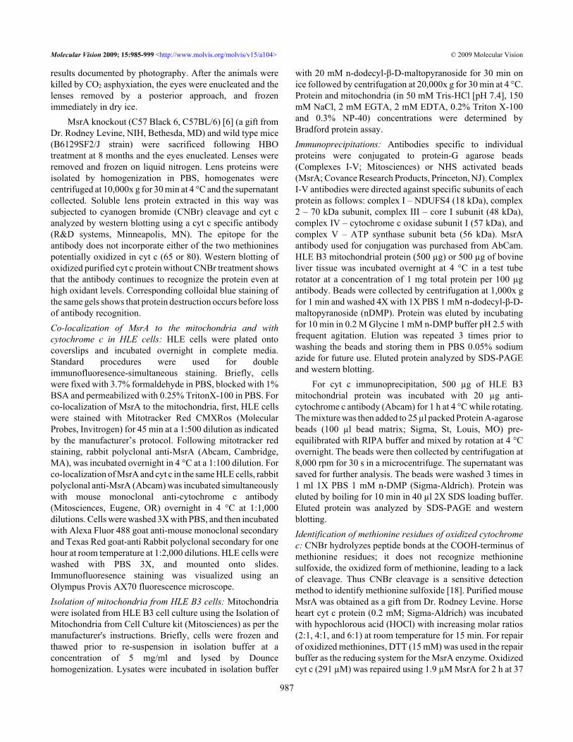
with 20 mM n-dodecyl-β-D-maltopyranoside for 30 min onice followed by centrifugation at 20,000x g for 30 min at 4 °C.Protein and mitochondria (in 50 mM Tris-HCl [pH 7.4], 150mM NaCl, 2 mM EGTA, 2 mM EDTA, 0.2% Triton X-100and 0.3% NP-40) concentrations were determined byBradford protein assay.Immunoprecipitations: Antibodies specific to individualproteins were conjugated to protein-G agarose beads(Complexes I-V; Mitosciences) or NHS activated beads(MsrA; Covance Research Products, Princeton, NJ). ComplexI-V antibodies were directed against specific subunits of eachprotein as follows: complex I – NDUFS4 (18 kDa), complex2 – 70 kDa subunit, complex III – core I subunit (48 kDa),complex IV – cytochrome c oxidase subunit I (57 kDa), andcomplex V – ATP synthase subunit beta (56 kDa). MsrAantibody used for conjugation was purchased from AbCam.HLE B3 mitochondrial protein (500 µg) or 500 µg of bovineliver tissue was incubated overnight at 4 °C in a test tuberotator at a concentration of 1 mg total protein per 100 µgantibody. Beads were collected by centrifugation at 1,000x gfor 1 min and washed 4X with 1X PBS 1 mM n-dodecyl-β-D-maltopyranoside (nDMP). Protein was eluted by incubatingfor 10 min in 0.2 M Glycine 1 mM n-DMP buffer pH 2.5 withfrequent agitation. Elution was repeated 3 times prior towashing the beads and storing them in PBS 0.05% sodiumazide for future use. Eluted protein analyzed by SDS-PAGEand western blotting.
For cyt c immunoprecipitation, 500 µg of HLE B3mitochondrial protein was incubated with 20 µg anti-cytochrome c antibody (Abcam) for 1 h at 4 °C while rotating.The mixture was then added to 25 µl packed Protein A-agarosebeads (100 µl bead matrix; Sigma, St, Louis, MO) pre-equilibrated with RIPA buffer and mixed by rotation at 4 °Covernight. The beads were then collected by centrifugation at8,000 rpm for 30 s in a microcentrifuge. The supernatant wassaved for further analysis. The beads were washed 3 times in1 ml 1X PBS 1 mM n-DMP (Sigma-Aldrich). Protein waseluted by boiling for 10 min in 40 µl 2X SDS loading buffer.Eluted protein was analyzed by SDS-PAGE and westernblotting.Identification of methionine residues of oxidized cytochromec: CNBr hydrolyzes peptide bonds at the COOH-terminus ofmethionine residues; it does not recognize methioninesulfoxide, the oxidized form of methionine, leading to a lackof cleavage. Thus CNBr cleavage is a sensitive detectionmethod to identify methionine sulfoxide [18]. Purified mouseMsrA was obtained as a gift from Dr. Rodney Levine. Horseheart cyt c protein (0.2 mM; Sigma-Aldrich) was incubatedwith hypochlorous acid (HOCl) with increasing molar ratios(2:1, 4:1, and 6:1) at room temperature for 15 min. For repairof oxidized methionines, DTT (15 mM) was used in the repairbuffer as the reducing system for the MsrA enzyme. Oxidizedcyt c (291 µM) was repaired using 1.9 µM MsrA for 2 h at 37
Molecular Vision 2009; 15:985-999 <http://www.molvis.org/molvis/v15/a104> © 2009 Molecular Vision
987
°C. Samples were dried on a vacuum centrifuge and dilutedin 70% formic acid to 10-20 mg/ml. CNBr (Sigma-Aldrich)was added in a 2:1 w/w ratio and the reaction mix incubatedat room temperature for 24 h in the dark. The reaction wasterminated by addition of 5 volumes ddH2O and 5 volumes 1M ammonium bicarbonate. Samples were then concentratedusing an Amicon stirred ultrafiltration cell (Amicon,Millipore, Bedford, MA). Each cyt c sample (5 µg) was runon a 16% Tricine-SDS-PAGE gel at 90 mV. The gels werestained with Colloidal blue for 4 h and de-stained in ddH2Oovernight.Enzyme assays: Cytochrome c oxidase activity was measuredusing a commercially available kit (Sigma-Aldrich) andfollowing the manufacturer’s instructions. The absorption ofcyt c changes with its oxidation state, this property is the basisof the cytochrome c oxidase assay. Cyt c (horse heart; Sigma-Aldrich) was treated with HOCl and subsequently MsrA/DTTas follows: cyt c was incubated with HOCl in a 4:1 ratio (0.8mM HOCl:0.2 mM cyt c) for 15 min at room temperature.After incubation, the excess HOCl was removed from theoxidized cyt c using ultrafiltration on an Amicon cell.Oxidized cyt c (291 µM) was incubated with MsrA (1.9 µM)for 2 h at 37 °C in the presence of DTT (15 mM). DTT alonein the repair buffer had no effect on oxidase activity usingoxidized cyt c.
The enzymatic peroxidase activity was measured byABTS oxidation. The effect of oxidation on the peroxidaticactivity of cyt c was assayed by adding 0.6 µM cyt c to 1 mlof the mixture containing 1.3 mM ABTS, 12 mM H2O2, and100 µM diethylenetriaminepentaacetic acid in 100 mMphosphate buffer (pH 7.4) at 23 °C, followed by measuringthe increase in absorbance at 420 nm over 60 s [19]. Cyt csamples were prepared exactly as described for the oxidaseassay. Some effect of decreasing peroxidase activity was seenwith DTT alone in the repair buffer; this is not shown but takeninto account when presenting the results of oxidized cyt crepaired by MsrA. Mass spectrometry used to confirmoxidation of cyt c was carried out by Dr. Rodney Levine at theNIH.SDS-PAGE, Tris-Tricine gel electrophoresis, and westernblotting: Unless specified, all electrophoresis reagents andapparatus were purchased from Bio-Rad (Richmond, CA).Protein samples were mixed with 2X sample buffer at a 1:1volume ratio and heated at 100 °C for 5 min. The samples wereseparated by electrophoresis on either 15%(immunoprecipitations) or 17% (CNBr samples) Tricine–sodium dodecyl sulfate (SDS) gels at room temperature for1.5 h at 120 volts. Tricine-SDS-PAGE gels were used forbetter separation of low molecular weight CNBr products ofcyt c. For Tricine-SDS-PAGE gel electrophoresis the methodof Schagger [20] was used.
Where appropriate, gels were transferred ontonitrocellulose membranes (Amersham-Pharmacia,
Piscataway, NJ) using a Mini Trans-Blot electrophoresistransfer cell apparatus (Bio–Rad). The transfers wereperformed at 100 volts in 25 mM Tris, 192 mM glycine, 20%methanol at room temperature for 2 h. The membrane wasequilibrated in Tris-buffered saline (TBS), pH 7.4, 0.05%Tween-20 for 15 min then blocked in TBS (pH 7.4), 5%Carnation nonfat milk, and 0.05% Tween-20, for 1 h. Themembrane was incubated with the appropriate antibodyovernight or for anti MsrA for 2 h, followed by incubation for1 h with the appropriate secondary antibodies (Sigma-Aldrich). In between incubations, three washes of 15 min andtwo of 5 min were performed with TBS, 0.05% Tween-20.The blot was washed and visualized using ECL westernblotting reagents (Amersham-Pharmacia) as specified by themanufacturer. The following antibody concentrations wereused where appropriate: primary anti-MsrA 1:2,500, primaryanti-cytochrome c 1:10,000 primary anti-NDUFS4 (ComplexI) 1:5,000, primary anti-Complex II 70 kDa subunit 1:10,000,primary anti-core I subunit (Complex III) 1:5,000, primaryanti-subunit I complex IV 1:1,000, primary anti-subunit betaComplex V 1:5,000, secondary anti-mouse (cytochrome c) oranti-rabbit (all others) 1:2,000. Gels were also stained withColloidal blue for 4 h and de-stained overnight in ddH2O.
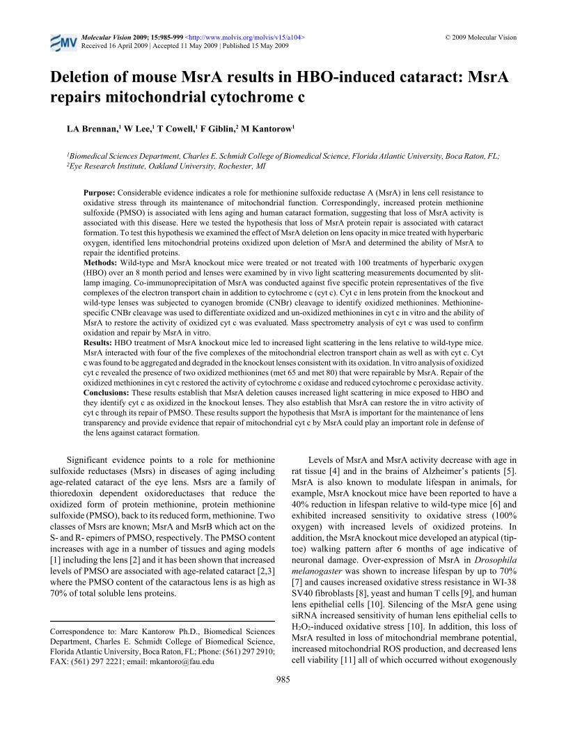
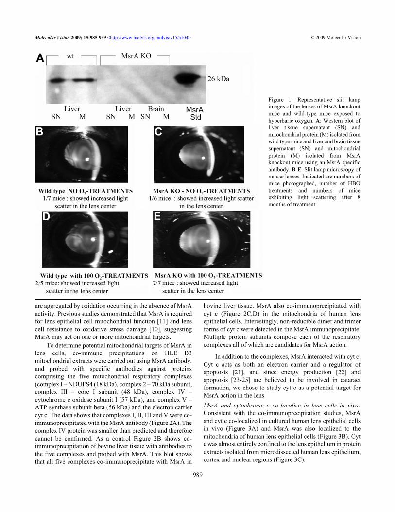
RESULTSDeletion of MsrA leads to increased light scattering in mouselenses treated with hyperbaric oxygen: Wild-type and MsrA-knockout mice were phenotyped to confirm lack of MsrAprotein expression by western analysis using an MsrA-specific antibody. As shown in Figure 1A, MsrA was detectedin wild-type liver supernatant (SN) and isolated mitochondrialfraction (M) but not in corresponding fractions of brain or liverisolated from the MsrA knockout mice. Knockout and wild-type mice were treated with 2.5 ATM of hyperbaric oxygenthree times per week, on alternate days at approximately thesame time of day, for a total of 100 treatments over a 8 monthperiod. The transparency of the lenses of control and HBO-treated mice was assessed by in vivo slit lamp microscopy andthe results documented by photography. Figures 1B-E showrepresentative lenses from wild-type and MsrA-knockoutmice treated or not with HBO. Light scattering was notsignificantly increased in the wild-type lenses with (Figure1D) or without (Figure 1B) HBO exposure. The knockoutlenses not exposed to HBO-induced oxidative stress (Figure1C) did not show increased light scatter compared to wild-type mice. By contrast, increased light scattering was detectedin the MsrA knockout mice exposed to HBO-inducedoxidative stress (Figure 1E) suggesting an increasedsensitivity of MsrA-knockout mouse lenses to HBO-inducedoxidative stress.MsrA co-immunoprecipitates with multiple lensmitochondrial complex proteins including cytochrome c invitro.: The presence of light scattering in the MsrA-knockoutmice treated with HBO indicates that one or more lens proteins
Molecular Vision 2009; 15:985-999 <http://www.molvis.org/molvis/v15/a104> © 2009 Molecular Vision
988
are aggregated by oxidation occurring in the absence of MsrAactivity. Previous studies demonstrated that MsrA is requiredfor lens epithelial cell mitochondrial function [11] and lenscell resistance to oxidative stress damage [10], suggestingMsrA may act on one or more mitochondrial targets.
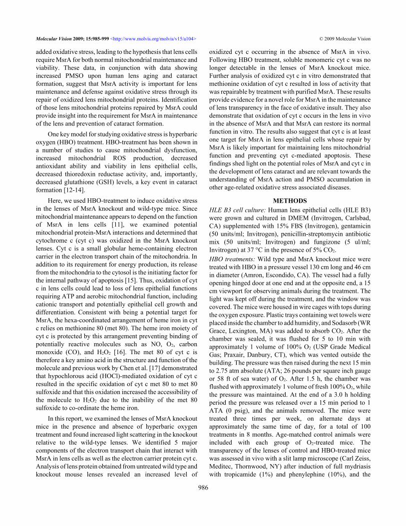
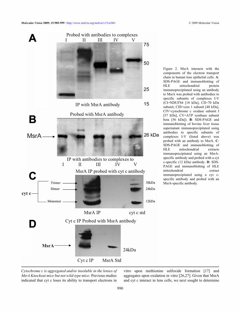
To determine potential mitochondrial targets of MsrA inlens cells, co-immune precipitations on HLE B3mitochondrial extracts were carried out using MsrA antibody,and probed with specific antibodies against proteinscomprising the five mitochondrial respiratory complexes(complex I – NDUFS4 (18 kDa), complex 2 – 70 kDa subunit,complex III – core I subunit (48 kDa), complex IV –cytochrome c oxidase subunit I (57 kDa), and complex V –ATP synthase subunit beta (56 kDa) and the electron carriercyt c. The data shows that complexes I, II, III and V were co-immunoprecipitated with the MsrA antibody (Figure 2A). Thecomplex IV protein was smaller than predicted and thereforecannot be confirmed. As a control Figure 2B shows co-immunoprecipitation of bovine liver tissue with antibodies tothe five complexes and probed with MsrA. This blot showsthat all five complexes co-immunoprecipitate with MsrA in
bovine liver tissue. MsrA also co-immunoprecipitated withcyt c (Figure 2C,D) in the mitochondria of human lensepithelial cells. Interestingly, non-reducible dimer and trimerforms of cyt c were detected in the MsrA immunoprecipitate.Multiple protein subunits compose each of the respiratorycomplexes all of which are candidates for MsrA action.
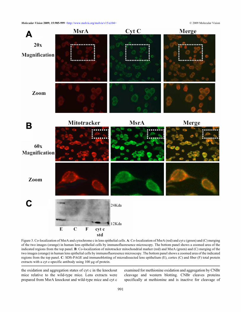
In addition to the complexes, MsrA interacted with cyt c.Cyt c acts as both an electron carrier and a regulator ofapoptosis [21], and since energy production [22] andapoptosis [23-25] are believed to be involved in cataractformation, we chose to study cyt c as a potential target forMsrA action in the lens.MsrA and cytochrome c co-localize in lens cells in vivo:Consistent with the co-immunoprecipitation studies, MsrAand cyt c co-localized in cultured human lens epithelial cellsin vivo (Figure 3A) and MsrA was also localized to themitochondria of human lens epithelial cells (Figure 3B). Cytc was almost entirely confined to the lens epithelium in proteinextracts isolated from microdissected human lens epithelium,cortex and nuclear regions (Figure 3C).
Figure 1. Representative slit lampimages of the lenses of MsrA knockoutmice and wild-type mice exposed tohyperbaric oxygen. A: Western blot ofliver tissue supernatant (SN) andmitochondrial protein (M) isolated fromwild type mice and liver and brain tissuesupernatant (SN) and mitochondrialprotein (M) isolated from MsrAknockout mice using an MsrA specificantibody. B-E. Slit lamp microscopy ofmouse lenses. Indicated are numbers ofmice photographed, number of HBOtreatments and numbers of miceexhibiting light scattering after 8months of treatment.
Molecular Vision 2009; 15:985-999 <http://www.molvis.org/molvis/v15/a104> © 2009 Molecular Vision
989
Cytochrome c is aggregated and/or insoluble in the lenses ofMsrA Knockout mice but not wild-type mice: Previous studiesindicated that cyt c loses its ability to transport electrons in
vitro upon methionine sulfoxide formation [17] andaggregates upon oxidation in vitro [26,27]. Given that MsrAand cyt c interact in lens cells, we next sought to determine
Figure 2. MsrA interacts with thecomponents of the electron transportchain in human lens epithelial cells. A:SDS-PAGE and immunoblotting ofHLE mitochondrial proteinimmunoprecipitated using an antibodyto MsrA was probed with antibodies tospecific subunits of complexes I-V(CI=NDUFS4 [18 kDa], CII=70 kDasubunit, CIII=core 1 subunit [48 kDa],CIV=cytochrome c oxidase subunit I[57 kDa], CV=ATP synthase subunitbeta [56 kDa]). B: SDS-PAGE andimmunoblotting of bovine liver tissuesupernatant immunoprecipitated usingantibodies to specific subunits ofcomplexes I-V (listed above) wasprobed with an antibody to MsrA. C:SDS-PAGE and immunoblotting ofHLE mitochondrial extractsimmunoprecipitated using an MsrA-specific antibody and probed with a cytc-specific (12 kDa) antibody. D: SDS-PAGE and immunoblotting of HLEmitochondrial extractimmunoprecipitated using a cyt c-specific antibody and probed with anMsrA-specific antibody.
Molecular Vision 2009; 15:985-999 <http://www.molvis.org/molvis/v15/a104> © 2009 Molecular Vision
990
the oxidation and aggregation states of cyt c in the knockoutmice relative to the wild-type mice. Lens extracts wereprepared from MsrA knockout and wild-type mice and cyt c
examined for methionine oxidation and aggregation by CNBrcleavage and western blotting. CNBr cleaves proteinsspecifically at methionine and is inactive for cleavage of
Figure 3. Co-localization of MsrA and cytochrome c in lens epithelial cells. A: Co-localization of MsrA (red) and cyt c (green) and (C) mergingof the two images (orange) in human lens epithelial cells by immunofluoresence microscopy. The bottom panel shows a zoomed area of theindicated regions from the top panel. B: Co-localization of mitotracker mitochondrial marker (red) and MsrA (green) and (C) merging of thetwo images (orange) in human lens epithelial cells by immunofluoresence microscopy. The bottom panel shows a zoomed area of the indicatedregions from the top panel. C: SDS-PAGE and immunoblotting of microdissected lens epithelium (E), cortex (C) and fiber (F) total proteinextracts with a cyt c-specific antibody using 100 µg of protein.
Molecular Vision 2009; 15:985-999 <http://www.molvis.org/molvis/v15/a104> © 2009 Molecular Vision
991
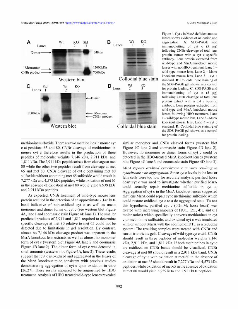
methionine sulfoxide. There are two methionines in mouse cytc at positions 65 and 80. CNBr cleavage of methionines inmouse cyt c therefore results in the production of threepeptides of molecular weights 7,146 kDa, 2,911 kDa, and1,811 kDa. The 2,911 kDa peptide arises from cleavage at met80 while the other two peptides result from cleavage at met65 and met 80. CNBr cleavage of cyt c containing met 80sulfoxide without containing met 65 sulfoxide would result in7,277 kDa and 4,573 kDa peptides; while oxidation of met 65in the absence of oxidation at met 80 would yield 8,939 kDaand 2,911 kDa peptides.
As expected, CNBr treatment of wild-type mouse lensprotein resulted in the detection of an approximate 7,146 kDaband indicative of non-oxidized cyt c as well as uncutmonomer and dimer forms of cyt c (see western blot Figure4A, lane 1 and coomassie stain Figure 4B lane 1). The smallerpredicted products of 2,911 and 1,811 required to determinespecific cleavage at met 80 relative to met 65 could not bedetected due to limitations in gel resolution. By contrast,almost no 7,146 kDa cleavage product was apparent in theMsrA knockout lens extracts as well as almost no monomerform of cyt c (western blot Figure 4A lane 2 and coomassieFigure 4B lane 2). The dimer form of cyt c was detected insmall amounts (western blot Figure 4A, lane 2). These resultssuggest that cyt c is oxidized and aggregated in the lenses ofthe MsrA knockout mice consistent with previous studiesdemonstrating aggregation of cyt c upon oxidation in vitro[26,27]. These results appeared to be augmented by HBOtreatment. Analysis of HBO treated wild-type lenses revealed
similar monomer and CNBr cleaved forms (western blotFigure 4C lane 2 and coomassie stain Figure 4D lane 2).However, no monomer or dimer forms of cyt c could bedetected in the HBO-treated MsrA knockout lenses (westernblot Figure 4C lane 3 and coomassie stain Figure 4D lane 3).MsrA repairs oxidized cytochrome c in vitro resulting incytochrome c de-aggregation: Since cyt c levels in the lens orlens cells were too low for accurate analysis, purified horseheart cyt c was used to investigate whether purified MsrAcould actually repair methionine sulfoxide in cyt c.Aggregation of cyt c in the MsrA knockout lenses suggestedthat lens MsrA could repair cyt c methionine sulfoxide whichcould restore oxidized cyt c to a de-aggregated state. To testthis hypothesis, purified cyt c (0.2mM; horse heart) wastreated with increasing amounts of HOCl (2:1, 4:1, and 6:1molar ratios) which specifically converts methionines in cytc to methionine sulfoxide, and oxidized cyt c was incubatedwith or without MsrA with the addition of DTT as a reducingsystem. The resulting samples were treated with CNBr andrun on tris-tricine gels. Cleavage of wild-type cyt c with CNBrshould result in three peptides of molecular weights 7,146kDa, 2,911 kDa, and 1,811 kDa. If both methionines in cyt care oxidized no CNBr bands should be visualized. CNBrcleavage at met 80 should result in a 2,911 kDa band. CNBrcleavage of cyt c with oxidation at met 80 in the absence ofoxidation at met 65 should result in 7,277 kDa and 4,573 kDapeptides; while oxidation of met 65 in the absence of oxidationat met 80 would yield 8,939 kDa and 2,911 kDa peptides.
Figure 4. Cyt c in MsrA deficient mouselenses shows evidence of oxidation andaggregation. A: SDS-PAGE andimmunoblotting of cyt c (5 µg)following CNBr cleavage of total lensprotein extract with a cyt c specificantibody. Lens protein extracted fromwild-type and MsrA knockout mouselenses with no HBO treatment. Lane 1 –wild type mouse lens, Lane 2 – MsrAknockout mouse lens, Lane 3 – cyt cstandard. B: Colloidal blue staining ofthe SDS-PAGE gel shown as a controlfor protein loading. C: SDS-PAGE andimmunoblotting of cyt c (5 µg)following CNBr cleavage of total lensprotein extract with a cyt c specificantibody. Lens proteins extracted fromwild-type and MsrA knockout mouselenses following HBO treatment. Lane1 – wild type mouse lens, Lane 2 – MsrAknockout mouse lens, Lane 3 – cyt cstandard. D: Colloidal blue staining ofthe SDS-PAGE gel shown as a controlfor protein loading.
Molecular Vision 2009; 15:985-999 <http://www.molvis.org/molvis/v15/a104> © 2009 Molecular Vision
992
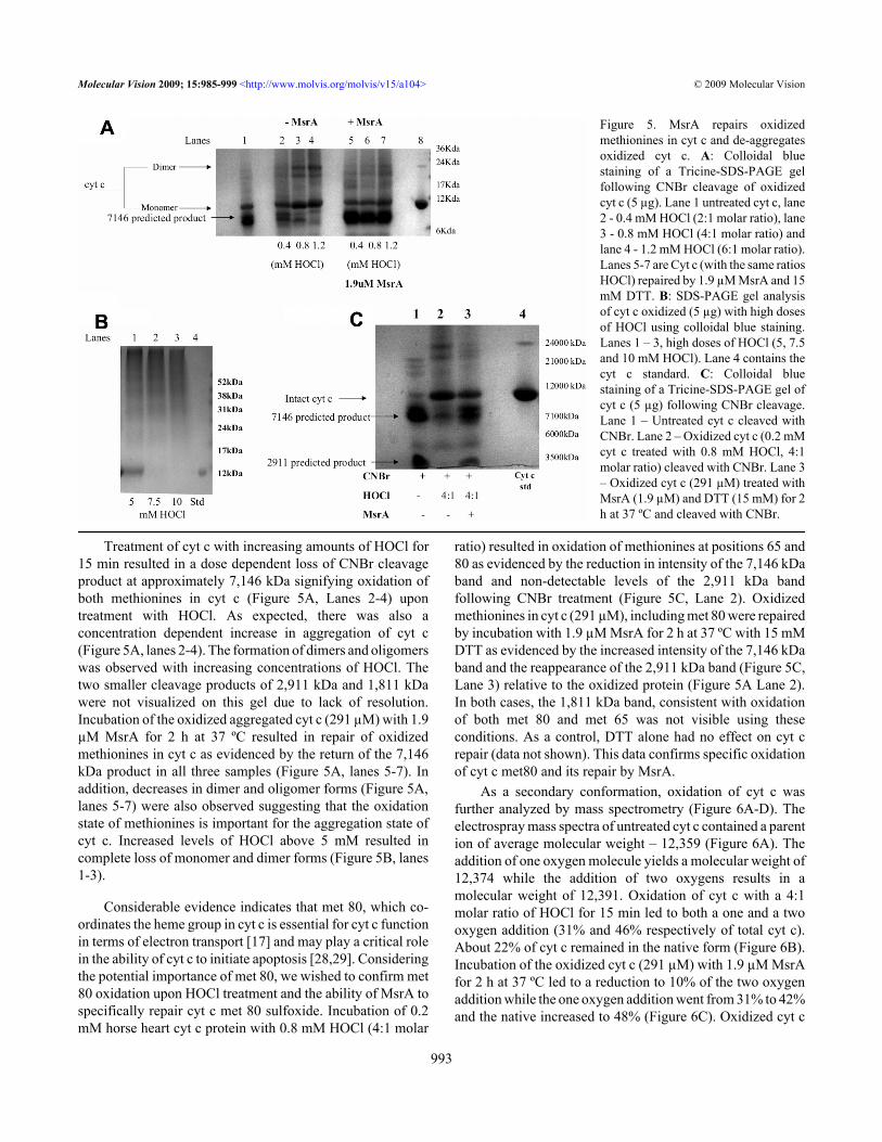
Treatment of cyt c with increasing amounts of HOCl for15 min resulted in a dose dependent loss of CNBr cleavageproduct at approximately 7,146 kDa signifying oxidation ofboth methionines in cyt c (Figure 5A, Lanes 2-4) upontreatment with HOCl. As expected, there was also aconcentration dependent increase in aggregation of cyt c(Figure 5A, lanes 2-4). The formation of dimers and oligomerswas observed with increasing concentrations of HOCl. Thetwo smaller cleavage products of 2,911 kDa and 1,811 kDawere not visualized on this gel due to lack of resolution.Incubation of the oxidized aggregated cyt c (291 µM) with 1.9µM MsrA for 2 h at 37 ºC resulted in repair of oxidizedmethionines in cyt c as evidenced by the return of the 7,146kDa product in all three samples (Figure 5A, lanes 5-7). Inaddition, decreases in dimer and oligomer forms (Figure 5A,lanes 5-7) were also observed suggesting that the oxidationstate of methionines is important for the aggregation state ofcyt c. Increased levels of HOCl above 5 mM resulted incomplete loss of monomer and dimer forms (Figure 5B, lanes1-3).
Considerable evidence indicates that met 80, which co-ordinates the heme group in cyt c is essential for cyt c functionin terms of electron transport [17] and may play a critical rolein the ability of cyt c to initiate apoptosis [28,29]. Consideringthe potential importance of met 80, we wished to confirm met80 oxidation upon HOCl treatment and the ability of MsrA tospecifically repair cyt c met 80 sulfoxide. Incubation of 0.2mM horse heart cyt c protein with 0.8 mM HOCl (4:1 molar
ratio) resulted in oxidation of methionines at positions 65 and80 as evidenced by the reduction in intensity of the 7,146 kDaband and non-detectable levels of the 2,911 kDa bandfollowing CNBr treatment (Figure 5C, Lane 2). Oxidizedmethionines in cyt c (291 µM), including met 80 were repairedby incubation with 1.9 µM MsrA for 2 h at 37 ºC with 15 mMDTT as evidenced by the increased intensity of the 7,146 kDaband and the reappearance of the 2,911 kDa band (Figure 5C,Lane 3) relative to the oxidized protein (Figure 5A Lane 2).In both cases, the 1,811 kDa band, consistent with oxidationof both met 80 and met 65 was not visible using theseconditions. As a control, DTT alone had no effect on cyt crepair (data not shown). This data confirms specific oxidationof cyt c met80 and its repair by MsrA.
As a secondary conformation, oxidation of cyt c wasfurther analyzed by mass spectrometry (Figure 6A-D). Theelectrospray mass spectra of untreated cyt c contained a parention of average molecular weight – 12,359 (Figure 6A). Theaddition of one oxygen molecule yields a molecular weight of12,374 while the addition of two oxygens results in amolecular weight of 12,391. Oxidation of cyt c with a 4:1molar ratio of HOCl for 15 min led to both a one and a twooxygen addition (31% and 46% respectively of total cyt c).About 22% of cyt c remained in the native form (Figure 6B).Incubation of the oxidized cyt c (291 µM) with 1.9 µM MsrAfor 2 h at 37 ºC led to a reduction to 10% of the two oxygenaddition while the one oxygen addition went from 31% to 42%and the native increased to 48% (Figure 6C). Oxidized cyt c
Figure 5. MsrA repairs oxidizedmethionines in cyt c and de-aggregatesoxidized cyt c. A: Colloidal bluestaining of a Tricine-SDS-PAGE gelfollowing CNBr cleavage of oxidizedcyt c (5 µg). Lane 1 untreated cyt c, lane2 - 0.4 mM HOCl (2:1 molar ratio), lane3 - 0.8 mM HOCl (4:1 molar ratio) andlane 4 - 1.2 mM HOCl (6:1 molar ratio).Lanes 5-7 are Cyt c (with the same ratiosHOCl) repaired by 1.9 µM MsrA and 15mM DTT. B: SDS-PAGE gel analysisof cyt c oxidized (5 µg) with high dosesof HOCl using colloidal blue staining.Lanes 1 – 3, high doses of HOCl (5, 7.5and 10 mM HOCl). Lane 4 contains thecyt c standard. C: Colloidal bluestaining of a Tricine-SDS-PAGE gel ofcyt c (5 µg) following CNBr cleavage.Lane 1 – Untreated cyt c cleaved withCNBr. Lane 2 – Oxidized cyt c (0.2 mMcyt c treated with 0.8 mM HOCl, 4:1molar ratio) cleaved with CNBr. Lane 3– Oxidized cyt c (291 µM) treated withMsrA (1.9 µM) and DTT (15 mM) for 2h at 37 ºC and cleaved with CNBr.
Molecular Vision 2009; 15:985-999 <http://www.molvis.org/molvis/v15/a104> © 2009 Molecular Vision
993
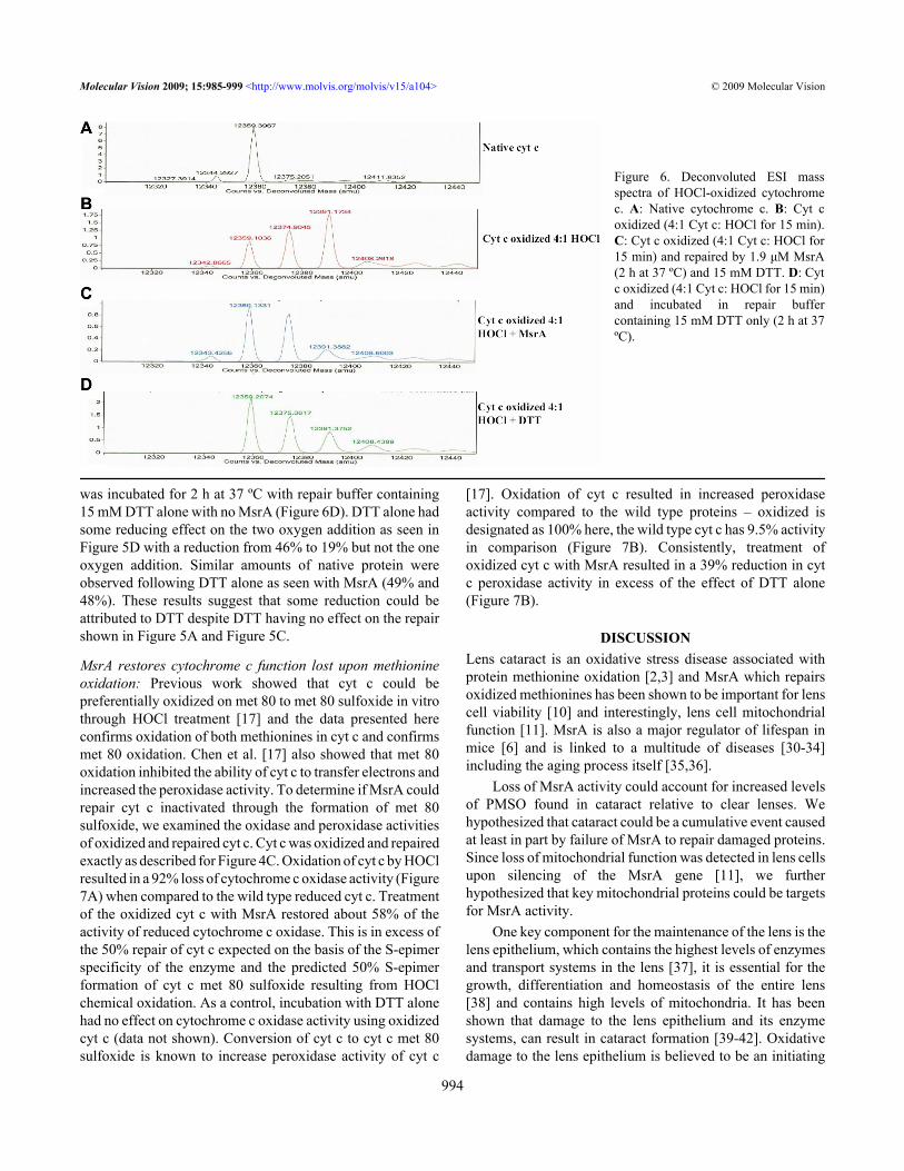
was incubated for 2 h at 37 ºC with repair buffer containing15 mM DTT alone with no MsrA (Figure 6D). DTT alone hadsome reducing effect on the two oxygen addition as seen inFigure 5D with a reduction from 46% to 19% but not the oneoxygen addition. Similar amounts of native protein wereobserved following DTT alone as seen with MsrA (49% and48%). These results suggest that some reduction could beattributed to DTT despite DTT having no effect on the repairshown in Figure 5A and Figure 5C.
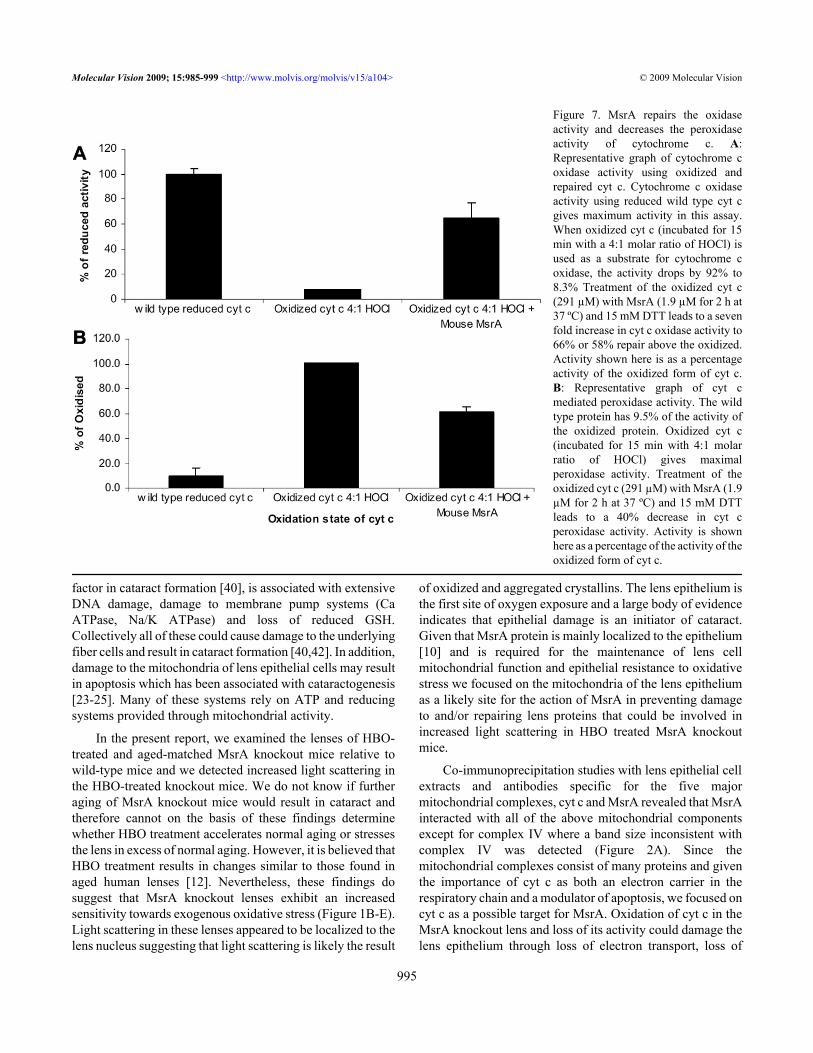
MsrA restores cytochrome c function lost upon methionineoxidation: Previous work showed that cyt c could bepreferentially oxidized on met 80 to met 80 sulfoxide in vitrothrough HOCl treatment [17] and the data presented hereconfirms oxidation of both methionines in cyt c and confirmsmet 80 oxidation. Chen et al. [17] also showed that met 80oxidation inhibited the ability of cyt c to transfer electrons andincreased the peroxidase activity. To determine if MsrA couldrepair cyt c inactivated through the formation of met 80sulfoxide, we examined the oxidase and peroxidase activitiesof oxidized and repaired cyt c. Cyt c was oxidized and repairedexactly as described for Figure 4C. Oxidation of cyt c by HOClresulted in a 92% loss of cytochrome c oxidase activity (Figure7A) when compared to the wild type reduced cyt c. Treatmentof the oxidized cyt c with MsrA restored about 58% of theactivity of reduced cytochrome c oxidase. This is in excess ofthe 50% repair of cyt c expected on the basis of the S-epimerspecificity of the enzyme and the predicted 50% S-epimerformation of cyt c met 80 sulfoxide resulting from HOClchemical oxidation. As a control, incubation with DTT alonehad no effect on cytochrome c oxidase activity using oxidizedcyt c (data not shown). Conversion of cyt c to cyt c met 80sulfoxide is known to increase peroxidase activity of cyt c
[17]. Oxidation of cyt c resulted in increased peroxidaseactivity compared to the wild type proteins – oxidized isdesignated as 100% here, the wild type cyt c has 9.5% activityin comparison (Figure 7B). Consistently, treatment ofoxidized cyt c with MsrA resulted in a 39% reduction in cytc peroxidase activity in excess of the effect of DTT alone(Figure 7B).
DISCUSSIONLens cataract is an oxidative stress disease associated withprotein methionine oxidation [2,3] and MsrA which repairsoxidized methionines has been shown to be important for lenscell viability [10] and interestingly, lens cell mitochondrialfunction [11]. MsrA is also a major regulator of lifespan inmice [6] and is linked to a multitude of diseases [30-34]including the aging process itself [35,36].
Loss of MsrA activity could account for increased levelsof PMSO found in cataract relative to clear lenses. Wehypothesized that cataract could be a cumulative event causedat least in part by failure of MsrA to repair damaged proteins.Since loss of mitochondrial function was detected in lens cellsupon silencing of the MsrA gene [11], we furtherhypothesized that key mitochondrial proteins could be targetsfor MsrA activity.
One key component for the maintenance of the lens is thelens epithelium, which contains the highest levels of enzymesand transport systems in the lens [37], it is essential for thegrowth, differentiation and homeostasis of the entire lens[38] and contains high levels of mitochondria. It has beenshown that damage to the lens epithelium and its enzymesystems, can result in cataract formation [39-42]. Oxidativedamage to the lens epithelium is believed to be an initiating
Figure 6. Deconvoluted ESI massspectra of HOCl-oxidized cytochromec. A: Native cytochrome c. B: Cyt coxidized (4:1 Cyt c: HOCl for 15 min).C: Cyt c oxidized (4:1 Cyt c: HOCl for15 min) and repaired by 1.9 μM MsrA(2 h at 37 ºC) and 15 mM DTT. D: Cytc oxidized (4:1 Cyt c: HOCl for 15 min)and incubated in repair buffercontaining 15 mM DTT only (2 h at 37ºC).
Molecular Vision 2009; 15:985-999 <http://www.molvis.org/molvis/v15/a104> © 2009 Molecular Vision
994
factor in cataract formation [40], is associated with extensiveDNA damage, damage to membrane pump systems (CaATPase, Na/K ATPase) and loss of reduced GSH.Collectively all of these could cause damage to the underlyingfiber cells and result in cataract formation [40,42]. In addition,damage to the mitochondria of lens epithelial cells may resultin apoptosis which has been associated with cataractogenesis[23-25]. Many of these systems rely on ATP and reducingsystems provided through mitochondrial activity.
In the present report, we examined the lenses of HBO-treated and aged-matched MsrA knockout mice relative towild-type mice and we detected increased light scattering inthe HBO-treated knockout mice. We do not know if furtheraging of MsrA knockout mice would result in cataract andtherefore cannot on the basis of these findings determinewhether HBO treatment accelerates normal aging or stressesthe lens in excess of normal aging. However, it is believed thatHBO treatment results in changes similar to those found inaged human lenses [12]. Nevertheless, these findings dosuggest that MsrA knockout lenses exhibit an increasedsensitivity towards exogenous oxidative stress (Figure 1B-E).Light scattering in these lenses appeared to be localized to thelens nucleus suggesting that light scattering is likely the result
of oxidized and aggregated crystallins. The lens epithelium isthe first site of oxygen exposure and a large body of evidenceindicates that epithelial damage is an initiator of cataract.Given that MsrA protein is mainly localized to the epithelium[10] and is required for the maintenance of lens cellmitochondrial function and epithelial resistance to oxidativestress we focused on the mitochondria of the lens epitheliumas a likely site for the action of MsrA in preventing damageto and/or repairing lens proteins that could be involved inincreased light scattering in HBO treated MsrA knockoutmice.
Co-immunoprecipitation studies with lens epithelial cellextracts and antibodies specific for the five majormitochondrial complexes, cyt c and MsrA revealed that MsrAinteracted with all of the above mitochondrial componentsexcept for complex IV where a band size inconsistent withcomplex IV was detected (Figure 2A). Since themitochondrial complexes consist of many proteins and giventhe importance of cyt c as both an electron carrier in therespiratory chain and a modulator of apoptosis, we focused oncyt c as a possible target for MsrA. Oxidation of cyt c in theMsrA knockout lens and loss of its activity could damage thelens epithelium through loss of electron transport, loss of
Figure 7. MsrA repairs the oxidaseactivity and decreases the peroxidaseactivity of cytochrome c. A:Representative graph of cytochrome coxidase activity using oxidized andrepaired cyt c. Cytochrome c oxidaseactivity using reduced wild type cyt cgives maximum activity in this assay.When oxidized cyt c (incubated for 15min with a 4:1 molar ratio of HOCl) isused as a substrate for cytochrome coxidase, the activity drops by 92% to8.3% Treatment of the oxidized cyt c(291 µM) with MsrA (1.9 µM for 2 h at37 ºC) and 15 mM DTT leads to a sevenfold increase in cyt c oxidase activity to66% or 58% repair above the oxidized.Activity shown here is as a percentageactivity of the oxidized form of cyt c.B: Representative graph of cyt cmediated peroxidase activity. The wildtype protein has 9.5% of the activity ofthe oxidized protein. Oxidized cyt c(incubated for 15 min with 4:1 molarratio of HOCl) gives maximalperoxidase activity. Treatment of theoxidized cyt c (291 µM) with MsrA (1.9µM for 2 h at 37 ºC) and 15 mM DTTleads to a 40% decrease in cyt cperoxidase activity. Activity is shownhere as a percentage of the activity of theoxidized form of cyt c.
Molecular Vision 2009; 15:985-999 <http://www.molvis.org/molvis/v15/a104> © 2009 Molecular Vision
995
cationic exchange and apoptotic induction, all of which couldultimately contribute to increased light scattering and cataractformation.
Consistent with a potential role for MsrA repair of cyt cin the lens, analysis of the MsrA knockout lenses revealedincreased oxidation and aggregation of cyt c that was notfound in wild-type lenses (Figure 4A) this was exacerbatedfollowing HBO treatment where cyt c was no longer detectedin the soluble protein (Figure 4C). Indeed, little soluble cyt ccould be detected at all in these lenses consistent with previousstudies showing that oxidized cyt c forms higher molecularweight aggregates and that oxidized cyt c degrades in vitro[17,26,27]. To further explore whether MsrA can repairoxidized cyt c, the ability of MsrA to repair cyt c was evaluatedusing purified cyt c and MsrA. CNBr treatment, whichspecifically cuts reduced but not oxidized methionine in cyt crevealed specific methionine oxidation that could be repairedby MsrA treatment (Figure 4A). Consistent with theaggregation observed in the knockout lenses, high levels ofoxidant (HOCl) resulted in increased aggregation anddegradation of cyt c in vitro (Figure 5B). Further analysis ofthe oxidized cyt c protein by CNBr treatment revealed twosites of methionine oxidation at met 80 and met 65 (Figure5C). Oxidation levels were further confirmed by mass specanalysis (Figure 6A-D). Consistent with these oxidationsbeing important for cyt c function, methionine oxidation ofcyt c reduced its activity as an electron carrier by 92% (Figure7A) and increased cyt c peroxidase activity tenfold (Figure7B) as previously reported [17]. MsrA treatment of oxidizedcyt c restored cyt c oxidase activity to 58% of the wild-typereduced protein and reduced the oxidized cyt c peroxidaseactivity by 39% (Figure 7A,B). Repair by MsrA of cyt coxidase activity was in excess of the expected 50% based onS-epimer specificity and the expected random 50-50distribution of S- and R- epimers in the oxidized cyt c whilefor cyt c peroxidase activity repair in terms of percent activitywas slightly lower than expected. In effect, the R- and S-epimer distribution is unlikely to be strictly 50-50 and theamount of repair found here reflects that situation. This dataindicates that MsrA efficiently repairs oxidized methioninesat low enzyme concentrations, here the ratio of oxidizedprotein to enzyme was 153:1.
With this HBO model the levels of oxygen are likely tobe higher in the epithelial and periphery than in the center ofthe lens through diffusion of oxygen through the cornea asshown by Shui et al. [43], potentially targeting the cells of thelens epithelium which as stated above can be the initiating sitefor cataractogenesis. Damage following HBO treatment ofguinea pigs or rabbits involves disulfide formation, membranedamage and loss of cytoskeletal proteins as well as decreasedGSH and ascorbate levels in the nucleus and cortex and β- andγ-crystallin aggregation, all ultimately leading to nuclearcataract [12,44-46]. HLE B3 cells treated with HBO showeda 30% decrease in ATP levels after 3 h of treatment consistent
with loss of mitochondrial function. Indeed, transmissionelectron microscopy analysis of those cells at 16 h indicateda decrease in the total number of mitochondria [14]. Haung etal. [13] cultured HLE B3 cells in hyperoxia conditions andshowed increased mitochondrial ROS (43%), decreasedmitochondrial membrane potential and loss of cardiolipin, akey player in the regulation of the mitochondrial apoptosis[47]. All of these studies point to loss of mitochondrialfunction as an important characteristic of lens oxidation andsuggest that MsrA repair of oxidized lens mitochondrialproteins is a key factor in lens maintenance.
In addition to serving as targets for MsrA, methionineresidues are thought to act as antioxidants by directlyscavenging ROS, lack of MsrA to reverse this process mayincrease not only PMSO but endogenous ROS that can nolonger be scavenged. This may explain the oxidation andaggregation of cyt c in vivo even without HBO treatment.These results correlate well with the findings of Marchetti etal. [11] where loss of cell viability, decreased mitochondrialmembrane potential and increased ROS levels were found inHLE B3 cells silenced for MsrA, even in the absence ofoxidative stress. Since HBO treatment results in changessimilar to aged human lens [12] the model used here couldreflect events in aged human lenses in the presence or absenceof MsrA, highlighting the importance of this enzyme as botha repair system and antioxidant scavenger. Interestingly,Shur-Perek and Avi-Dor [48] showed that the dimer form ofcyt c, found here in MsrA knockout mice, was relativelyineffective as an electron carrier in the respiratory system.Upon HBO treatment of MsrA knockout mice the cyt c proteinappears to be completely degraded or aggregated to aninsoluble form which was also observed in Figure 4B withhigh concentrations of HOCl (5-10 mM) and observed byChen et al. [17] when ratios of HOCl:cyt c above 8:1 wereused. It could be that in the absence of MsrA activity, themitochondrial system is overwhelmed with ROS, damagingnot just cyt c but other key mitochondrial proteins andeventually causing lens epithelial cell apoptosis andultimately cataract.
It has been proposed that the main function ofmitochondria in the lens is to keep oxygen tension low [49].Mitochondria consume about 90% of the oxygen entering thecell and when uncoupled, the lens sharp focus is lost [50].Without MsrA functioning as both an antioxidant and repairsystem, oxygen consumption may decrease, leading tomitochondrial dysfunction, increased ROS production andultimately apoptosis. In bovine lenses at least 30% of theenergy used in the form of ATP is derived from mitochondrialrespiration [51]. Loss of this energy is likely destructive to thelens.
In this report, we show that one key protein likely to beoxidized under these conditions is cyt c. In vitro methionineoxidation in cyt c led to decreased electron transport ability
Molecular Vision 2009; 15:985-999 <http://www.molvis.org/molvis/v15/a104> © 2009 Molecular Vision
996
and increased peroxidase activity. Decreased electrontransport ability leads to mitochondrial dysfunction which isseen in a number of studies using hyperoxia and cultured lensepithelial cells. Previous studies have shown that only theoxidized form of cyt c has the ability to initiate apoptosis[52].
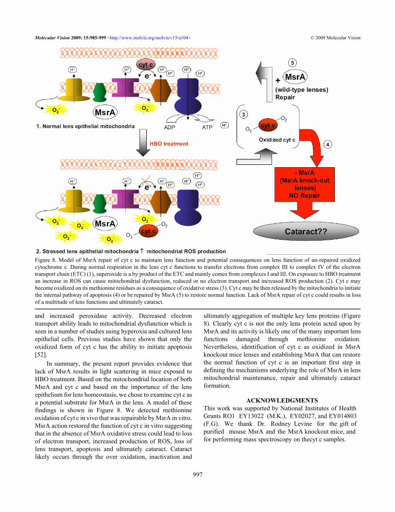
In summary, the present report provides evidence thatlack of MsrA results in light scattering in mice exposed toHBO treatment. Based on the mitochondrial location of bothMsrA and cyt c and based on the importance of the lensepithelium for lens homeostasis, we chose to examine cyt c asa potential substrate for MsrA in the lens. A model of thesefindings is shown in Figure 8. We detected methionineoxidation of cyt c in vivo that was repairable by MsrA in vitro.MsrA action restored the function of cyt c in vitro suggestingthat in the absence of MsrA oxidative stress could lead to lossof electron transport, increased production of ROS, loss oflens transport, apoptosis and ultimately cataract. Cataractlikely occurs through the over oxidation, inactivation and
ultimately aggregation of multiple key lens proteins (Figure8). Clearly cyt c is not the only lens protein acted upon byMsrA and its activity is likely one of the many important lensfunctions damaged through methionine oxidation.Nevertheless, identification of cyt c as oxidized in MsrAknockout mice lenses and establishing MsrA that can restorethe normal function of cyt c is an important first step indefining the mechanisms underlying the role of MsrA in lensmitochondrial maintenance, repair and ultimately cataractformation.
ACKNOWLEDGMENTSThis work was supported by National Institutes of Health Grants RO1 EY13022 (M.K.), EY02027, and EY014803(F.G). We thank Dr. Rodney Levine for the gift ofpurified mouse MsrA and the MsrA knockout mice, andfor performing mass spectroscopy on thecyt c samples.
Figure 8. Model of MsrA repair of cyt c to maintain lens function and potential consequences on lens function of un-repaired oxidizedcytochrome c. During normal respiration in the lens cyt c functions to transfer electrons from complex III to complex IV of the electrontransport chain (ETC) (1), superoxide is a by product of the ETC and mainly comes from complexes I and III. On exposure to HBO treatmentan increase in ROS can cause mitochondrial dysfunction, reduced or no electron transport and increased ROS production (2). Cyt c maybecome oxidized on its methionine residues as a consequence of oxidative stress (3). Cyt c may be then released by the mitochondria to initiatethe internal pathway of apoptosis (4) or be repaired by MsrA (5) to restore normal function. Lack of MsrA repair of cyt c could results in lossof a multitude of lens functions and ultimately cataract.
Molecular Vision 2009; 15:985-999 <http://www.molvis.org/molvis/v15/a104> © 2009 Molecular Vision
997
REFERENCES1. Stadtman ER, Van Remmen H, Richardson A, Wehr NB,
Levine RL. Methionine oxidation and aging. BiochimBiophys Acta 2005; 1703:135-40. [PMID: 861252]
2. Truscott RJ, Augusteyn RC. Oxidative changes in human lensproteins during senile nuclear cataract formation. BiochimBiophys Acta 1977; 492:43-52. [PMID: 861252]
3. Garner MH, Spector A. Selective oxidation of cysteine andmethionine in normal and senile cataractous lenses. Proc NatlAcad Sci USA 1980; 77:1274-7. [PMID: 6929483]
4. Petropoulos I, Mary J, Perichon JMM, Friguet B. Rat peptidemethionine sulphocide reductase: cloning of the cDNA, anddown-regulation of gene expression and enzyme activityduring aging. Biochem J 2001; 355:819-25. [PMID:11311146]
5. Gabbita SP, Aksenov MY, Lovell MA, Markesbery WR.Decrease in peptide methionine sulfoxide reductase inAlzheimer’s disease brain. J Neurochem 1999; 73:1660-6.[PMID: 10501213]
6. Moskovitz J, Bar-Noy S, Williams WM, Requena J, Berlett BS,Stadtman ER. Methionine sulfoxide reductase (MsrA) is aregulator of antioxidant defense and lifespan in mammals.Proc Natl Acad Sci USA 2001; 98:12920-5. [PMID:11606777]
7. Ruan H, Tang XD, Chen ML, Joiner ML, Sun G, Brot N,Weissbach H, Heinemann SH, Iverson L, Wu CF, Hoshi T.High-quality life extension by the enzyme peptide methioninesulfoxide reductase. Proc Natl Acad Sci USA 2002;99:2748-53. [PMID: 11867705]
8. Picot CR, Petropoulos I, Perichon M, Moreau M, Nizard C,Friguet B. Overexpression of MsrA protects WI-38 SV40fibroblasts against H2O2-mediated oxidative stress. FreeRadic Biol Med 2005; 39:1332-41. [PMID: 16257642]
9. Moskovitz J, Flescher E, Berlett BS, Azare J, Poston JM,Stadtman ER. Overexpression of peptide-methioninesulfoxide reductase in Saccharomyces cerevisiae and humanT cells provides them with high resistance to oxidative stress.Proc Natl Acad Sci USA 1998; 95:14071-5. [PMID:9826655]
10. Kantorow M, Hawse JR, Cowell TL, Benhamed S, Pizarro GO,Reddy VN, Hejtmancik JF. Methionine sulfoxide reductaseA is important for lens cell viability and resistance tooxidative stress. Proc Natl Acad Sci USA 2004;101:9654-9. [PMID: 15199188]
11. Marchetti MA, Lee W, Cowell TL, Wells TM, Weissbach H,Kantorow M. Silencing of the methionine sulfoxide reductaseA gene results in loss of mitochondrial membrane potentialand increased ROS production in human lens cells. Exp EyeRes 2006; 83:1281-6. [PMID: 16934804]
12. Padgaonkar VA, Leverenz VR, Fowler KE, Reddy VN, GiblinFJ. The effects of hyperbaric oxygen on the crystallins ofcultured rabbit lenses: a possible catalytic role for copper. ExpEye Res 2000; 71:371-83. [PMID: 10995558]
13. Huang L, Tang D, Yappert MC, Borchman D. Oxidation-induced changes in human lens epithelial cells. 2.Mitochondria and the generation of reactive oxygen species.Free Radic Biol Med 2006; 41:926-36. [PMID: 16934675]
14. Padgaonkar VA, Leverenz VR, Dang L, Chen SC, Palliccia S,Giblin FJ. Thioredoxin reductase may be essential for the
normal growth of hyperbaric oxygen-treated human lensepithelial cells. Exp Eye Res 2004; 79:847-57. [PMID:15642322]
15. Ow YP, Green DR, Hao Z, Mak TW. Cytochrome c: Functionsbeyond respiration. Nat Rev Mol Cell Biol 2008; 9:532-42.[PMID: 18568041]
16. Kagan VE, Borisenko GG, Tyurina YY, Tyurin VA, Jiang J,Potapovich AI, Kini V, Amoscato AA, Fujii Y. Oxidativelipidomics of apoptosis: Redox catalytic interactions ofcytochrome c with cardolipin and phosphatidylserine. FreeRadic Biol Med 2004; 37:1963-85. [PMID: 15544916]
17. Chen YR, Deterding LJ, Sturgeon BE, Tomer KB, Mason RP.Protein oxidation of cytochrome c by reactive halogen speciesenhances its peroxidase activity. J Biol Chem 2002;277:29781-91. [PMID: 12050149]
18. Simpson RJ. Peptide Mapping and Sequence Analysis of Gel-Resolved Proteins. In: Simpson, R.J., editor. Proteins andProteomics. Cold Spring Harbor (NY): Cold Spring HarborLaboratory Press; 2003. p. 385-387.
19. Cassina AM, Hodara R, Souza JM, Thomson L, Castro L,Ischiropoulos H, Freeman BA, Radi R. Cytochrome cnitration by peroxynitrite. J Biol Chem 2000; 275:21409-15.[PMID: 10770952]
20. Schägger H. Tricine-SDS-PAGE. Nat Protoc 2006; 1:16-22.[PMID: 17406207]
21. Brown GC, Borutaite V. Regulation of apoptosis by the redoxstate of cytochrome c. Biochim Biophys Acta 2008;1777:877-81. [PMID: 18439415]
22. Delamere NA, Tamiya S. Lens ion transport: from basicconcepts to regulation of Na,K-ATPase activity. Exp Eye Res2009; 88:140-3. [PMID: 18614168]
23. Li WC, Spector A. Lens epithelial cell apoptosis is an earlyevent in the development of UVB-induced cataract. FreeRadic Biol Med 1996; 20:301-11. [PMID: 8720900]
24. Li WC, Kuszak JR, Dunn K, Wang R, Ma W, Wang GM,Spector A, Leib M, Cotliar AM, Weiss M, Espy J, Howard G,Farris RL, Auran J, Donn A, Hofeldt A, Mackay C, MerriamJ, Mittl R, Smith TR. Lens Epithelial Cell Apoptosis Appearsto Be A Common Cellular Basis for Non-Congenital CataractDevelopment in Humans and Animals. J Cell Biol 1995;130:169-81. [PMID: 7790371]
25. Long AC, Colitz CM, Bomser JA. Apoptotic and necroticmechanisms of stress-induced human lens epithelial celldeath. Exp Biol Med (Maywood) 2004; 229:1072-80. [PMID:15522844]
26. Hashimoto M, Takeda A, Hsu HJ, Takenouchi T, Masliah E.Role of cytochrome c as a stimulator of alpha-synucleinaggregation in Lewy body disease. J Biol Chem 1999;274:28849-52. [PMID: 10506125]
27. Chen YR, Chen CL, Liu X, Li H, Zweier JL, Mason RP.Involvement of protein radical, protein aggregation andeffects on NO metabolism in the hypochlorite-mediatedoxidation of mitochondrial cytochrome c. Free Radic BiolMed 2004; 37:1591-603. [PMID: 15477010]
28. Balakrishnan G, Hu T, Oyerinde OF, Su J, Groves JT, Spiro TG.A conformational switch to beta-sheet structure incytochrome c leads to heme exposure. Implications forcardiolipin peroxidation and apoptosis. J Am Chem Soc 2007;129:504-5. [PMID: 17227009]
Molecular Vision 2009; 15:985-999 <http://www.molvis.org/molvis/v15/a104> © 2009 Molecular Vision
998
29. Vladimirov YA, Proskurnina EV, Izmailov DY, Novikov AA,Brusnichkin AV, Osipov AN, Kagan VE. Cardiolipinactivates cytochrome c peroxidase activity since it facilitatesH2O2 access to heme. Biochemistry (Mosc) 2006;71:998-1005. [PMID: 17009954]
30. Wassef R, Haenold R, Hansel A, Brot N, Heinemann SH, ToshiT. Methionine sufloxide reductase A and a dietary supplementS-methyl-L-cysteine prevent Parkinson’s-like symptoms. JNeurosci 2007; 27:12808-16. [PMID: 18032652]
31. Pal R, Oien DB, Ersen FY, Moskovitz J. Elevated levels of brainpathologies associated with neurodegenerative diseases inmethionine sulfoxide reductase A knockout mice. Exp BrainRes 2007; 180:765-74. [PMID: 17333008]
32. Yermolaieva O, Xu R, Schinstock C, Brot N, Weissbach H,Heinemann SH, Hoshi T. Methionine sulfoxide reducatase Aprotects neuronal cells against brief hypoxia/reoxygenation.Proc Natl Acad Sci USA 2004; 101:1159-64. [PMID:14745014]
33. Liu F, Hindupur J, Nguyen JL, Ruf KL, Zhu J, Schieler JL,Bonham CC, Wood KV, Davisson VJ, Rochet JC. Methioninesulfoxide reductase A protects dopaminergic cells fromParkinson’s disease-related insults. Free Radic Biol Med2008; 45:242-55. [PMID: 18456002]
34. Oien DB, Osterhaus GL, Latif SA, Pinkerton JW, Fulks J,Johnston M, Fowler SC, Moskowitz J. MsrA knockout miceexhibits abnormal behavior and brain dopamine levels. FreeRadic Biol Med 2008; 45:193-200. [PMID: 18466776]
35. Wood JM, Decker H, Hartmann H, Chavan B, Rokos H,Spencer JD, Hasse S, Thornton MJ, Shalbaf M, Paus R,Schallreuter KU. Senile hair graying: H2O2-mediatedoxidative stress affects human hair color by bluntingmethionine sulfoxide repair. FASEB J. 2009 [PMID:18456002]
36. Koc A, Gladyshev VN. Methionine sulfoxide reduction and theaging process. Ann N Y Acad Sci 2007; 1100:383-6. [PMID:17460202]
37. Reddy VN. Metabolism of glutathione in the lens. Exp Eye Res1971; 11:310-28. [PMID: 4399363]
38. Bloemendal H, ed. Molecular and cellular biology of the eyelens. New York: Wiley; 1981. p. 1-47.
39. Brown NP, Bron NJ, editors. Lens Disorders: A Clinical Manualof Cataract Diagnosis. Boston: Butterworth-Heinemann;1996.
40. Spector A. Oxidative stress induced cataract: mechanism ofaction. FASEB J 1995; 9:1173-82. [PMID: 7672510]
41. Harding JJ, Crabb MJ. The Lens: Development, Proteins,Metabolism and Cataract. In: Davson, H. editor. The Eye. Vol1B. Orlando, FL: Academic Press; 1984. p.207-492.
42. Hightower KR. The role of lens epithelium in development ofUV cataract. Curr Eye Res 1995; 14:71-8. [PMID: 7720407]
43. Shui YB, Fu JJ, Garcia C, Dattilo LK, Rajagopal R, McMillanS, Mak G, Holekamp NM, Lewis A, Beebe DC. Oxygendistribution in the rabbit eye and oxygen consumption by thelens. Invest Ophthalmol Vis Sci 2006; 47:1571-80. [PMID:16565394]
44. Padgaonkar VA, Lin LR, Leverenz VR, Rinke A, Reddy VN,Giblin FJ. Hyperbaric oxygen in vivo accelerates the loss ofcytoskeletal proteins and MIP26 in guinea pig lens nucleus.Exp Eye Res 1999; 68:493-504. [PMID: 10192807]
45. Giblin FJ, Padgaonkar VA, Leverenz VR, Lin LR, Lou MF,Unakar NJ, Dang L, Dickerson JE, Reddy VN. Nuclear lightscattering, disulfide formation and membrane damage inlenses of older guinea pigs treated with hyperbaric oxygen.Exp Eye Res 1995; 60:219-35. [PMID: 7789403]
46. Simpanya MF, Ansari RR, Sub KI, Leverenz VR, Giblin FJ.Aggregation of lens crystallins in an in vivo hyperbaricoxygen guinea pig model of nuclear cataract: dynamic lightscattering and HPLC analysis. Invest Ophthalmol Vis Sci2005; 46:4641-51. [PMID: 16303961]
47. Orrenius S, Zhivotovsky B. Cardiolipin oxidation setscytochrome c free. Nat Chem Biol 2005; 1:188-9. [PMID:16408030]
48. Shur-Perek T, Avi-Dor Y. The Effect of Cytochrome c and its‘Dimer’ on Electron Transfer and Energy Transformation.Biochem J 1972; 126:709-16. [PMID: 4263039]
49. McNulty R, Wang H, Mathias RT, Ortwerth BJ, Truscott RJ,Bassnett S. Regulation of tissue oxygen levels in themammalian lens. J Physiol 2004; 559:883-98. [PMID:15272034]
50. Bantseev V, Youn HY. Mitochondrial “movement” and lensoptics following oxidative stress from UV-B Irradiation. AnnN Y Acad Sci 2006; 1091:17-33. [PMID: 17341599]
51. Trayhurn P, Van Heyningen R. The role of respiration in theenergy metabolism of the bovine lens. Biochem J 1972;129:507-9. [PMID: 4643337]
52. Pan Z, Voehringer DW, Meyn RE. Analysis of redox regulationof cytochrome c-induced apoptosis in a cell free system. CellDeath Differ 1999; 6:683-8. [PMID: 10453079]
Molecular Vision 2009; 15:985-999 <http://www.molvis.org/molvis/v15/a104> © 2009 Molecular Vision
The print version of this article was created on 12 May 2009. This reflects all typographical corrections and errata to the articlethrough that date. Details of any changes may be found in the online version of the article.
999