Embed Size (px)
Citation preview

Romanian Reports in Physics, Vol. 66, No. 1, P. 223–246, 2014
EYE PROTONTHERAPY: PROPOSED FEASIBILITY PLAN
N. VERGA1*, I. URSU2, LIVIU CRACIUN2, D.A. MIREA1, R. VASILESCU3, GH. CATA-DANIL2,4, ANDREEA GROZA5, M. GANCIU5, F. SCARLAT5,6, C.A. STAN7,8, S.F. ZARMA9
1“Carol Davila” University of Medicine and Pharmacy Bucharest, Romania, Colţea Oncological Radiotherapy and Medical Imaging Discipline, Bucharest, Romania
2“Horia Hulubei” National Institute of Physics and Nuclear Engineering - IFIN HH, Măgurele, Romania
3Canberra Packard Romania 4University Politehnica of Bucharest, Romania
5National Institute of Lasers, Plasma and Radiation Physics, Bucharest-Magurele, Romania 6Valahia University Targoviste, Romania
7“Mina Minovici” National Forensic Institute, Bucharest, Romania 8Titu Maiorescu University Bucharest, Romania
9Brăila County Emergency Hospital, Romania *Corresponding author at “Carol Davila” University of Medicine and Pharmacy Bucharest, Romania,
Colţea Clinical Hospital, Oncological Radiotherapy and Medical Imaging Discipline, E-mail: [email protected]
Received February 24, 2014
Abstract. Accelerated proton radiotherapy of tumors and ocular adnexa is required in addition to brachytherapy as a valuable tool in the destruction of these tumors and preservation of vision. The survival up to 5 years without the tumor may reach values as high as 90%. Although medical needs require it, Romania does not have this feature, which is why this article is trying to suggest a plan of medical feasibility, an organizational and technological plan that could be implemented in collaboration with organizations and companies from European Community. In this way, it is the present proposal to use the existing cyclotron, TR-19, to generate a beam of protons of 19MeV and, with a sufficient intensity, to provide a linear accelerator of protons the proton beam sufficiently accelerated to 90-120MeV to be used in the treatment of eye tumors. This project was originally proposed by the Italian Foundation for dissemination of the hadrontherapy, TERA, who called the hybrid cyclotron-LIBO, Cyclinac and applied this idea in the treatment of eye tumors. Technical facilities and real estate in Romania allow its implementation and it need for a medical facility, also possible.
Keywords: Proton therapy of the eye, Nuclear Medicine, Feasibility Plan, CYCLINAC, TR 19 cyclotron, LIBO.
1. INTRODUCTION
The current article represents a summary of the pre-feasibility study performed in order to implement hadrontherapy through the treatment of eye tumors via protontherapy in Romania.

N. Verga et al. 2 224
Radiation therapy has been used for more than 100 years with X-rays (photons). X-rays were successful in destroying tumors but also destroy healthy tissue around the tumor. Protontherapy is the method of conformational radiotherapy which treats a malignant tumor avoiding irradiation of the body located between tumor target volume and the output radiations from the body.
For the case of eye tumors, this area is mostly represented by the brain of the patient, a vital organ.
Proton energy does not decrease exponentially with depth (as do X-rays) but deposit more energy as they slow down, culminating in a peak (called the Bragg peak). Accelerated proton beams can be very precisely controlled to where they exert maximum energy. More than 90000 patients worldwide have been successfully treated with protons or carbon ions as yet.
In Romania, from a total of 440 933 cancer patients in evidence in 2012, were reported as new cases of cancer a number of 58632 patients. From these, a total of 2047 new cases of eye cancer have been reported in the period 2008–2012, from whom 249 patients were reported deceased, alive remaining a number of 1798 patients with malignant tumor of the eye and adnexa. For the reported period of for years, a total number of 53 patients have benefited from brachytherapy.
Table 1
Romania, the situation of location cancers of the eye
2008 2009 2010 2011 2012 TOTAL 46161 47155 47337 48070 48843
New Cases 521 411 398 458 259 2047 Died 53 38 45 54 59 249
Remaining 468 373 353 404 200 1798
It is known from literature that the eye protontherapy provides a degree of
cure of malignant tumor of the eye by about 90% (86%–93%), this, in the conditions in which patient’s function of the eye remains functional.
If the eye cancer were to be treated by protontherapy, the percentages above would look like this: number of deaths from cancer of the eye would have been about 204 patients, remaining 1843 patients being able to be declared healthy with vision preserved without disabilities, a burden for them, their families and the Romanian state, continuously having the perspective of dying.
The treatment of approximately 150-200 patients / year would make this facility to be economically feasible; as shown in the table above, the needs are far greater.
Inexistent in Romania, the native patients who want to be treated using protontherapy are addressed to the foreign centers where prices for such treatment vary between 12000 and 24000 euro.

3 Eye protontherapy: proposed feasibility plan 225
Fig. 1 – A proposal for the Cyclinac based on the TR-cyclotron and LIBO who can be built
at IFIN HH, Magurele, Bucharest for eye protontherapy.
In this way there is an economic differentiation between patients and a loss for the national health insurance in favor of the countries that have such a facility (Poland, Czech Republic, Switzerland, Germany, France, Russia, Sweden, Italy, England).
The Romanian Society of Hadrontherapy (RSH) has been founded in the year 2006. The RSH is a professional, scientific organization of physicians and other high-educated specialists (physicists, biologists, chemists, biophysicists, engineers, etc.) who are working in domains regarding the use of hadrons in diagnostic and medical treatment and who action for studying, implementing and developing of hadronotherapy in Romania. RSH is an independent society which can affiliate to others societies with the same profile and can cooperate with other medical and academic societies from Romania and abroad. RSH’s main aim is the implementation of hadrontherapy in Romania, as this treatment represents a very significant step ahead for cancer treatment. Since protontherapy is considered to be a hadrontherapeutical treatment technique, SRH actively tries to implement this treatment technique as a first step to the successful implementation of hadrontherapy in Romania.
In addition the difference in it is widening scientific expertise both professional and in research between us and those countries.
Currently three countries are on the verge of developing this treatment facility: Greece, Turkey and Austria.

N. Verga et al. 4 226
This type of treatment, in addition to the advantages brought to health, would save and bring to the country's budget approximately 1000000 euro annually.
The success of radiotherapy with accelerated proton beams is impressive, up to about 3500 patients, local developments without tumor at five years is 95% - 98% for ocular melanoma. The overall survival at 5 years without metastases is 80%, with a better outcome for smaller lesions (95%) and a worse outcome (60%) for larger lesions. Improving vision depends on the initial state of the eye, tumor size, location and if there is a retinal detachment [13].
2. ORGANIZATIONAL STRUCTURE
Our center must be organized optimally to meet the objectives while maintaining responsibility quality, and efficiency.
Fig. 2 – Organizational chart of accelerator department [8].
2.1. DEPARTMENT OF ACCELERATOR
Accelerator Department has operational responsibilities, maintenance and upgrade required for cyclotron and LIBO.
The group in charge of maintaining infrastructure for proton beam, their magnets and other devices [8].
Computer: From intensive data acquisition network, storage, processing and supply conclusions.

5 Eye protontherapy: proposed feasibility plan 227
Design Office: Office design translates research concepts and intentions of scientists in drawings, models and instruments necessary for production and services [8].
Group for detectors: The Group develops and implements the best technology to record the particles and how they interact [8].
Diagnostic Group: This team monitors and controls the proton beam [8].
Fig. 3 – Organizational chart of engineering department [8].
2.2. ENGINEERING DEPARTMENT
From design to construction to operation and maintenance engineering team is involved. Engineering Department has overall responsibility for engineering, design and manufacturing mechanical components, structural and electronic.
The department is also responsible for electrical and mechanical services and maintenance of the facility [8].
The operations team: In every hour of every day of the year, the operations team provides performance of safe and optimal conditions of the accelerator, collateral facilities everywhere in the center [8]. It deals with the detection, monitoring, control and protection of people and the environment from ionizing radiation [8].

N. Verga et al. 6 228
Radiation Protection Group:
Fig. 4 – Organizational chart of science department [8].
2.3. SCIENCES DEPARTMENT
Science department is responsible for experiments approved and programmed as to the proper application of proton radiotherapy. Science department is also responsible for the design, installation, operation and maintenance of components, systems and subsystems for all experimental operations. Science department is also responsible for coordinating support infrastructure for external programs [8].
General presentation: Our center must establish a project-oriented a management system. In this system, the allocation of any resource in our center will be directed to a specific project or undertaking official engagements on a list to be approved.
Criteria for project approval: For a project to be approved should fulfill a set of criteria set out in the document containing the criteria for approval of these projects and programs [8].
Group supervision: This group solves the problems of the projects, is responsible for the general implementation and monitoring the project management system [8].
Industrial Innovation and Partnerships: Carries out activities of knowledge and technology transfer between our center and other organizations.

7 Eye protontherapy: proposed feasibility plan 229
Romanian Society of Hadrontherapy (SRH) is a non-profit scientific society incorporated into our center partner in the development and commercialization of technologies [8].
2.4. MEDICAL DEPARTMENT
Fig. 5 – Organizational chart of medical department [8].
The medical department is triple subordinate. Methodological reports to University of Medicine and Pharmacy “Carol Davila” (UMF CD), administratively the Institute of Nuclear Physics and Technology "Horia Hulubei" (IFIN HH) and from the point of view of scientific policy is subordinated to the National Research Council (CNCS).
2.5. DEPARTMENT OF RADIOTHERAPY
It deals with the organization of radiotherapy, treatment schedule, consultation, therapeutically obeying instructions, proper performance of radiotherapy, follow up post therapeutic monitoring and periodical control of

N. Verga et al. 8 230
patients. It has a general medical laboratory as a sub department. The Department of Radiobiology, will be working closely with the Nuclear Medicine Department. It has the load of the study efficiency of proton radiotherapy of the eye; radiation therapy efficiency analyzes proposing to continue optimization trials with proton radiotherapy of the eye. Keep in touch with ophthalmology and oncology centers to participate in diagnostic and therapeutic indications Commissions, in protontherapy of eye.
2.6. DEPARTMENT OF NUCLEAR MEDICINE
Department of Nuclear Medicine is responsible for supporting projects approved and supports collaborations with other research and educational institutions using radionuclides produced in our center.
This department is also responsible for the design, installation, operation and maintenance of components, systems and subsystems for radioisotope production and processing facilities for diagnosis in our center [8].
2.7. ADMINISTRATIVE DEPARTMENT
Administrative Department has general supervisory duties for the following business and administrative functions:
� general administration, � human resources � risk managment and site security � quality assurance. Supplementary supervision shall be for Innovation and Industrial
Partnerships, proton therapy, and nuclear medicine specialist group that focus on the production of radioactive isotopes [8]. Director Office: The overall responsibility for the operation and development of our center and its scientific program at national and international level.
Director General has oversight responsibilities for the following: health and safety, finance, project management, strategy planning, communication.
All projects and ongoing commitments of our center are on the list of commitments. This is a controlled document approved by the Director. Only commitments on this list can access resources of our center.
2.8. DEPARTMENT OF BUSINESS AND MANAGEMENT ACCOUNTING
In charge with distribution of information about the Centre accounting, control, monitoring, planning, etc.
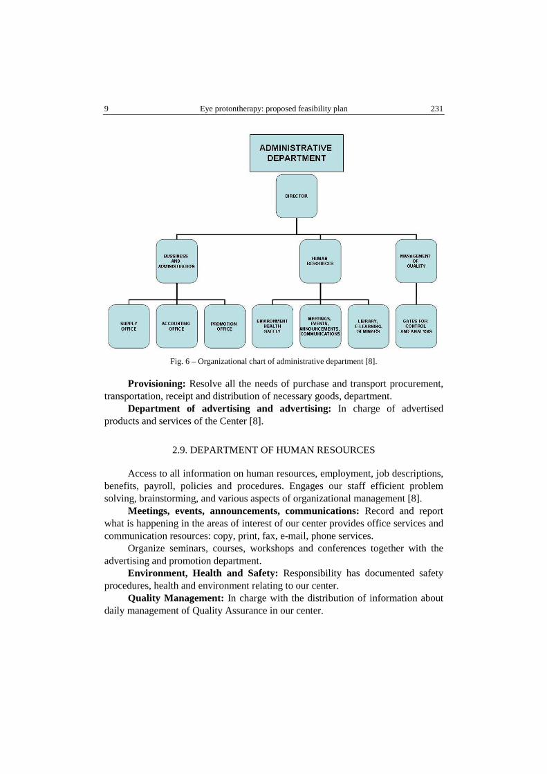
9 Eye protontherapy: proposed feasibility plan 231
Fig. 6 – Organizational chart of administrative department [8].
Provisioning: Resolve all the needs of purchase and transport procurement, transportation, receipt and distribution of necessary goods, department. Department of advertising and advertising: In charge of advertised products and services of the Center [8].
2.9. DEPARTMENT OF HUMAN RESOURCES
Access to all information on human resources, employment, job descriptions, benefits, payroll, policies and procedures. Engages our staff efficient problem solving, brainstorming, and various aspects of organizational management [8].
Meetings, events, announcements, communications: Record and report what is happening in the areas of interest of our center provides office services and communication resources: copy, print, fax, e-mail, phone services.
Organize seminars, courses, workshops and conferences together with the advertising and promotion department.
Environment, Health and Safety: Responsibility has documented safety procedures, health and environment relating to our center. Quality Management: In charge with the distribution of information about daily management of Quality Assurance in our center.
N. Verga et al. 10 232
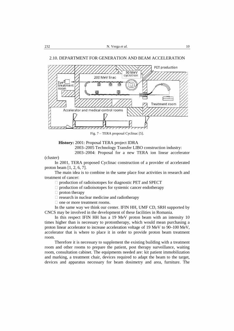
2.10. DEPARTMENT FOR GENERATION AND BEAM ACCELERATION
Fig. 7 – TERA proposal Cyclinac [5].
History: 2001: Proposal TERA project IDRA 2003–2005 Technology Transfer LIBO construction industry: 2003–2004: Proposal for a new TERA ion linear accelerator
(cluster) In 2001, TERA proposed Cyclinac construction of a provider of accelerated
proton beam [1, 2, 6, 7]. The main idea is to combine in the same place four activities in research and
treatment of cancer: � production of radioisotopes for diagnostic PET and SPECT � production of radioisotopes for systemic cancer endotherapy � proton therapy � research in nuclear medicine and radiotherapy � one or more treatment rooms. In the same way we think our center. IFIN HH, UMF CD, SRH supported by
CNCS may be involved in the development of these facilities in Romania. In this respect IFIN HH has a 19 MeV proton beam with an intensity 10
times higher than is necessary to protontherapy, which would mean purchasing a proton linear accelerator to increase acceleration voltage of 19 MeV to 90–100 MeV, accelerator that is where to place it in order to provide proton beam treatment room. Therefore it is necessary to supplement the existing building with a treatment room and other rooms to prepare the patient, post therapy surveillance, waiting room, consultation cabinet. The equipments needed are: kit patient immobilization and marking, a treatment chair, devices required to adapt the beam to the target, devices and apparatus necessary for beam dosimetry and area, furniture. The
11 Eye protontherapy: proposed feasibility plan 233
personnel required will be activated as described above. The three institutions above IFIN HH, UMF CD, SRH, could provide recruitment, contacts necessary for specialization abroad quality procedures, business continuity in this interdisciplinary unit, research and development of a method.
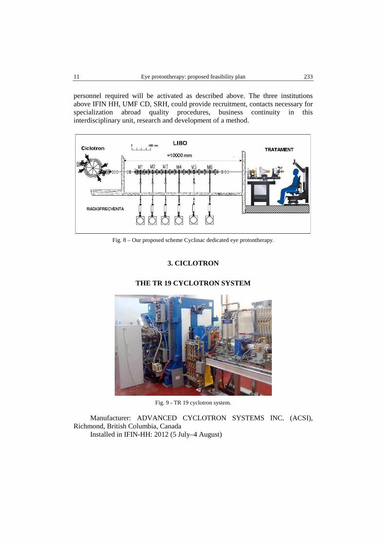
Fig. 8 – Our proposed scheme Cyclinac dedicated eye protontherapy.
3. CICLOTRON
THE TR 19 CYCLOTRON SYSTEM
Fig. 9 - TR 19 cyclotron system.
Manufacturer: ADVANCED CYCLOTRON SYSTEMS INC. (ACSI), Richmond, British Columbia, Canada
Installed in IFIN-HH: 2012 (5 July–4 August)

N. Verga et al. 12 234
Site Acceptance Tests (SAT): 2012 Oct 24; SAT report: All equipment of TR19 were responding very well and acting
very stable. During the tests we witness that the cyclotron was performing better than “performance guarantee” required by contract.
Description
TR 19 is a negative ion cyclotron ion source external and generating two beams leaving at 180°. Since the energy extraction can be varied the cyclotron TR19 by varying the the radius of extraction extracted beam energy can be varied at the choice of the operator. At TR19, it can extract protons at an energy of 14 MeV to 19 MeV. There are two simultaneous beams with intensities independent variables up to a maximum beam current. TR-series cyclotron magnet 19 are designed to generate such a strong focus and avoid resonated frequencies horizontal and vertical focus. This is done by sector geometry. The magnetic poles are nickel-plated to maintain a clean, a low gassing rate. TR-19 using an external ion source. This technology allows for higher beam currents, easier maintenance and flexibility for future upgrades. TR-19 provides more than 300 µA beam of protons on target and has the ability to increase the intensity of 2mA. TR-19 functions to ~ 5x10⁻⁷ Torr an order of magnitude better than machinery having internal ion source. At 18.5 MeV, only 1% of the proton beam is lost in the vacuum tank. To maintain vacuum TR-19, both the ion source and the main tank is a vacuum pump that provides clean cryopumps [14, 15].
Beam extraction system
Because the extraction probe is inserted radially in the plane of acceleration, extracted beam energy can be varied by adjusting the radius of the anterior edge of the probe.TR-19 can extract 14-19 MeV proton beam with a 0.1MeV step. The radial movement of the extraction probes allows the extraction of two beams simultaneously in any report of division.
It is not necessary to control the RF magnetic field; cyclotron maintaining optimal set does not affect the beam extraction [14, 15].
High stability due to proper cooling system
As a result, the derived frequency RF system is small, and the isochronous frequency of circulating particles does not deviate in time.Therefore, the system is extremely easy to start and reaching a stable operating mode can be accomplished in a very short time.
It is necessary to adjust the RF or magnet system during the startup process. System deviations are kept to a minimum due to the limited temperature variations from critical components [14].

13 Eye protontherapy: proposed feasibility plan 235
The main parameters of TR 19 cyclotron
Table 2
Cyclotron Type [14]
Negative Hydrogen External Ion Source
Local Shielding 2 Extracted Beams 8 External Targets
Beam Current Nominal - 300µA H-
Beam Energy (Variable Energy) Energy Range 14 MeV to 19 MeV H-
Table 3
Magnet [14]
Weight 22 Tons Geometry 4 Sectors (Closed) Hill Angle 45 degrees Hill Field 1.9 Wb/m2
Valley Field 0.5Wb/m2 Magnet Power 19 kW
Hill Gap 4 cm (nominal) Valley Gap 20 cm Pole Radius 57 cm Max Energy 19 MeV Amp Turns 8.4 x 10⁴
Table 4
RF System [14]
Number of Dees 2 (45 degrees) Dee Voltage 50 kV
RF Frequency 74 MHz Power Req’d 13 kW
Energy per Rev 200 keV
Table 5
Ion Source (External) [14]
Type Multicusp Output Current 2.8 mA
Emittance < 0.3 pi mm mrad (normalized) Injection Line 1 Quadrupole doublet, tilted
spiral inflector Bias Voltage 25 kV

N. Verga et al. 14 236
Table 6
Vacuum System [14]
Operating Pressure 5 x 10⁻⁷ Torr Base Pressure 2 x 10⁻⁷ Torr
Pumps 2 Cryopumps (4000 l/s H2O speed each)
2 Turbomolecular 2 Two Stage Rotary Vane
Table 7
Computer System [14]
Computer System Intel based Controllers Allen- Bradley Industrial PLC
Modules Interface Graphical User Interface
(RSVIEW)
LIBO
LIBO is a proton accelerator used for radiotherapy. The main advantages of a medical linear accelerator for protons are: � energy output varies as the synchrotron; � simple injection and extraction � modular construction � structure can be built in stages
Table 8
The main parameters of LIBO [2]
Accelerated particles p+1 Type of Linac CCL RF Frequency (MHz) 2998.5 Input energy (MeV) 30 Output energy (MeV) 230 The total length of the linac (m) 18.5 Cells per tank / tanks on the module 16/2 Number of accelerating modules 20 The thickness of half of cell it in a container (mm) 6.3–14.6 The diameter of the beam hole (mm) 7.0 Normalized transversal acceptance (mm mrad) 1.8 π Number of permanent magnetic quadrupoles 41 The length of PMQ (mm) 30 PMQ Gradients (T / m) 130–153 Synchronous phase (degrees) −15 Peak power per module (20% loss) (MW) 3.0 Shunt effective impedance ZT2 (inj.-extr.) 30–90 (MQ / m)

15 Eye protontherapy: proposed feasibility plan 237
Table 8 (continued)
Axial electric field (inj.-extr.) (MV / m) 15–17 Number of klystrons (peak power = 7.5MW) 10 Total peak power all RF klystrons (MW) 60 RF klystron efficiency 0.42 Repetition rate (Hz) ≤ 200 The duration of a pulse of protons (pS) 1.5 Max. Number of protons in 1.5 pS 4 · 107(2 Gy L-1 min-1) The actual length of each RF pulse (SP) 3.2 RF cycle 3.2 · 10−4 Power at 100 Hz + 100 auxiliary kW (kW) 150
Table 9
Clinical requirements for hadrontherapy [2]
Clinical requirement Has influence on Beam range in patient 2-20 cm continuously
adjustable Energy: 180MeV + 20 MeV accounting for energy loss in the spreading devices to obtain a 20 x 20 cm2 field with a throw of 3 m.
Field size 20 x 20 cm2 Beam spreading – beam intensity Range modulation Range adjustment
continuously adjustable continuously adjustable
Dose rate large fields >2 Gy/min Beam intensity small fields >> 10 Gy/min Beam intensity Field homogeneity Longitudinal <= 111% Beam delivery system Orthogonal <= 105% Beam spreading Field symmetry <= 105% Beam spreading – beam delivery
system Lateral penumbra < 2 mm at the entrance Multiple scattering spreading
devices Source site – source to axis distance Distal fall-off < 2 mm Range straggling (energy) – energy
spread Source to axis distance
Beam delivery system
The beam has two dipole magnets, five quadripolar magnet for focusing and three centering steering magnets. There are also safety locks in this section. If one of them detects that something is wrong, beam is turned off instantly [2].
Table 10
Cooling system of LIBO-62 prototype [2]
Applied peak power 4.2 MW Duty cycle 0.2% Average power inside LIBO 8400 W
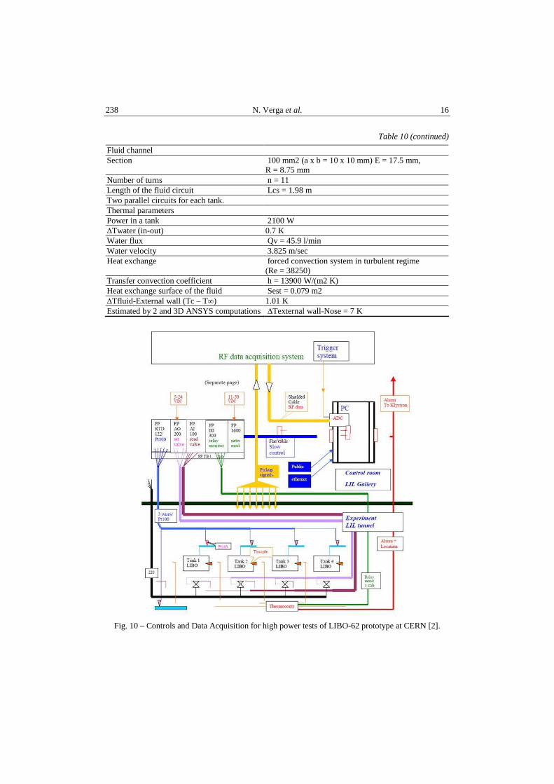
N. Verga et al. 16 238
Table 10 (continued)
Fluid channel Section 100 mm2 (a x b = 10 x 10 mm) E = 17.5 mm,
R = 8.75 mm Number of turns n = 11 Length of the fluid circuit Lcs = 1.98 m Two parallel circuits for each tank. Thermal parameters Power in a tank 2100 W ∆Twater (in-out) 0.7 K Water flux Qv = 45.9 l/min Water velocity 3.825 m/sec Heat exchange forced convection system in turbulent regime
(Re = 38250) Transfer convection coefficient h = 13900 W/(m2 K) Heat exchange surface of the fluid Sest = 0.079 m2 ∆Tfluid-External wall (Tc – T∞) 1.01 K Estimated by 2 and 3D ANSYS computations ∆Texternal wall-Nose = 7 K
Fig. 10 – Controls and Data Acquisition for high power tests of LIBO-62 prototype at CERN [2].
17 Eye protontherapy: proposed feasibility plan 239
4. MEDICAL PHYSICS DEPARTMENT
Physico-medical facility with accelerated protons for eye tumor radiotherapy
Beam depth range reached
Eye treatments require a variable interval between 2.5 cm and 3.2 cm depth [2].
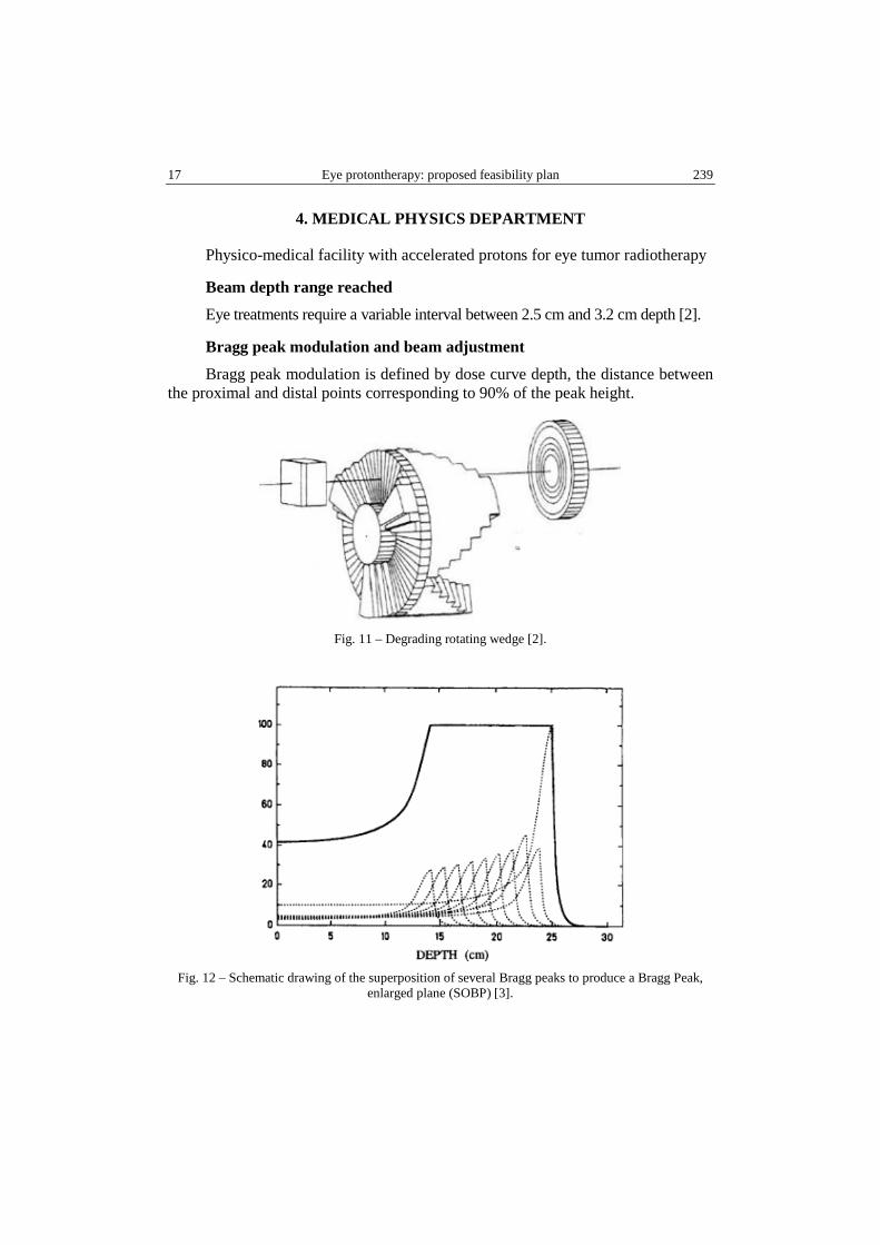
Bragg peak modulation and beam adjustment
Bragg peak modulation is defined by dose curve depth, the distance between the proximal and distal points corresponding to 90% of the peak height.
Fig. 11 – Degrading rotating wedge [2].
Fig. 12 – Schematic drawing of the superposition of several Bragg peaks to produce a Bragg Peak,
enlarged plane (SOBP) [3].

N. Verga et al. 18 240
Normally, the adjustment range refers to the ability to translate SOBP depth. The modulation of the Bragg peak should be variable in steps of 0.05 cm for the expansion to ≤ 5 cm and by 0.1 cm for the expansion in> 5 cm. The depth of treatment can be varied through the degrading rotating wedge which is located between the first and second collimator.
Modulator wheel rotates at 240 RPM and has four modulations per revolution. A number of wheel modulators are processed to vary the width of the Bragg peak covering the treatment required depth of 5 to 25 mm in 1 mm steps.
The dose rate
For ocular treatments, were a very short irradiation time (≤ 20-30 sec) is needed, requires high rates of SOBP dose (30-45 Gy / min), as greater accuracy in dose deposition is necessary [12].
Output power distribution LIBO-62
These parameters are required for volumes smaller than 50 cm3 of cancer.
Field size
For a fixed horizontal beam, the field size should range from 2 by 2 to 15 by 15 cm2 cm2 on the beam entry area projected on the patient.
The dimensions of the irradiated region can be increased by the translation of chair treatment. In all cases, the dimensions of the field must be adjustable in steps of 1 mm (± 0.05 mm).
Field homogeneity and symmetry
The homogenity of on the axis of the beam is defined as:
Rl = (P max / P min) x 100 Eq. 1
where Pmax is the maximum absorbed dose throughout the the field of radiation, averaged over an area not more than 1cm2, and Pmin is the minimum absorbed dose homogeneity region, averaged over an area not more than 1cm2.
Given a plane transverse to the beam axis, field symmetry is defined as the maximum value (in percent) of the ratio between highest and lowest dose absorbed, averaged over an area no more than 1 cm2, for each pair of symmetrical positions in relation to the axis of the beam within the homogenity region. Uniformity across (Rt) and longitudinal (Rl) field must be: Rt ≤ 105%, Rl ≤ 111%.
The symmetry of the field is less than 105% [2].
Methods of alignment
A laser system is initially used as a rough guide to align the beam directly to the tumor.

19 Eye protontherapy: proposed feasibility plan 241
Fig. 13 – A laser system to align the beam [10].
This is done by a chair that is electronically controlled by servomotors. Seat position can be changed in six modes.
Fig. 14 – Treatment chair [8].
Distal dose at stopping power
Considering the depth dose curve beam axis, distal dose at stopping power is defined as the distance between the 80% and 20% absorbed dose points on the beam axis beyond the Bragg peak.
100% maximum corresponding to the SOBP. Distal dose at stopping power due to dispersion of the beam must be less 2-3 mm against the stopping power of the beam [2].
N. Verga et al. 20 242
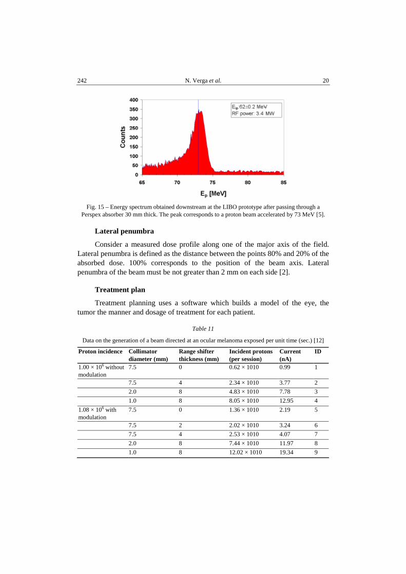
Fig. 15 – Energy spectrum obtained downstream at the LIBO prototype after passing through a
Perspex absorber 30 mm thick. The peak corresponds to a proton beam accelerated by 73 MeV [5].
Lateral penumbra
Consider a measured dose profile along one of the major axis of the field. Lateral penumbra is defined as the distance between the points 80% and 20% of the absorbed dose. 100% corresponds to the position of the beam axis. Lateral penumbra of the beam must be not greater than 2 mm on each side [2].
Treatment plan
Treatment planning uses a software which builds a model of the eye, the tumor the manner and dosage of treatment for each patient.
Table 11
Data on the generation of a beam directed at an ocular melanoma exposed per unit time (sec.) [12]
Proton incidence Collimator diameter (mm)
Range shifter thickness (mm)
Incident protons (per session)
Current (nA)
ID
1.00 × 10⁶ without modulation
7.5 0 0.62 × 10¹⁰ 0.99 1
7.5 4 2.34 × 10¹⁰ 3.77 2
2.0 8 4.83 × 10¹⁰ 7.78 3
1.0 8 8.05 × 10¹⁰ 12.95 4
1.08 × 10⁶ with modulation
7.5 0 1.36 × 10¹⁰ 2.19 5
7.5 2 2.02 × 10¹⁰ 3.24 6
7.5 4 2.53 × 10¹⁰ 4.07 7
2.0 8 7.44 × 10¹⁰ 11.97 8
1.0 8 12.02 × 10¹⁰ 19.34 9

21 Eye protontherapy: proposed feasibility plan 243
Fig. 16 – Steps for the treatment plan protontherapy eye dedicated [8].
Once a treatment plan is developed, it should adapt the beam energy especially for the treatment of the patient’s eye.
A thermoplastic facial mask and an immobilizer for head position (which will be „bitten” by the patient) will be adjusted, in such manner as to immobilize the patient during the treatment. The collimator is usually adapted to the tumor characteristics.
5. MEDICAL DEPARTMENT
Clinical indications of Ocular protontherapy
Choroidal melanomas are the most common primary intraocular tumor: estimated incidence rate is approximately 6-7 new cases per million inhabitants [11].
� Ocular melanoma: including those who only partially responded to other treatments
� Melanoma of the conjunctivae � Retinoblastoma � Metastatic tumors in the eye

N. Verga et al. 22 244
� Eye benign tumors: choroidal hemangioma and angioma � For patients with detached retina and other eye disorders � Age related macular degeneration. Not everyone is a candidate for protontherapy. Imaging studies are needed
and an evaluation by an eye specialist to ensure that the treatment is appropriate for each case.
Patient preparation
First, under general anesthesia, an ophthalmologist attaches small tantalum clips to the outer surface of the eye to define tumor boundaries. The patient has three to five markers attached around the base of the tumor.
Fig. 17 – Eye protontherapy technique [11].
This helps orientation and determination of the best treatment position (using a radiology device), so that the patient can be positioned properly – the error margin must be 0.1 to 0.2 mm at most. This includes the treatment angle (the direction in which the patient will look) and the isocenter of the tumor [11].
The treatment
Because the eye is not immobilized itself, it is crucial that patient does not move the eye during treatment. However, to help the patient, room lights are off and the patient is asked to look at a flashing light to determine the position of the eye.
Normally, the patient is allowed to blink, as treatment duration is short; in order to keep it to minimum, specific treatment should be applied.
To correctly position the affected eye, the patient fixes a light point on a coordinate grid. Ahead of each radiotherapy session, the eye position is verified through radiology. Only when the position deviates less than 0.2 mm from the correct position the radiotherapy session can be performed.

23 Eye protontherapy: proposed feasibility plan 245
Fig. 18 – Eye protontherapy technique [9].
The position of the of the eye is monitored during treatment with closed-circuit television. If for any reason the patient moves the eye, the beam is turned off to avoid damage to other parts of the eye. If the eye treated is fully blind, then the other eye can be used to fix the flashing light.
The total dose may vary for each radiotherapy center and is about 60 Gy (at 54.5 Gy in 4 Uppsala fractions; in other centers it may be 56.4 Gy). Therapeutic fractions can be single or multiple. Usually, the treatment consists of four identical treatment doses spread out over four days, with a duration of each session of approximately 30, 60 or 90 seconds. Sometimes, patients can see a blue light when a higher energy dose is used for a retrooculary tumor [8].
Patient follow up and control
Following treatment, patients receive usual care and monitoring from their doctors or by ophthalmologists carrying out inspections every six months for the rest of their life.
Secondary effects
There is a small risk of glaucoma, palpebral radiodermatitis, keratitis sicca, keratoconjunctivitis sicca or xerophthalmia (eye dryness if the lacrimal glands are hit). Loss of vision is a possibility, but errors are unlikely, because targeting the tumor is precise and neighboring tissue are protected [13].
6. CONCLUSIONS
Construction of the Cyclinac is techncally feasible at IFIN HH. Founding of a medical center for eye protontherapy at IFIN HH is possible, with the collaboration of „Carol Davila” University of Medicine and Pharmacy and the Romanian Society of Hadrontherapy, as long as financing of a feasibility plan will be accepted by the CNCS, in order to establish a theoretical basis to obtain funding from the European Community. Cooperation with specialized institutions from the EU is necessary, cooperation which SRH has already established.

N. Verga et al. 24 246
Acknowledgements. To our families and colleagues of the UMF “Carol Davila”, our colleagues from IFIN “Horia Hulubei” to Manjit Dosanj, Ugo Amaldi, Sarolta ILIESCU-HOLM, colleagues from Enlight + +, colleagues from the JINR Dubna, University Politehnica Bucharest, and last but not least colleagues from ICPE-CA Bucharest.
REFERENCES
1. Szeless, B. et al., Successful high power test of a proton linac booster (LIBO) prototype for hadrontherapy, CERN, Geneva, Switzerland; -Particle Accelerator Conference, 2001. PAC 2001. Proceedings of the 2001 (Volume 1) Chicago, ISBN: 0-7803-7191-7, DOI: 10.1109/PAC.2001.987592
2. Paolo Berra, Conception, construction et essai d’un accélérateur linéaire à protons impulsé à 3 GHz (LIBO) pour la thérapie du cancer, These, diplome de doctorat - Université Claude Bernard Lyon I. Institut de Physique Nucléaire - 14 Octobre 2005.
3. U. Amaldi et al., LIBO—a linac-booster for protontherapy: construction and tests of a prototype, Nuclear Instruments and Methods in Physics Research A 521 (2004) 512–529.
4. C. De Martinisand et al., Beam tests on a proton linac booster for hadrontherapy, Proceedings of EPAC 2002, Paris, France.
5. Ugo Amaldi, Saverio Braccini, Paolo Puggioni, High Frequency Linacs for Hadrontherapy, Reviews of Accelerator Science and Technology, Vol. 2 (2009) 111–131.
6. V. G. Vaccaro et al., Some Relevant Aspects in the Design and Construction of a 30-62 MeV Linac Booster for Proton Therapy, LINAC 2004, Lubeck, Germany (August 2004).
7. P. Berra et al., Study, construction and test of a 3 ghz proton linacbooster (libo) for cancer therapy, Proceedings of EPAC 2000, Vienna, Austria
8. http://www.triumf.ca 9. http://www.european-hospital.com 10. https://www.ucdmc.ucdavis.edu/ 11. http://www.dkfz.de 12. Marília Tavares Christóvăo, Tarcísio Passos Ribeiro de Campos, Spatial distribution analysis of
absorbed dose in ocular proton radiation therapy, Radiol Bras. 2010;43(4):249–254. 13. http://p-therapie.web.psi.ch 14. Advanced Cyclotron Systems, Inc, Advanced Cyclotron Systems, Inc, 7851, Alderbridge Way,
Richmond, B.C. V6X 2A4 “TR19 Technical Overview”. 15. http://www.advancedcyclotron.com/cyclotron-solutions/tr19