Embed Size (px)
Citation preview
Eye, magnetism and magnetsMankind has always been familiar with the magnetism ordrawing power of the eyes. Eye contact between mother andchild is a strong force in bonding between the two. Eye contactis also the initial signal of attraction between two individuals,triggering special emotions especially if the attraction is mutual.Paramhansa Yongnanda describes magnetism, which originatesin the ‘Infinite Spirit’, as the power by which one draws thingsto oneself like the right partner, friends, business associates andothers. ‘Each human being is a medium through which God’smagnetism flows. ‘All parts of the body that come in pairs’ suchas eyes, ears, hands ‘form magnets’. ‘Soul magnetism isexpressed through the eyes, weakly or strongly, depending onone’s spiritual development. Some highly developed people areable to spiritualise or heal others solely by the magnetism oftheir eyes’.
Magnetism transcends from the spiritual to the physical. Ironis an essential element for most living animals and iron com-pounds abound in Nature. Magnetite, an oxide of iron is themost magnetic mineral on Earth. The ability of naturally occur-ring pieces of ‘rock’, termed ‘lodestone’ to attract iron particles–‘magnetism’ was known to ancient Man. Crystals of magnetiteare distributed extensively in cells and in some species theyenable ‘magnetoreception’, a special sense with which theorganism is able to be aware of the Earth’s magnetic field anduse it to navigate across the globe. Biomagnetism is also manifestin other ways. The front of the eye is electropositive comparedto the back. The current that flows from front to back cancreate a magnetic field around the eye ball which in turn couldinfluence the behaviour of cells. Various ‘lines’ formed by thedeposition of iron compounds on the corneal surface have beendescribed—Hudson Stahli line, Stocker’s line, Ferry’s line,Fliescher ring and other un-named deposits that are associatedwith angles on the corneal surface created by scars and laserrefractive surgery. The vortex or whorl pattern formed by
corneal epithelial cells—‘hurricane keratopathy’ has been attrib-uted to the effect of the electromagnetic field of the eyeball onmigrating epithelial cells that may contain magnetite.
It is not surprising therefore that magnets have had a role toplay in medicine. There are many reports on the use of magnetsto heal tissue like bone and treat disease.
Lodestone was used in India before the Christian era. Thefirst mention of the removal of a foreign body from the eye bymagnetic attraction is attributed to the physician HieronymousBrunschwyck of Strasburg in 1497. He removed iron filingswhich had hit the surface of the eye of a patient by placing lode-stone against the open lids. Various attempts were made toextract metal foreign bodies from within the eye in the secondhalf of the 19th century with more powerful magnets. The firstperson to insert a magnetic probe within the eye was Dr W AMacKeown (1844–1924) of Belfast. He successfully removed aforeign body from within the vitreous in 1874 by using a longslender terminal in contact with the foreign body. The instru-ment shown on the front cover of this issue is a magnetic devicedesigned in 1881 by the ophthalmologist Simeon Snell. Themagnet was designed to be used for the removal of ferrousmetal fragments near to or on the surface of the eye. This handheld instrument contrasts with the much larger ‘giant magnets’.
It was Julius Hirschberg (1843–1925), the incomparable oph-thalmic historian who, in 1879, was the first to introduce anelectromagnet into ophthalmic practice. Amongst his 109 pub-lished works including the monumental History ofOphthalmology volumes there are two classical monographs(1885, 1899) on operations with his electromagnet which forthe first time produced a more powerful ‘pull’ on a foreignbody. Snell’s instrument is very similar to Hirschberg’s with thesame wide choice of terminals. The strength of a magnet, mea-sured in Gauss, is determined by the shape and length of theterminal. The blunter the end the stronger the magnetic pull.For the extraction of foreign bodies in the eye a blunt end tothe magnet limited the ability to place it close to the site wherethe metal particle is lodged in the eye. Although longer slenderterminals were far less powerful they were more practical. Forthe most efficient extraction the terminal was placed as close aspossible, if not in contact with the tissue in front of the foreignbody.
Simeon Snell was born in Launceston, Cornwall in 1851. Hestudied medicine in Leeds and then moved to Guy’s Hospital inLondon before spending time at The Royal London OphthalmicHospital (Moorfields). He achieved his MRCS in 1872. Hethen moved to Sheffield where he set up practice and becamethe first ophthalmologist at the Royal Infirmary in 1879, a pos-ition he held for the rest of his career. Sheffield was an indus-trial city and because of the frequency of eye injuries andforeign bodies in the eye Snell became an authority on opera-tions with the magnet. He also became an expert on the preven-tion of occupational diseases. In 1892 he was made a Fellow ofthe Royal College of Surgeons of Edinburgh (FRCS Ed.). Hefounded the Medical Faculty in Sheffield and became its firstProfessor of Ophthalmology there.
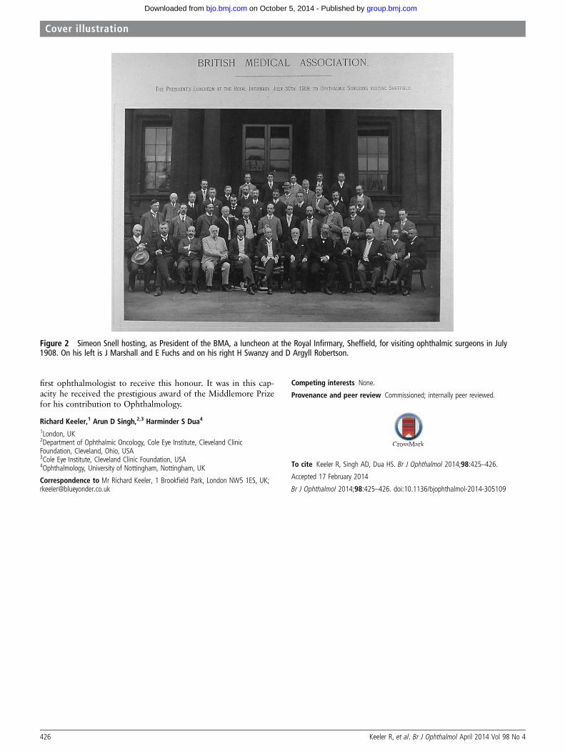
Like Hirschberg he wrote a monograph (1883) entitled TheElectro magnet and its employment in ophthalmic surgery. Hegave a full description of his instrument in this monograph.Snell wrote on a variety of subjects but mostly on the effects ofindustry on the eye. Included in his writings are articles onMiner’s nystagmus 1884 (he frequently went down the mines toget first hand experience), Glass blower’s cataract 1907 andSchool life and Eye sight. Snell’s crowning achievement wasbeing elected President of the British Medical Association, theFigure 1 Simeon Snell (1851–1909).
Keeler R, et al. Br J Ophthalmol April 2014 Vol 98 No 4 425
Cover illustration
group.bmj.com on October 5, 2014 - Published by bjo.bmj.comDownloaded from
first ophthalmologist to receive this honour. It was in this cap-acity he received the prestigious award of the Middlemore Prizefor his contribution to Ophthalmology.
Richard Keeler,1 Arun D Singh,2,3 Harminder S Dua4
1London, UK2Department of Ophthalmic Oncology, Cole Eye Institute, Cleveland ClinicFoundation, Cleveland, Ohio, USA3Cole Eye Institute, Cleveland Clinic Foundation, USA4Ophthalmology, University of Nottingham, Nottingham, UK
Correspondence to Mr Richard Keeler, 1 Brookfield Park, London NW5 1ES, UK;[email protected]
Competing interests None.
Provenance and peer review Commissioned; internally peer reviewed.
To cite Keeler R, Singh AD, Dua HS. Br J Ophthalmol 2014;98:425–426.
Accepted 17 February 2014
Br J Ophthalmol 2014;98:425–426. doi:10.1136/bjophthalmol-2014-305109
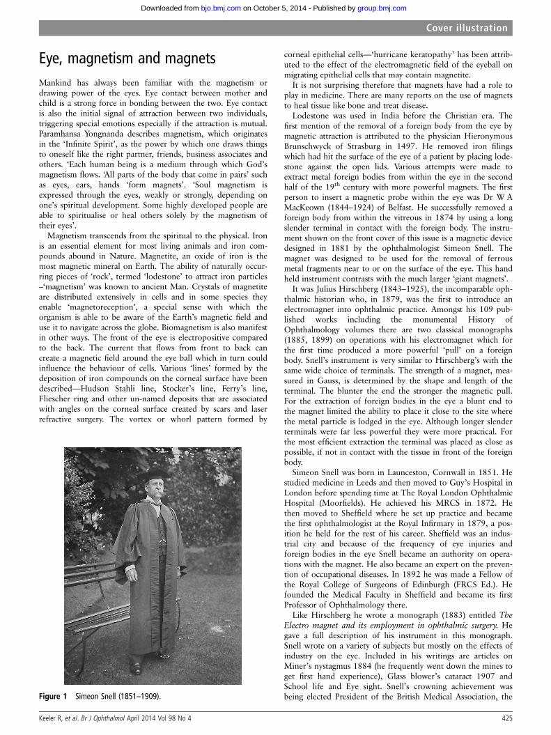
Figure 2 Simeon Snell hosting, as President of the BMA, a luncheon at the Royal Infirmary, Sheffield, for visiting ophthalmic surgeons in July1908. On his left is J Marshall and E Fuchs and on his right H Swanzy and D Argyll Robertson.
426 Keeler R, et al. Br J Ophthalmol April 2014 Vol 98 No 4
Cover illustration
group.bmj.com on October 5, 2014 - Published by bjo.bmj.comDownloaded from
doi: 10.1136/bjophthalmol-2014-305109 2014 98: 425-426Br J Ophthalmol
Richard Keeler, Arun D Singh and Harminder S Dua Eye, magnetism and magnets
http://bjo.bmj.com/content/98/4/425.full.htmlUpdated information and services can be found at:
These include:
serviceEmail alerting
the box at the top right corner of the online article.Receive free email alerts when new articles cite this article. Sign up in
CollectionsTopic
(327 articles)Paediatrics � (548 articles)Ocular surface �
(460 articles)Cornea � Articles on similar topics can be found in the following collections
Notes
http://group.bmj.com/group/rights-licensing/permissionsTo request permissions go to:
http://journals.bmj.com/cgi/reprintformTo order reprints go to:
http://group.bmj.com/subscribe/To subscribe to BMJ go to:
group.bmj.com on October 5, 2014 - Published by bjo.bmj.comDownloaded from


![1 L 27 Electricity & Magnetism [5] Magnets –permanent magnets –Electromagnets –The Earth’s magnetic field magnetic forces applications Magnetism](https://img.pdfslide.us/doc/110x75/56649d9c5503460f94a85bd1/1-l-27-electricity-magnetism-5-magnets-permanent-magnets-electromagnets.jpg)


![1 L 28 Electricity and Magnetism [5] magnetism magnetic forces applications Why are magnets magnets?](https://img.pdfslide.us/doc/110x75/56649f485503460f94c6a785/1-l-28-electricity-and-magnetism-5-magnetism-magnetic-forces-applications.jpg)