Embed Size (px)
Citation preview
EYE EDUCATION FOR EMERGENCY CLINICIANS 1
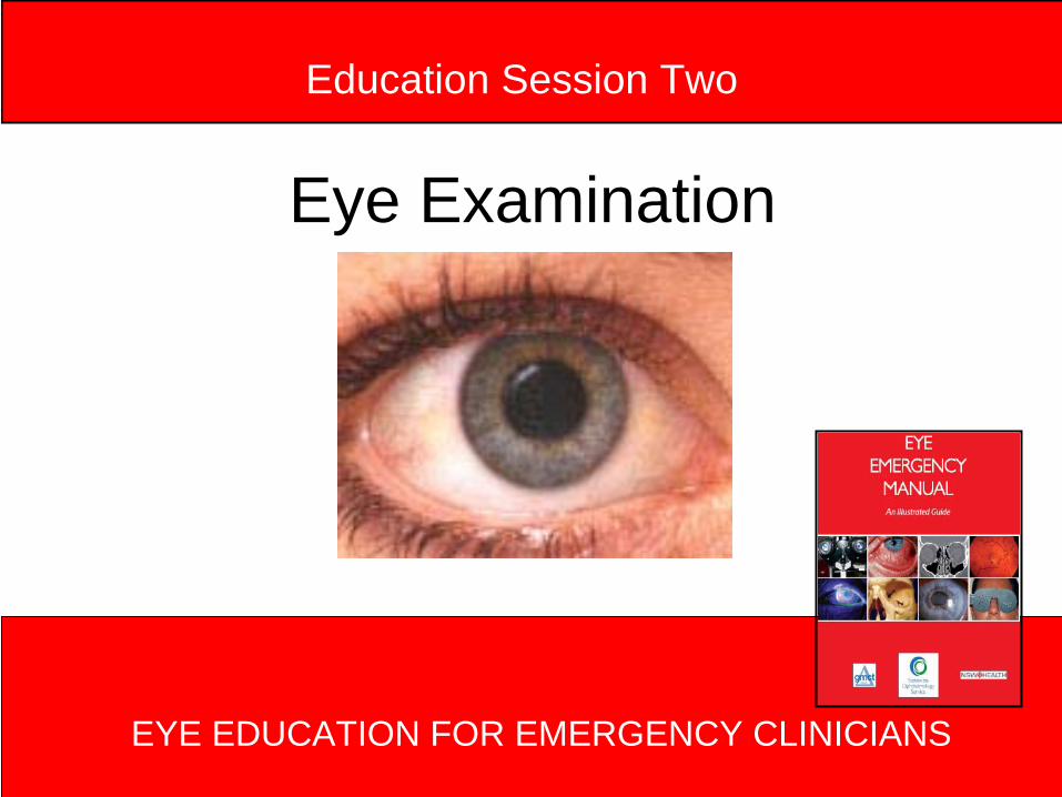
Eye Examination
EYE EDUCATION FOR EMERGENCY CLINICIANS
Education Session Two
EYE EDUCATION FOR EMERGENCY CLINICIANS 2
These presentations have been prepared by:
• Jillian Grasso, Clinical Nurse Consultant, Ophthalmology
• Janet Long, Clinical Nurse Consultant Community Liaison, Ophthalmology
• Joanna McCulloch, Transitional Nurse Practitioner, Ophthalmology
• Cheryl Moore, Nurse Educator, Ophthalmology
Further information contact us at Sydney Hospital & Sydney Eye Hospital: 02 9382 7111
Modules originally designed for emergency nurses as a component of the Eye Emergency Manual Project.
December 2008
EYE EDUCATION FOR EMERGENCY CLINICIANS 3
Aim and Objectives
Understand the fundamental principles and perform a systematic eye examination.
On completion of this session you will be able to:
• Recognise normal and abnormal anatomy
• Systematically examine an eye• Correctly document examination findings
EYE EDUCATION FOR EMERGENCY CLINICIANS 4
Equipment required to examine an eye
• Fine beamed torch (with optional blue filter for examination using Fluorescein)
• Cotton buds• Local anaesthetic eye drops, eg
Amethocaine 0.5%, Oxybuprocaine 0.4%• Fluorescein strips or Minims• Magnification – slit lamp, indirect
ophthalmoscope, loupes or Woods lamp
EYE EDUCATION FOR EMERGENCY CLINICIANS 5
Patient Assessment
• If injury is SELF EVIDENT – Eg, impaled object – OR totally closed, tightly swollen eyelid
associated with trauma– Do not try to examine this eye – Patient requires an immediate referral to
an Ophthalmologist
EYE EDUCATION FOR EMERGENCY CLINICIANS 6
Patient Preparation
• Head well supported (eg. chair back against the wall prevents head moving back and away)
• If using a slitlamp make sure patient is correctly positioned.
• Appropriate lighting for patient comfort– e.g. dim lights if photophobic
EYE EDUCATION FOR EMERGENCY CLINICIANS 7
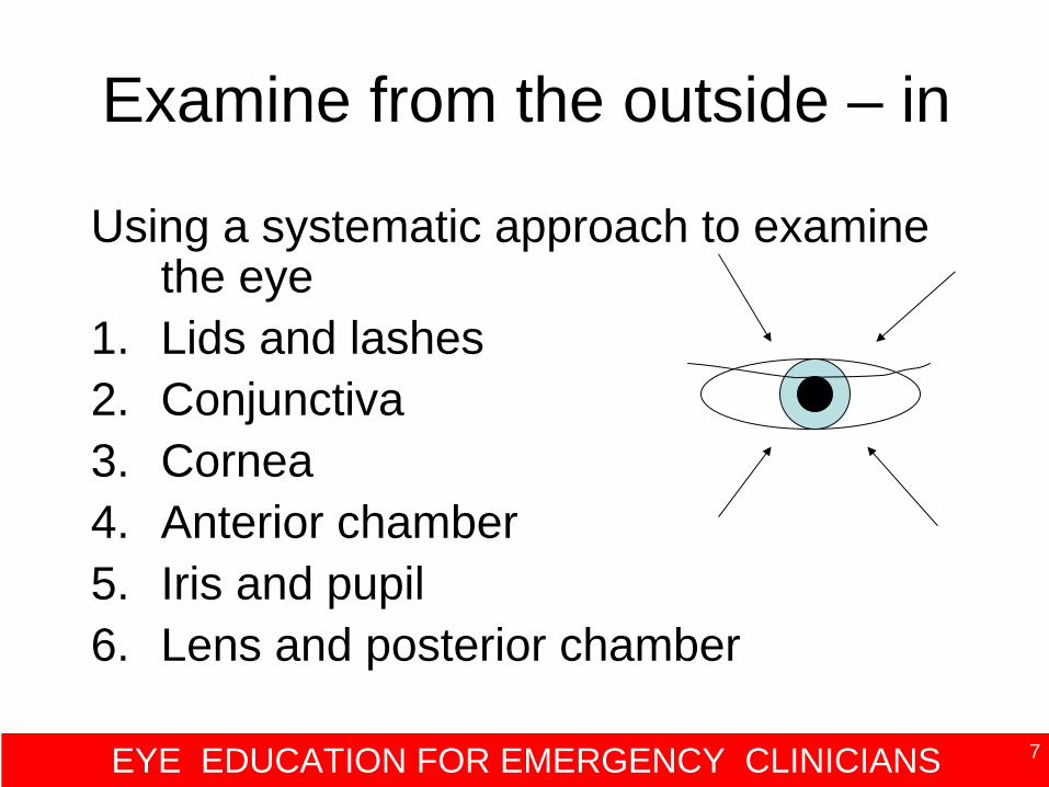
Examine from the outside – in
Using a systematic approach to examine the eye
1. Lids and lashes2. Conjunctiva3. Cornea4. Anterior chamber5. Iris and pupil6. Lens and posterior chamber
EYE EDUCATION FOR EMERGENCY CLINICIANS 8
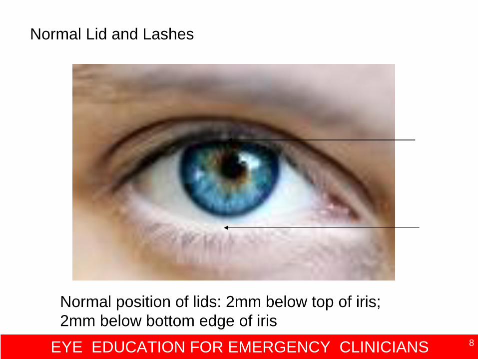
Normal position of lids: 2mm below top of iris; 2mm below bottom edge of iris
Normal Lid and Lashes
EYE EDUCATION FOR EMERGENCY CLINICIANS 9
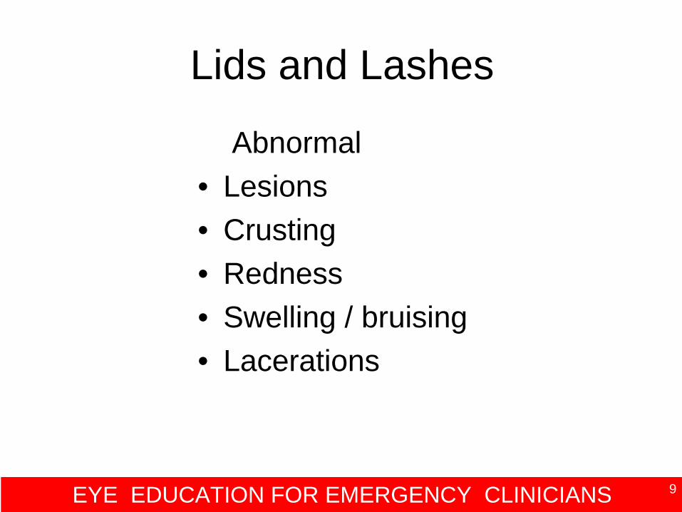
Lids and Lashes
Abnormal• Lesions• Crusting• Redness• Swelling / bruising• Lacerations
EYE EDUCATION FOR EMERGENCY CLINICIANS 10
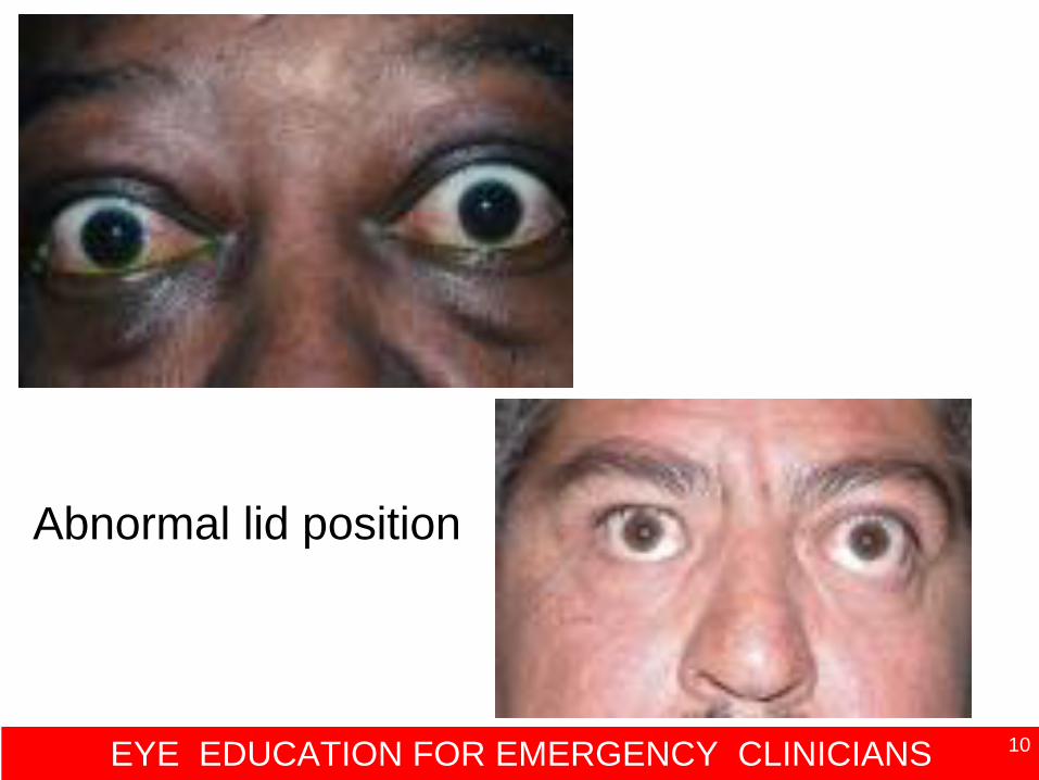
Abnormal lid position
EYE EDUCATION FOR EMERGENCY CLINICIANS 11
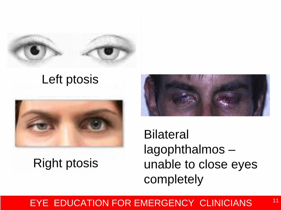
Left ptosis
Right ptosis
Bilateral lagophthalmos – unable to close eyes completely
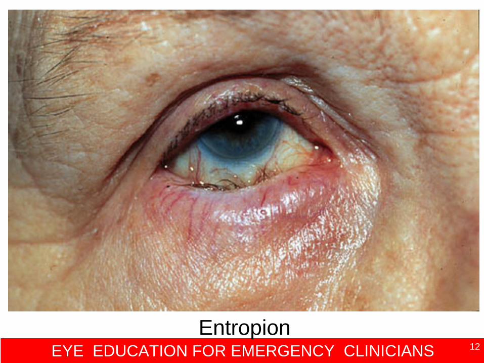
EYE EDUCATION FOR EMERGENCY CLINICIANS 12Entropion
EYE EDUCATION FOR EMERGENCY CLINICIANS 13
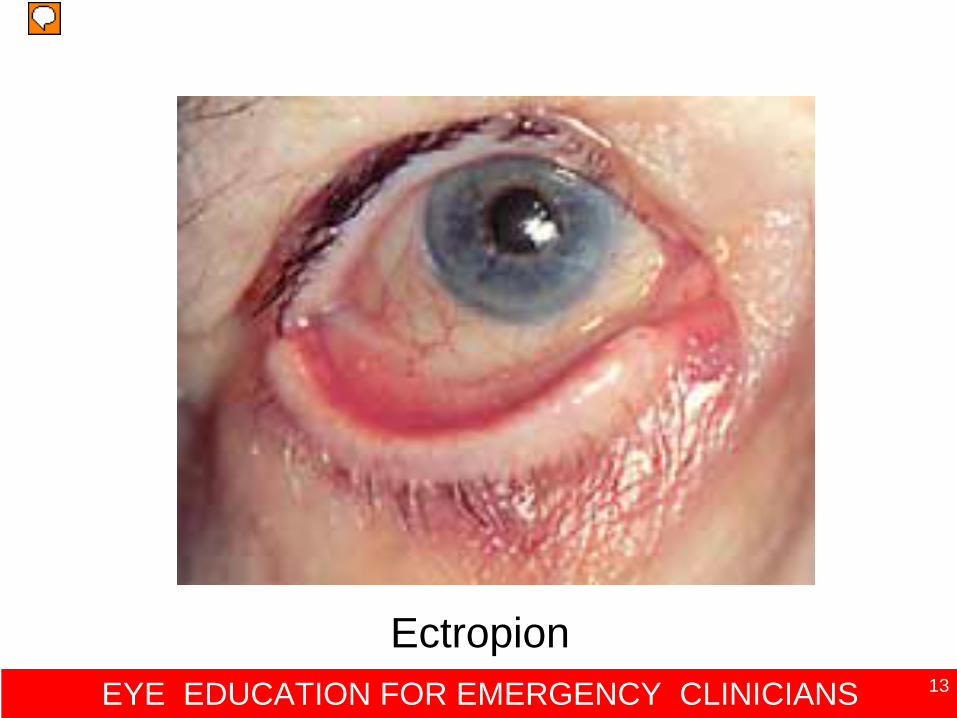
Ectropion
EYE EDUCATION FOR EMERGENCY CLINICIANS 14
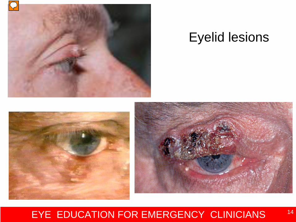
Eyelid lesions
EYE EDUCATION FOR EMERGENCY CLINICIANS 15
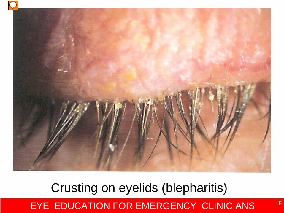
Crusting on eyelids (blepharitis)
EYE EDUCATION FOR EMERGENCY CLINICIANS 16
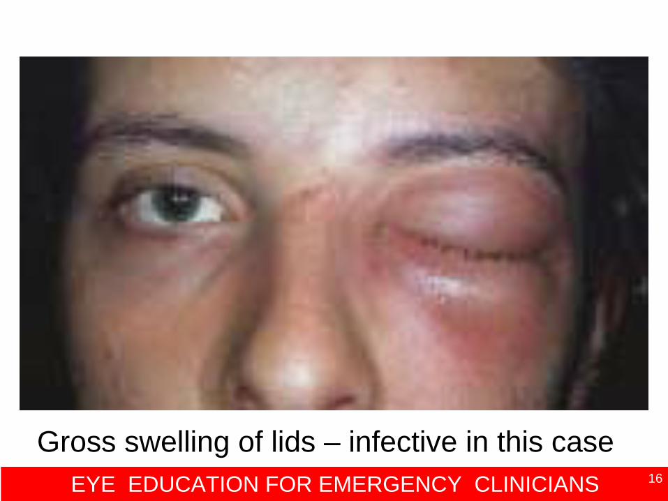
Gross swelling of lids – infective in this case
EYE EDUCATION FOR EMERGENCY CLINICIANS 17
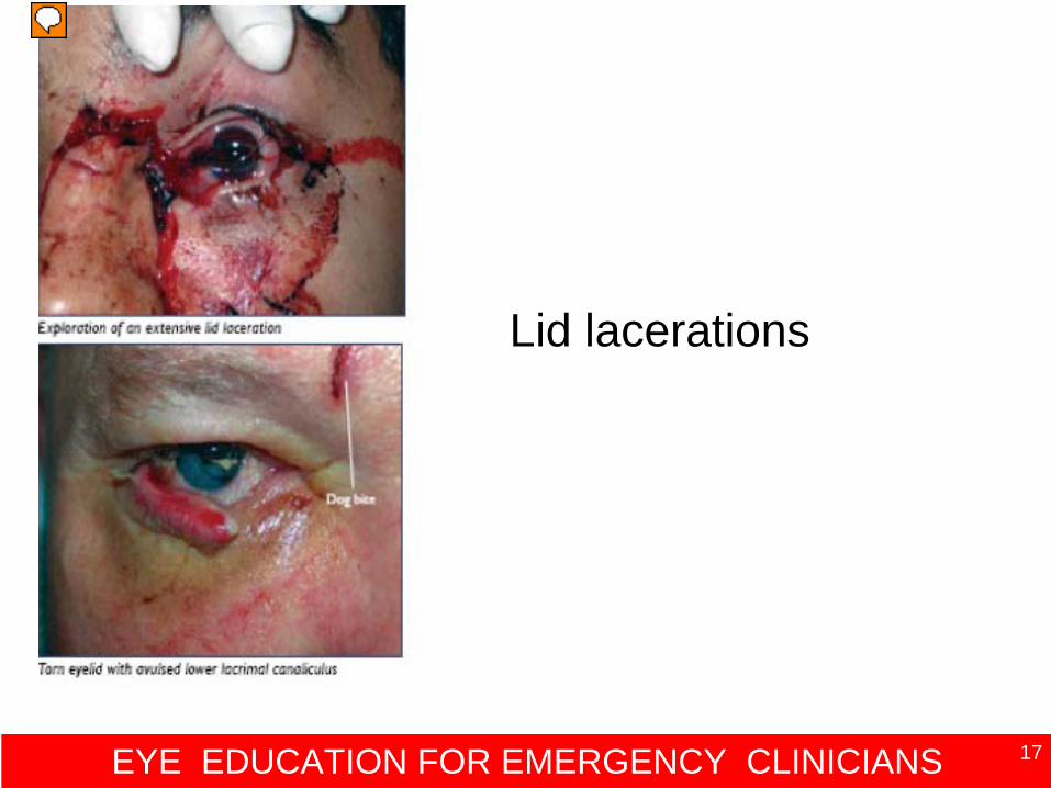
Lid lacerations EEM p.32
Lid lacerations
EYE EDUCATION FOR EMERGENCY CLINICIANS 18
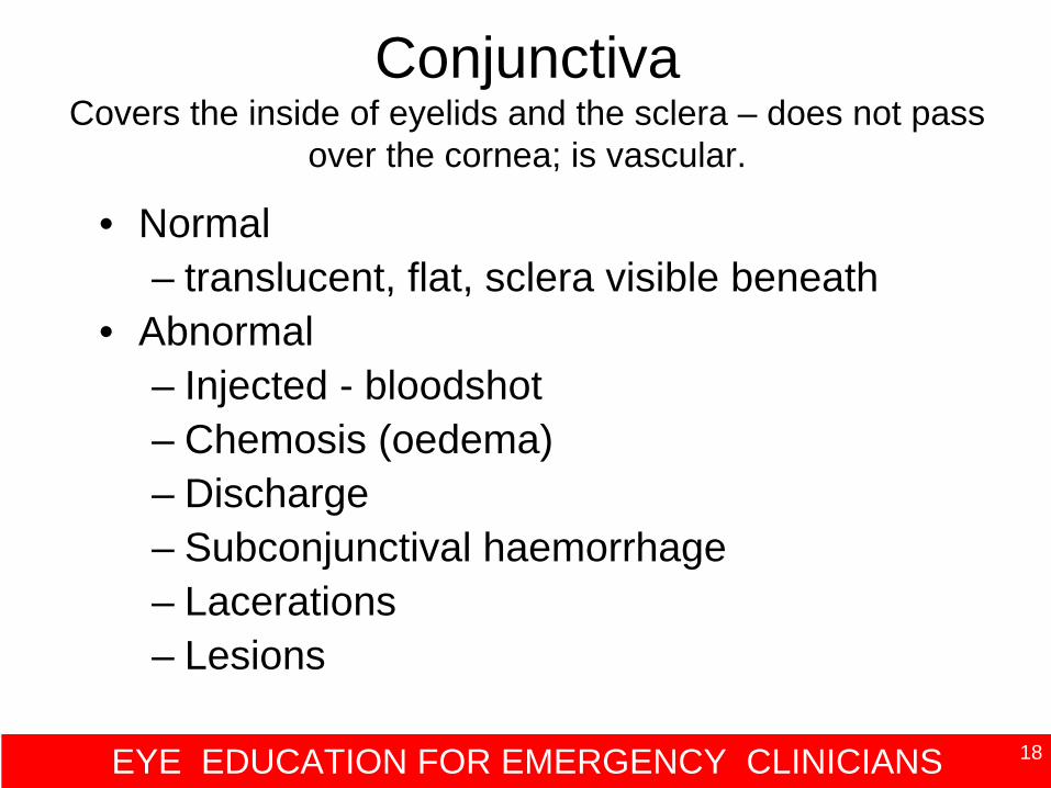
Conjunctiva Covers the inside of eyelids and the sclera – does not pass
over the cornea; is vascular.
• Normal– translucent, flat, sclera visible beneath
• Abnormal– Injected - bloodshot– Chemosis (oedema)– Discharge– Subconjunctival haemorrhage– Lacerations– Lesions
EYE EDUCATION FOR EMERGENCY CLINICIANS 19
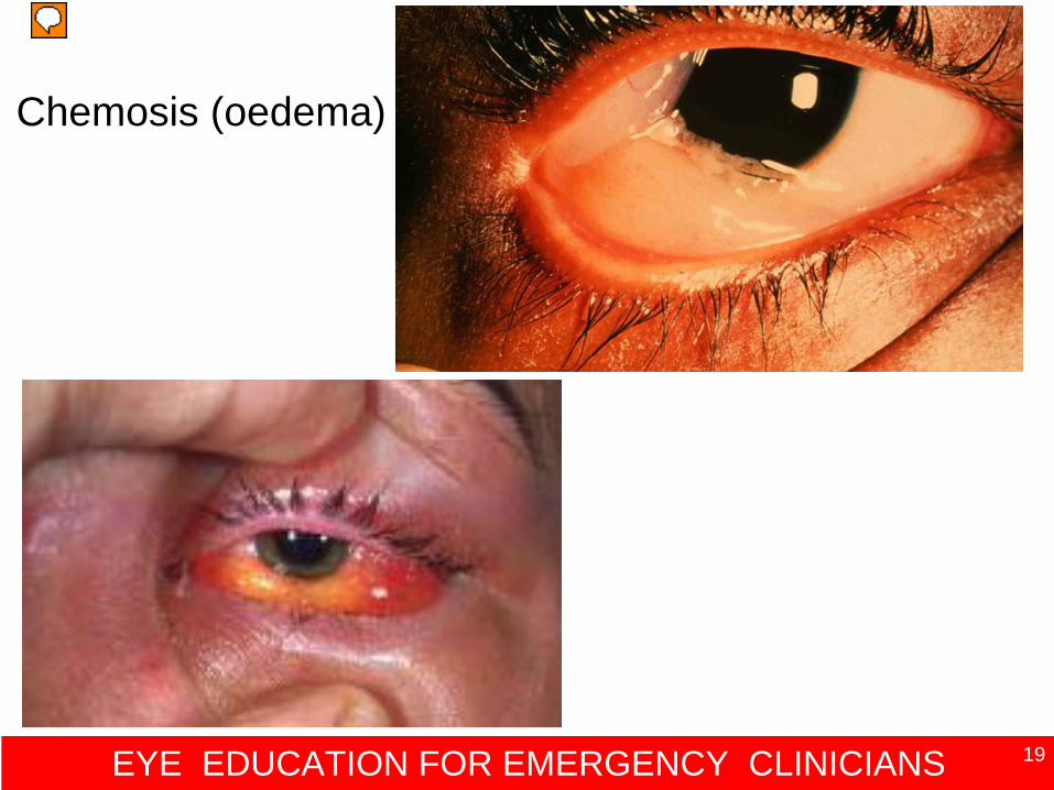
Chemosis (oedema)
EYE EDUCATION FOR EMERGENCY CLINICIANS 20
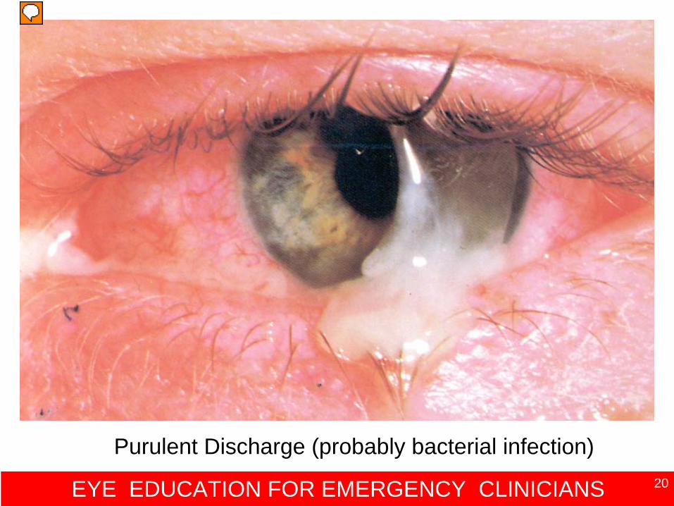
Purulent Discharge (probably bacterial infection)
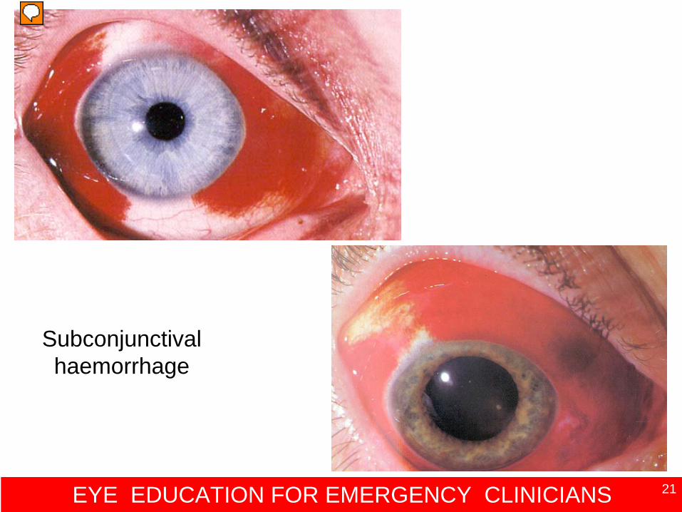
EYE EDUCATION FOR EMERGENCY CLINICIANS 21
Subconjunctival haemorrhage
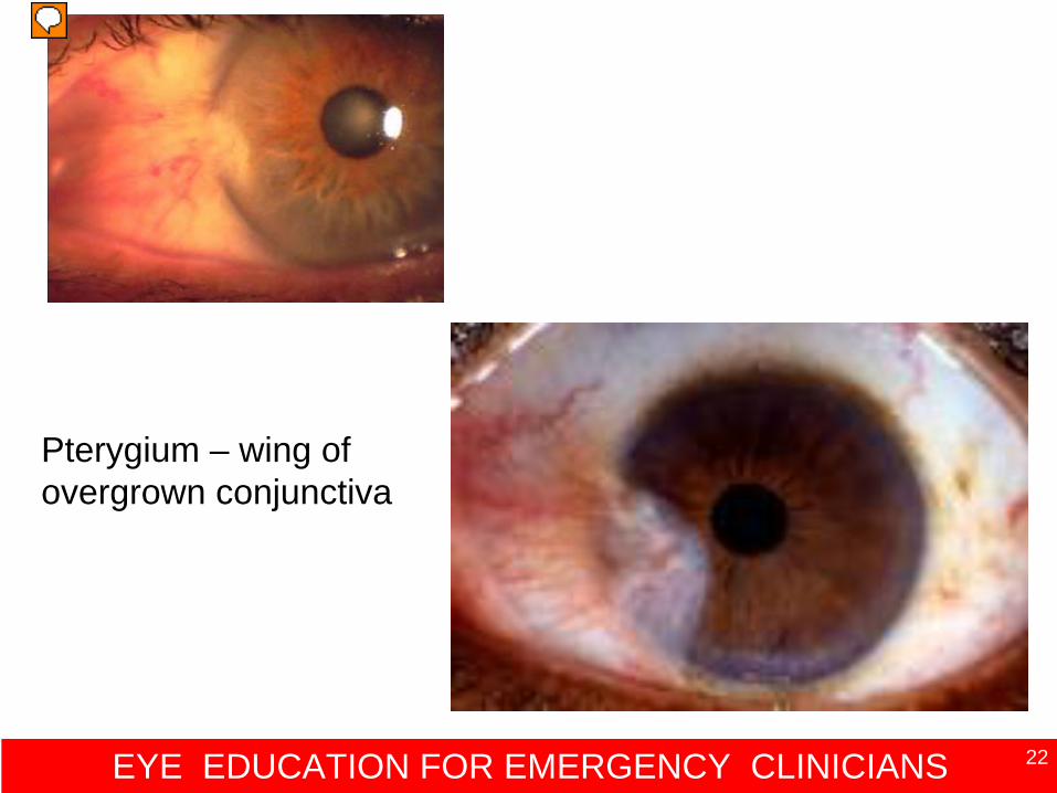
EYE EDUCATION FOR EMERGENCY CLINICIANS 22
Pterygium – wing of overgrown conjunctiva
EYE EDUCATION FOR EMERGENCY CLINICIANS 23
Cornea Avascular circular ‘window’ of the eye
• Normal – clear, bright, smooth surface
• Abnormal– Cloudy – iris may be difficult to see– Scarring - milky line, localised opacity– Foreign body– Rust ring– Abscess– Laceration
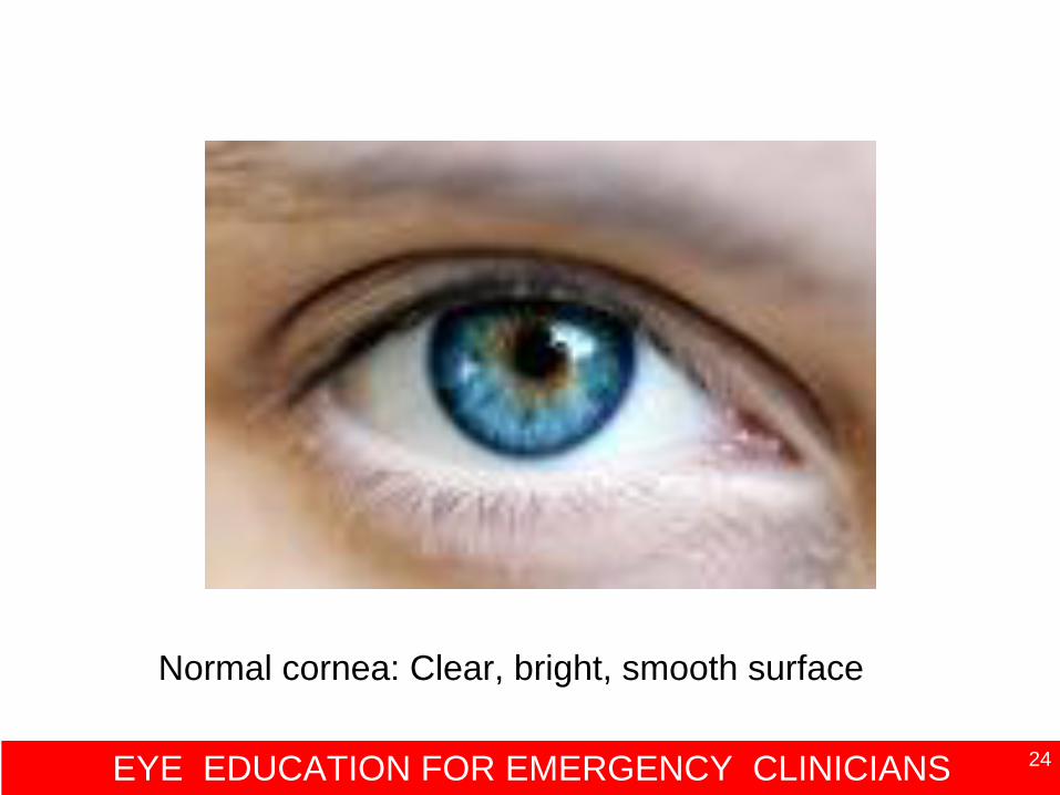
EYE EDUCATION FOR EMERGENCY CLINICIANS 24
Normal cornea: Clear, bright, smooth surface
EYE EDUCATION FOR EMERGENCY CLINICIANS 25
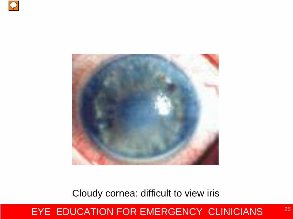
Cloudy cornea: difficult to view iris
EYE EDUCATION FOR EMERGENCY CLINICIANS 26
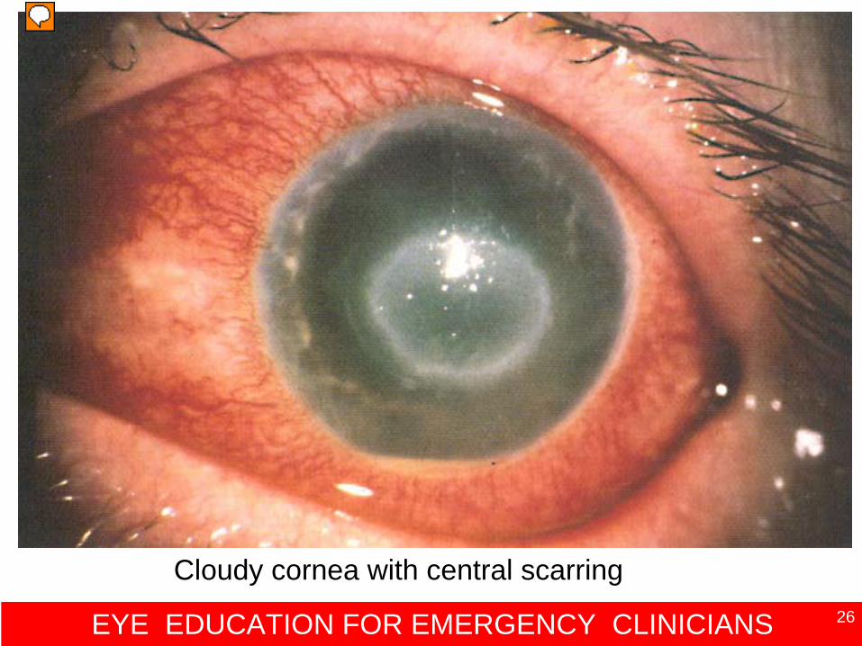
Cloudy cornea with central scarring
EYE EDUCATION FOR EMERGENCY CLINICIANS 27
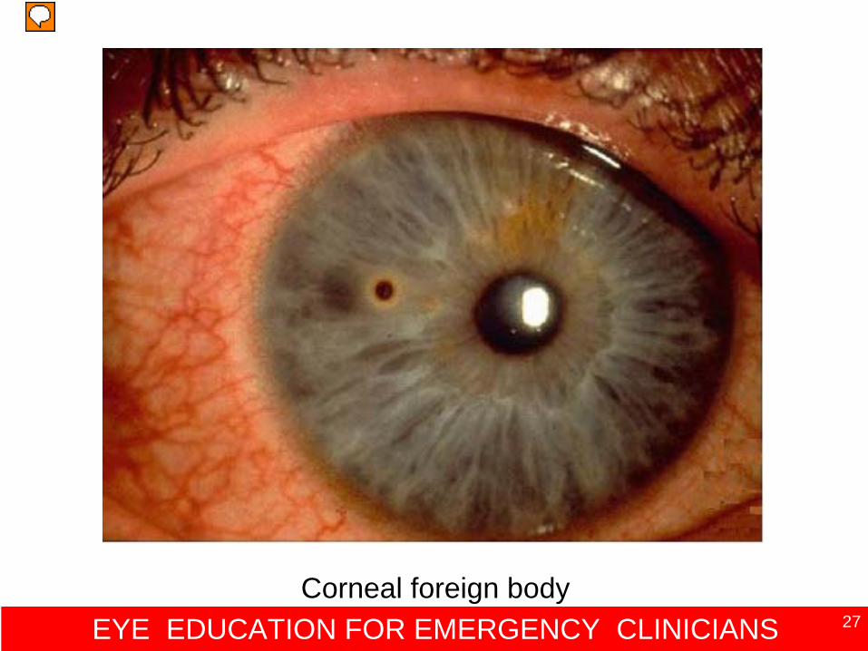
Corneal foreign body
EYE EDUCATION FOR EMERGENCY CLINICIANS 28
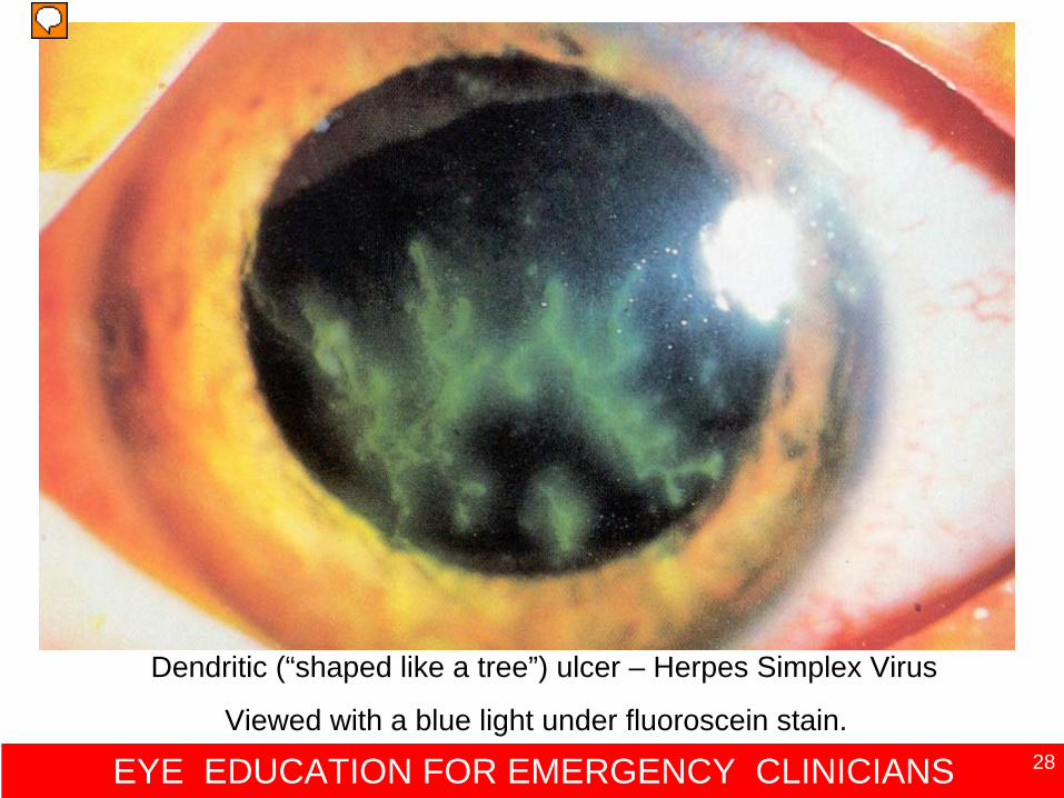
Dendritic (“shaped like a tree”) ulcer – Herpes Simplex Virus
Viewed with a blue light under fluoroscein stain.
EYE EDUCATION FOR EMERGENCY CLINICIANS 29
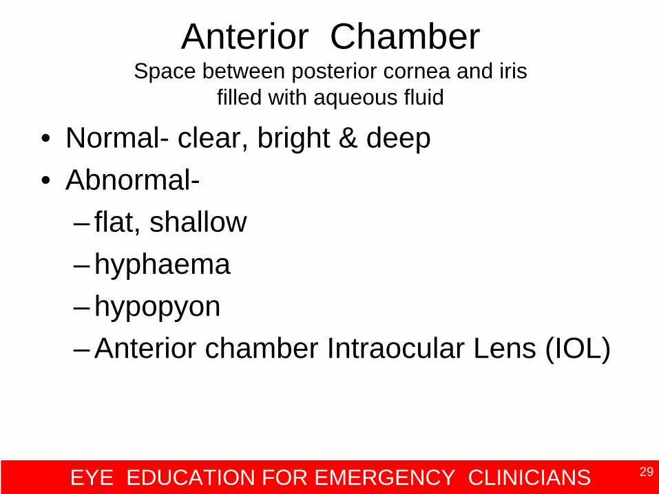
Anterior Chamber Space between posterior cornea and iris
filled with aqueous fluid
• Normal- clear, bright & deep• Abnormal-
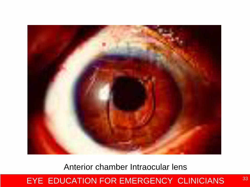
– flat, shallow– hyphaema– hypopyon– Anterior chamber Intraocular Lens (IOL)
EYE EDUCATION FOR EMERGENCY CLINICIANS 30
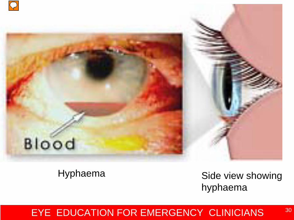
Hyphaema Side view showing hyphaema
EYE EDUCATION FOR EMERGENCY CLINICIANS 31
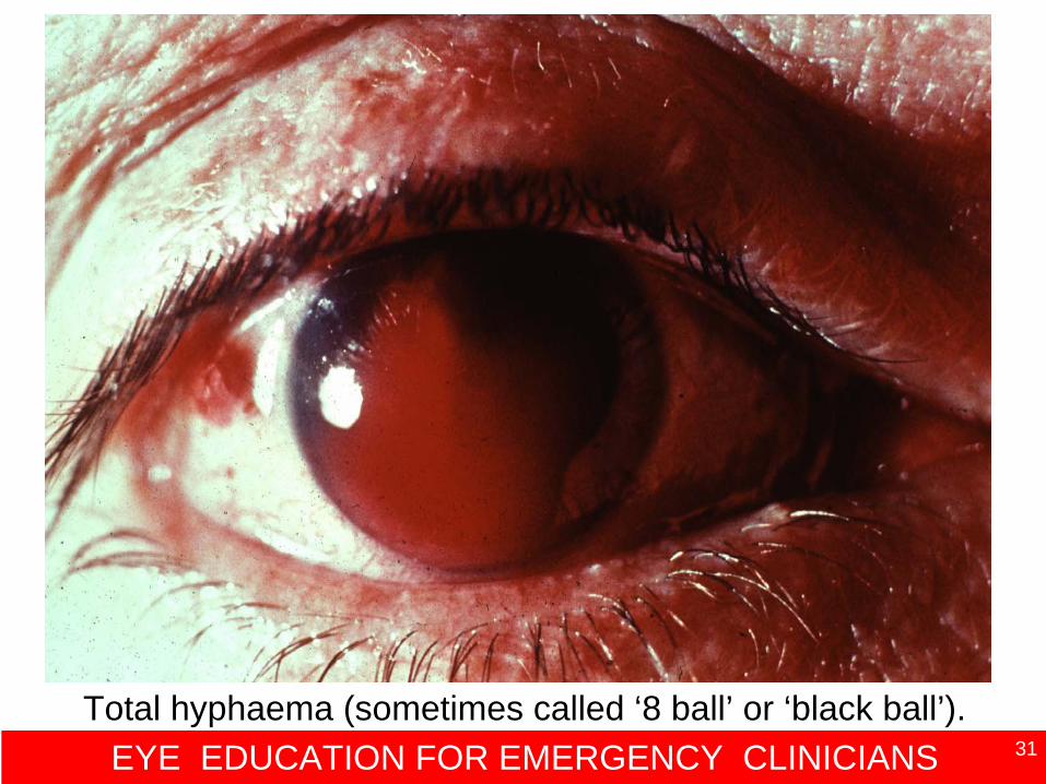
Total hyphaema (sometimes called ‘8 ball’ or ‘black ball’).
EYE EDUCATION FOR EMERGENCY CLINICIANS 32
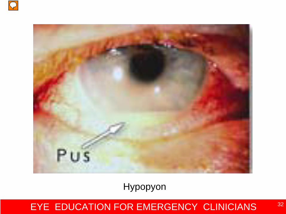
Hypopyon
EYE EDUCATION FOR EMERGENCY CLINICIANS 33
Anterior chamber Intraocular lens
EYE EDUCATION FOR EMERGENCY CLINICIANS 34
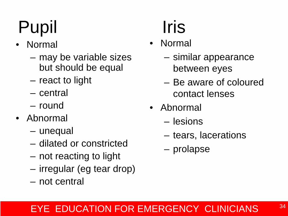
Pupil Iris• Normal
– may be variable sizes but should be equal
– react to light– central – round
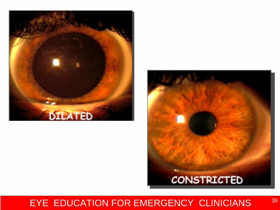
• Abnormal – unequal– dilated or constricted– not reacting to light– irregular (eg tear drop) – not central
• Normal– similar appearance
between eyes– Be aware of coloured
contact lenses• Abnormal
– lesions– tears, lacerations– prolapse
EYE EDUCATION FOR EMERGENCY CLINICIANS 35
EYE EDUCATION FOR EMERGENCY CLINICIANS 36
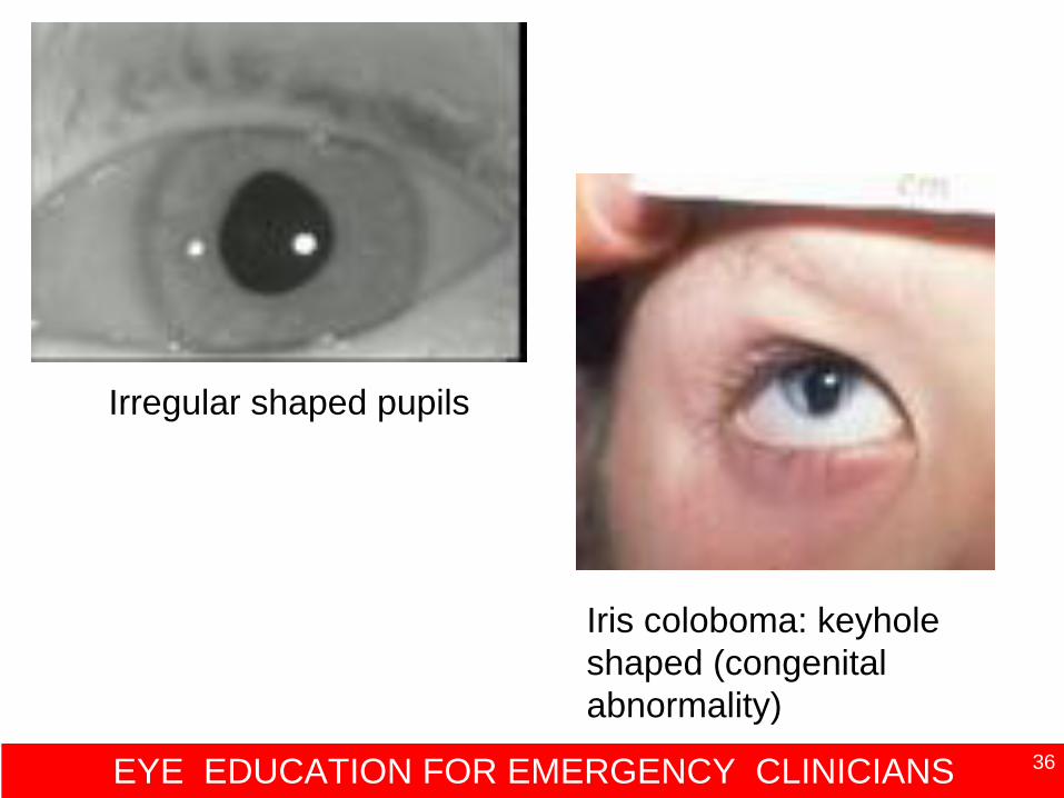
Irregular shaped pupils
Iris coloboma: keyhole shaped (congenital abnormality)
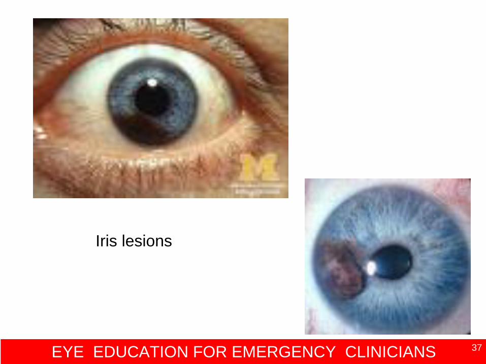
EYE EDUCATION FOR EMERGENCY CLINICIANS 37
Iris lesions
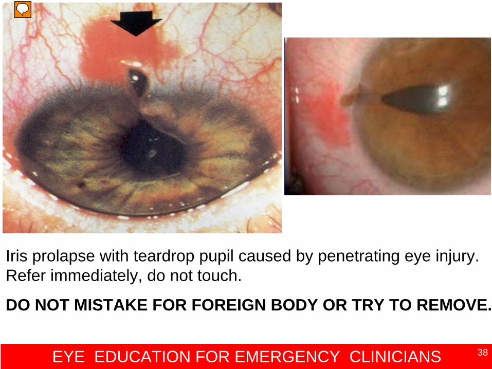
EYE EDUCATION FOR EMERGENCY CLINICIANS 38
Iris prolapse with teardrop pupil caused by penetrating eye injury. Refer immediately, do not touch.
DO NOT MISTAKE FOR FOREIGN BODY OR TRY TO REMOVE.
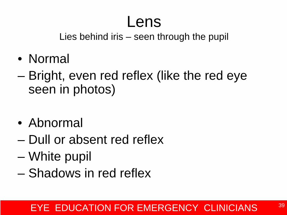
EYE EDUCATION FOR EMERGENCY CLINICIANS 39
Lens Lies behind iris – seen through the pupil
• Normal– Bright, even red reflex (like the red eye
seen in photos)
• Abnormal– Dull or absent red reflex– White pupil– Shadows in red reflex
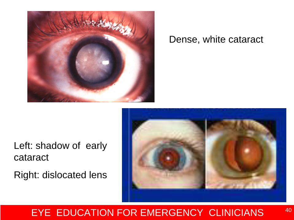
EYE EDUCATION FOR EMERGENCY CLINICIANS 40
Dense, white cataract
Left: shadow of early cataract
Right: dislocated lens
EYE EDUCATION FOR EMERGENCY CLINICIANS 41
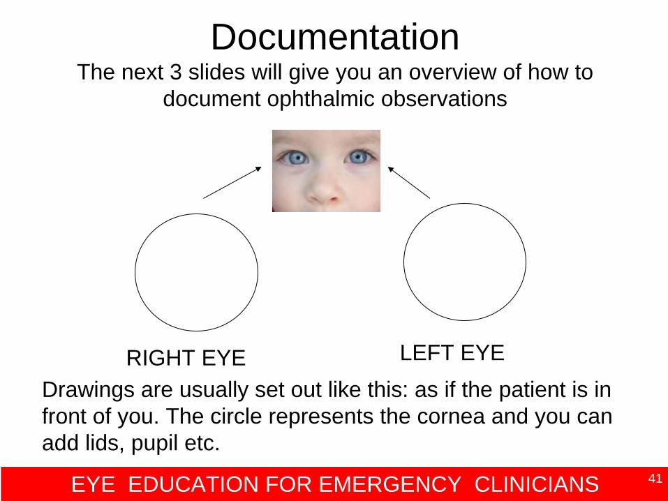
Documentation The next 3 slides will give you an overview of how to
document ophthalmic observations
RIGHT EYE LEFT EYEDrawings are usually set out like this: as if the patient is in front of you. The circle represents the cornea and you can add lids, pupil etc.
EYE EDUCATION FOR EMERGENCY CLINICIANS 42
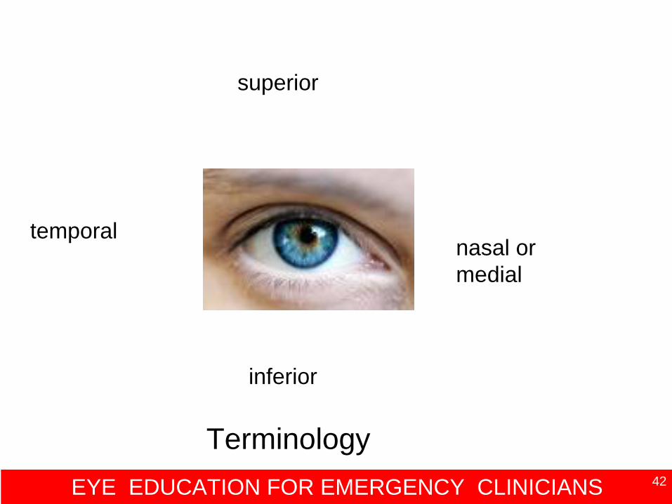
nasal or medial
temporal
superior
inferior
Terminology
EYE EDUCATION FOR EMERGENCY CLINICIANS 43
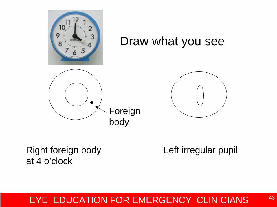
Foreign body
Right foreign body at 4 o’clock
Left irregular pupil
Draw what you see
EYE EDUCATION FOR EMERGENCY CLINICIANS 44
On completion of this session you will now be able to:
• Recognise normal and abnormal eye anatomy
• Perform a systematic eye examination• Correctly document examination
findings


![[Www.shamsology.net]Eye Essentials Routine Eye Examination](https://img.pdfslide.us/doc/110x75/553530504a79599c778b468b/wwwshamsologyneteye-essentials-routine-eye-examination.jpg)