Embed Size (px)
Citation preview
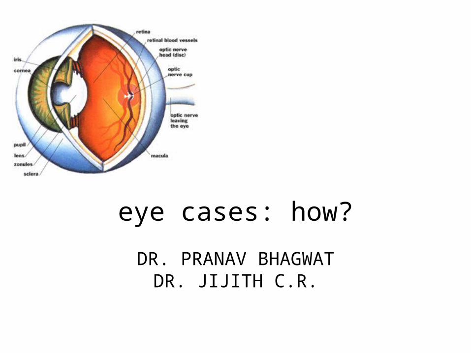
eye cases: how?
DR. PRANAV BHAGWATDR. JIJITH C.R.
HISTORY TAKING
– Allow patient to tell story.
Decreased vision
Ask for
• Onset
• Duration
• Uni / Bilateral
• Distant or near vision
• Whether the patient wears glasses
• Diurnal variation
Sudden unilateral loss of vision
• Acute congestive glaucoma• Acute optic neuritis• Acute iridocyclitis• Retinal detachment• Central retinal artery occlusion• Spasm of retinal artery• Vitreous haemorrhage• Injuries
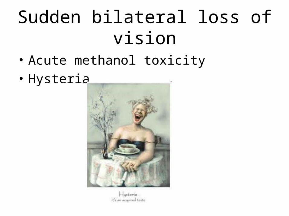
Sudden bilateral loss of vision
• Acute methanol toxicity
• Hysteria
Reduced vision in the morning, improved in afternoon
• Intermittent corneal edema
Gradual onset loss of vision
• Cataract• Refractory errors• Retinopathy-DM, HT• Retinal degenerations
and RP• Chronic iridocyclitis• Chronic simple
glaucoma
• Keratitis• K opacities• Chorio retinitis• Chronic optic neuritis• MS• Drug toxicity
Pain in the eye
• Type
• Onset
• Duration
• Diurnal variation
• Associated complaints e.g., nausea, vomiting, DV

Severe eye pain
• Adhimantha
• K abrasions and ulcers
• Acute iridocyclitis
• Panopthalmitis
• Acute glaucoma
• Scleritis
• Episcleritis
Dull Aching pain
• Cases with eye strain
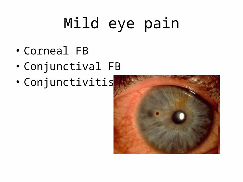
Mild eye pain
• Corneal FB
• Conjunctival FB
• Conjunctivitis
Pain around the eye
• Pathology of the lid and lacrimal apparatus (eg: stye)
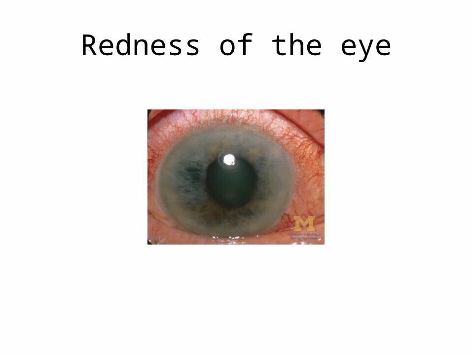
Redness of the eye
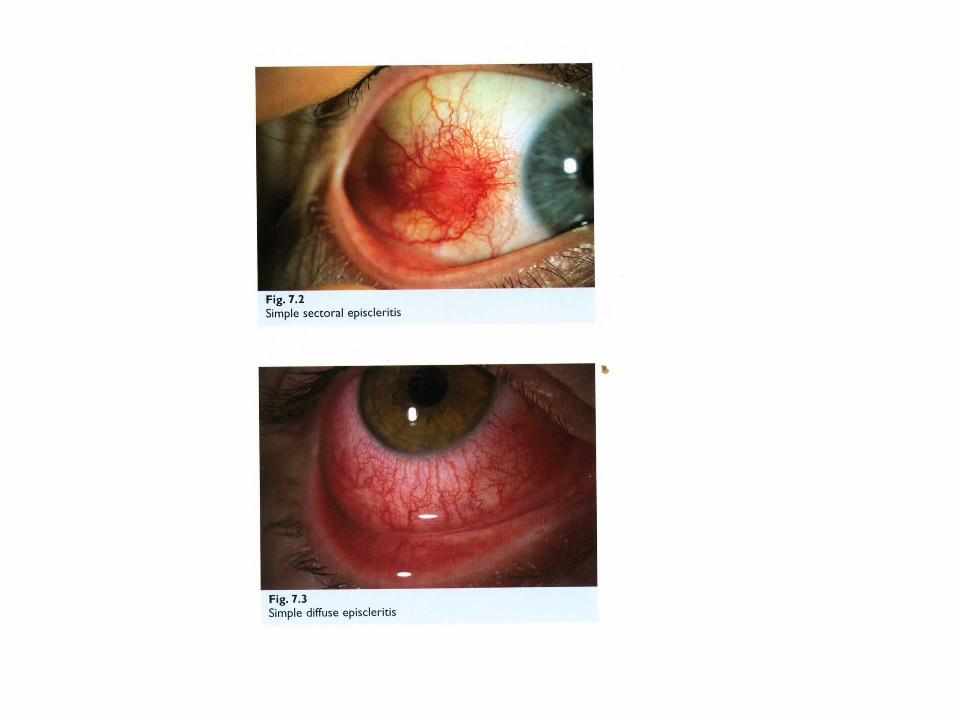
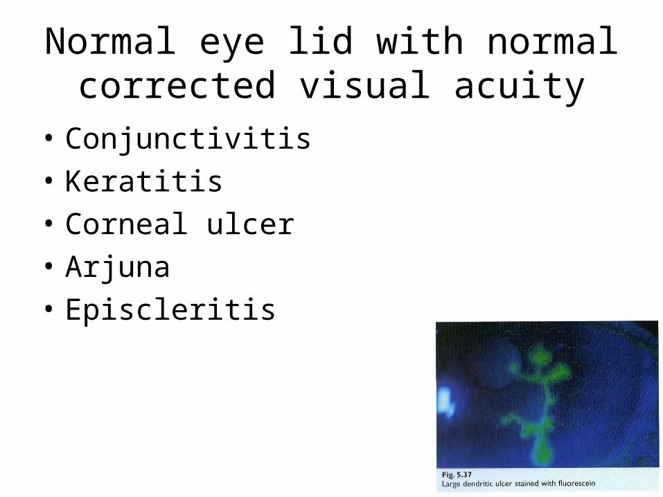
Normal eye lid with normal corrected visual acuity
• Conjunctivitis
• Keratitis
• Corneal ulcer
• Arjuna
• Episcleritis
Abnormal eye lid with normal corrected visual acuity
• Ectropion
• Entropion
• Stye
• Blepharitis
Reduced corrected visual acuity without diplopia
• Iritis
• ACG
• K foreign body
• K ulcer
Reduced corrected visual acuity with diplopia
• Carvernos sinus thrombosis
• Orbital cellulitis
• Caratico carvernos fistula
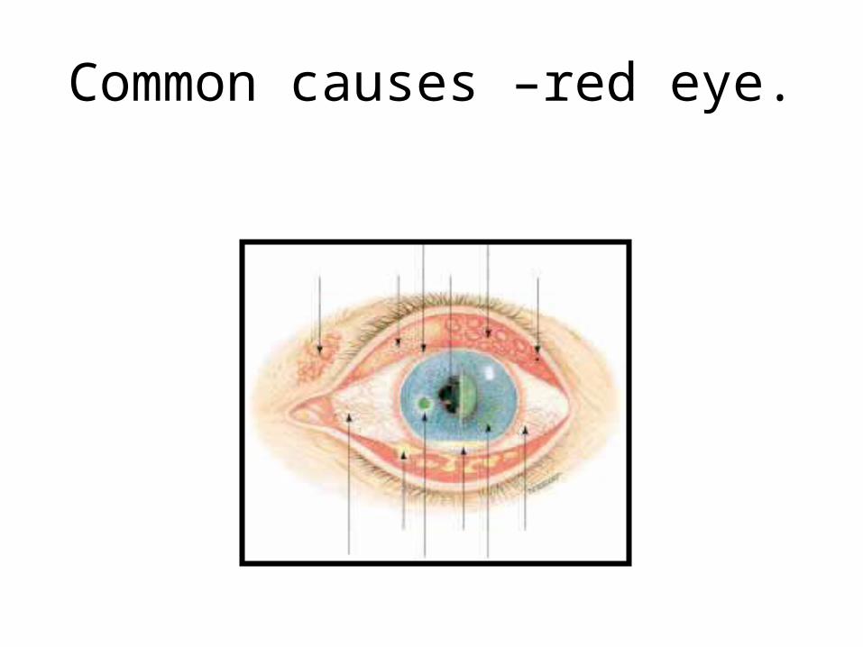
Common causes –red eye.
Photophobia
• Acute infective or inflammatory lesion of the anterior segment
• Recently operated eyes
Watering of the eyes
1)Excessive lacrimation• Keratitis• Uveitis• Glaucoma
2) Epiphora• Mechanical obstruction to drainage- stricture,
punctal stenosis & chronic dacryocistitis• Defective orbicularis action
Discharge
• Watery-Mild bacterial infection
• Serous -viral
• Mucoid -mild conjunctivitis
• Mucopurulent & purulent- acute pyogenic infection of anterior segment
• Serosanguinous- opthalmia neonatrum
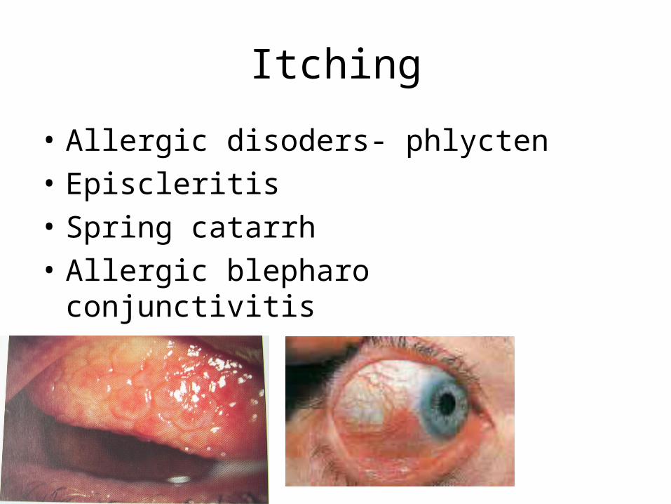
Itching
• Allergic disoders- phlycten
• Episcleritis
• Spring catarrh
• Allergic blepharo conjunctivitis

Netra daaha
• Pittotklishta
• Pittaja abhishyanda
• Pitta vidagdha drishti.
Foreign body sensation
• Presence of FB
• Distorted eye lashes- trichiasis, entropion
• Conjunctival concretion, calcification
• Contact lenses
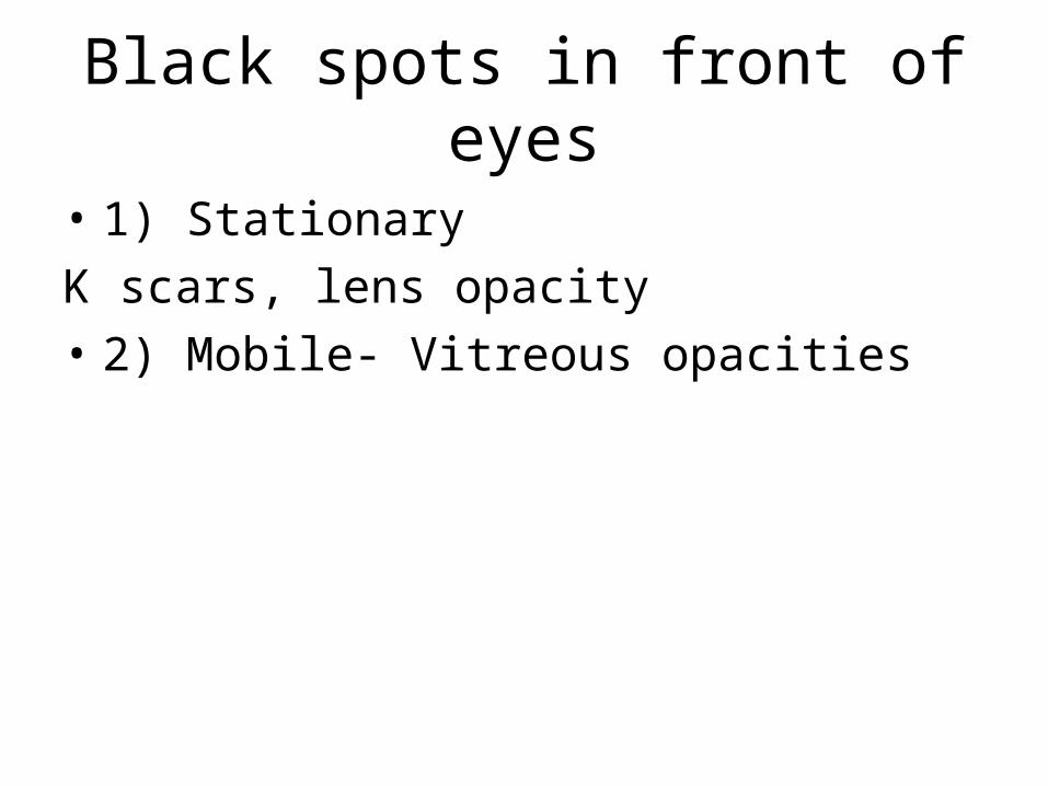
Black spots in front of eyes
• 1) Stationary
K scars, lens opacity
• 2) Mobile- Vitreous opacities
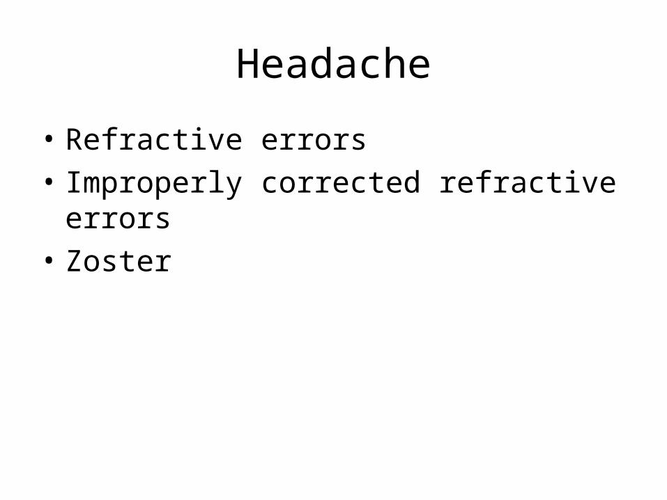
Headache
• Refractive errors
• Improperly corrected refractive errors
• Zoster
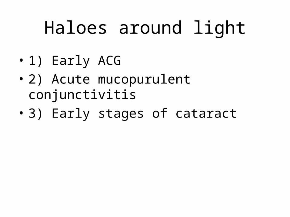
Haloes around light
• 1) Early ACG
• 2) Acute mucopurulent conjunctivitis
• 3) Early stages of cataract

Photopsiae
• Irritative lesions of retina
• Impending RD

Diplopia
• 1) Unioccular- high K astigmatism, subluxated or dislocated lens
• 2) Binocular- Squint
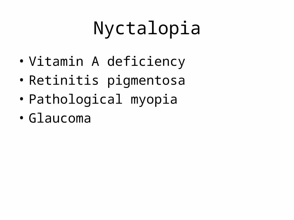
Nyctalopia
• Vitamin A deficiency
• Retinitis pigmentosa
• Pathological myopia
• Glaucoma

Occupation
• Welders
• Black smiths- foreign body in the eye
• Farmers- fungal keratitis
Medication
• 1) Gentamicin, miotics, Atropin- follicular response
• 2) Topical anasthetics for long time- severe corneal reactions
• 3) Topical and systemic steroids- K disease, cataract, glaucoma
• 4) Thiomersal- allergic conjunctivitis, epithelial Keratitis
• 5) Benzalkonium- toxic papillary reaction
Past history
• Systemic diseases- diabetis mellitus
• Arjuna-HT
• Iritis- ankylosing spondylitis
History of previous ocular disease
• Childhood squint- lazy eye
• Blunt injury- traumatic mydriasis ( could be confused with partial third nerve palsy)
Family history
• Chronic glaucoma- the incidence nearly 5 times greater in siblings and children of affected patients
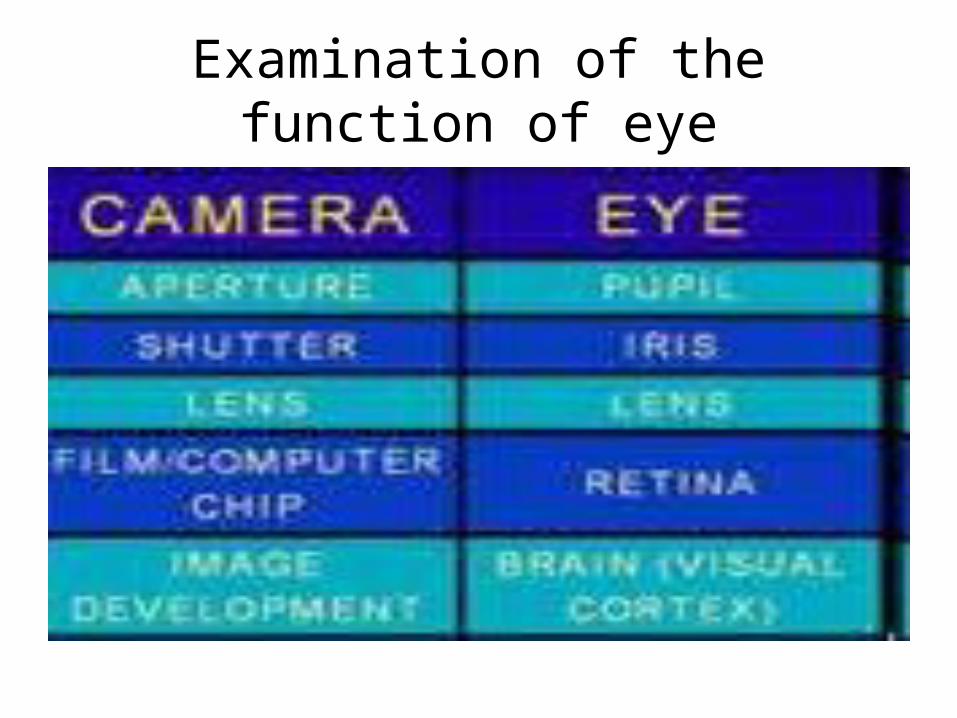
Examination of the function of eye
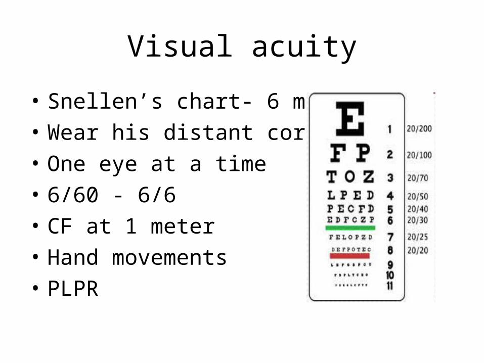
Visual acuity
• Snellen’s chart- 6 mtrs
• Wear his distant corretion
• One eye at a time
• 6/60 - 6/6
• CF at 1 meter
• Hand movements
• PLPR

• Jaeger’s test type
• N5 to N48

Visual field
• 1) Peripheral field- confrontation or perimeter
• 2) Central field by scotometery
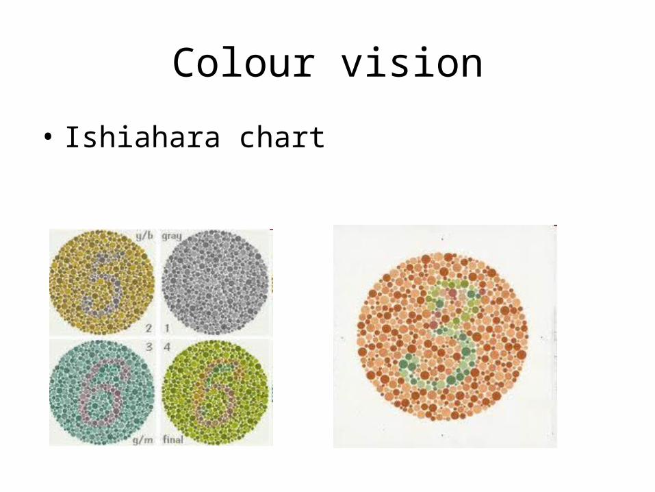
Colour vision
• Ishiahara chart
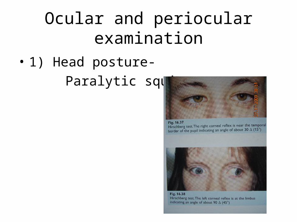
Ocular and periocular examination
• 1) Head posture-
Paralytic squint.
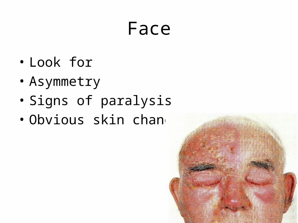
Face
• Look for
• Asymmetry
• Signs of paralysis
• Obvious skin changes
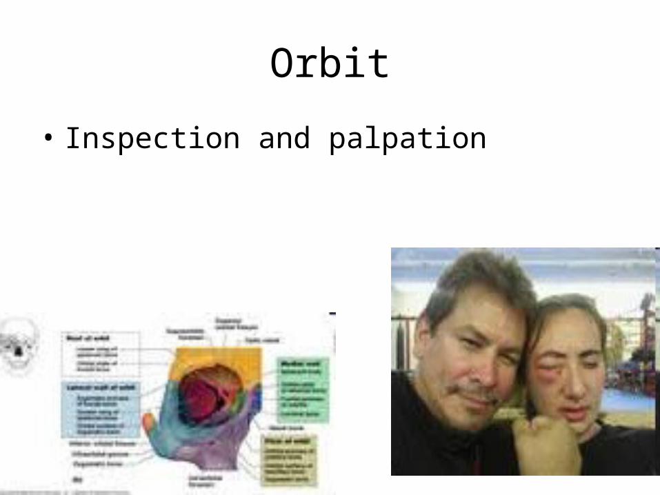
Orbit
• Inspection and palpation
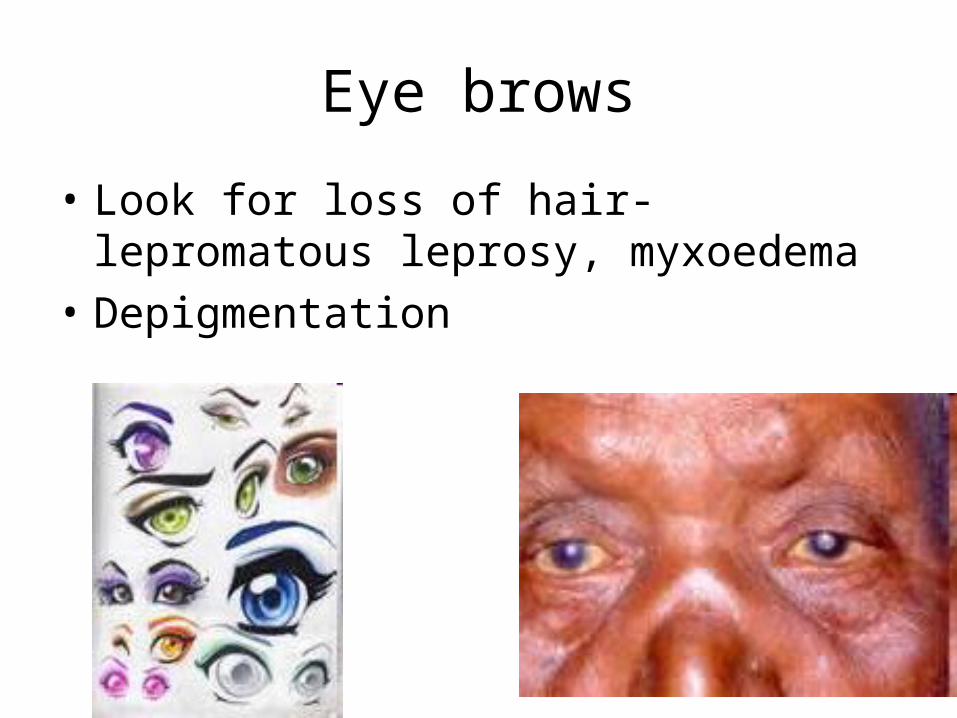
Eye brows
• Look for loss of hair- lepromatous leprosy, myxoedema
• Depigmentation
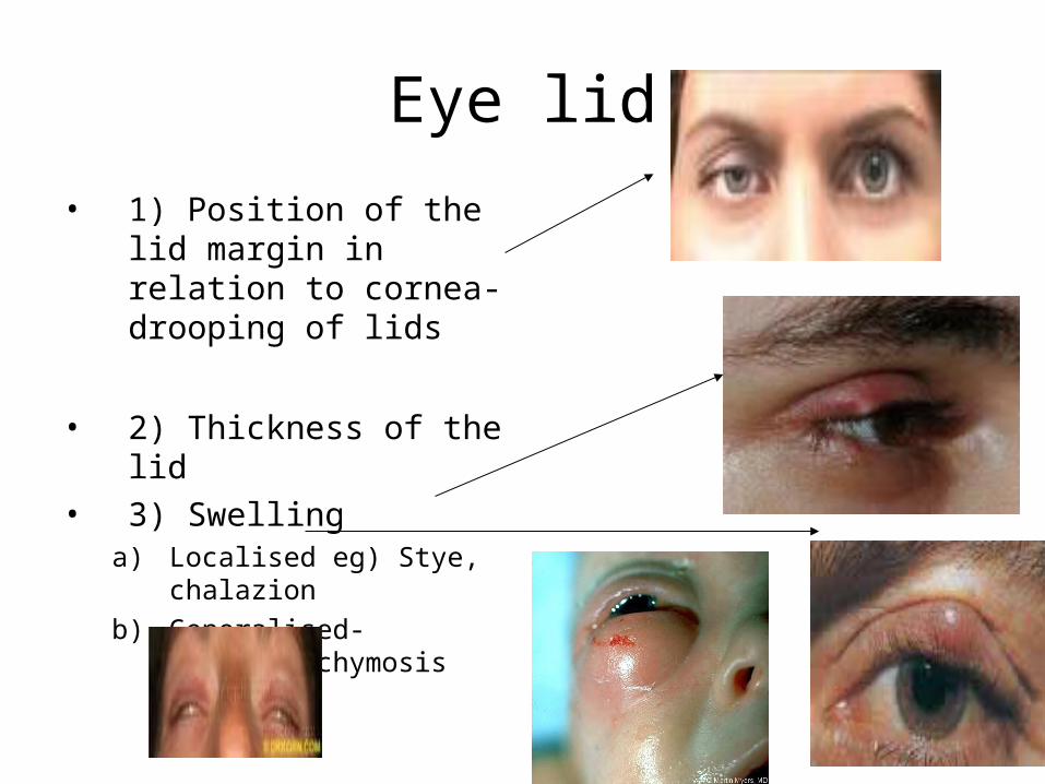
Eye lid
• 1) Position of the lid margin in relation to cornea- drooping of lids
• 2) Thickness of the lid• 3) Swelling
a) Localised eg) Stye, chalazion
b) Generalised- Oedema,ecchymosis
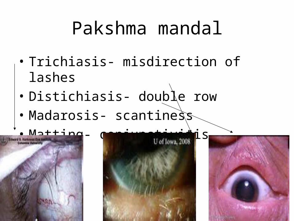
Pakshma mandal
• Trichiasis- misdirection of lashes
• Distichiasis- double row
• Madarosis- scantiness
• Matting- conjunctivitis
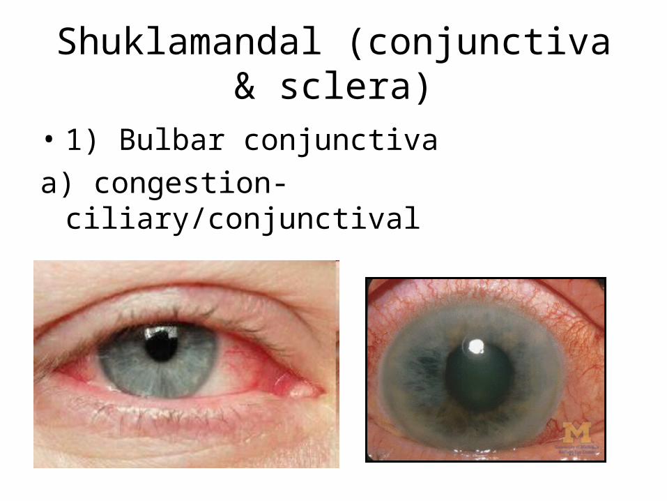
Shuklamandal (conjunctiva & sclera)
• 1) Bulbar conjunctiva
a) congestion- ciliary/conjunctival

b) Chemosis-
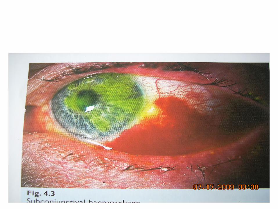
c) Subconjunctival haemorrhage
d) Pigmentation
e) Nodule

Upper tarsal conjunctiva
• Congestion
• Alteration of normal vertical vascular pattern
• Follicle/papilla
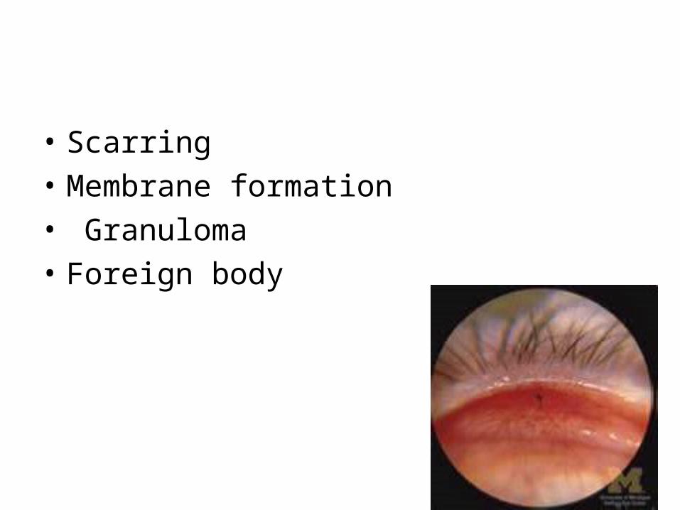
• Scarring
• Membrane formation
• Granuloma
• Foreign body
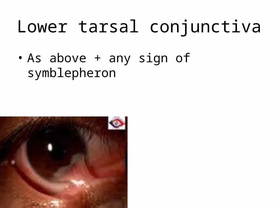
Lower tarsal conjunctiva
• As above + any sign of symblepheron
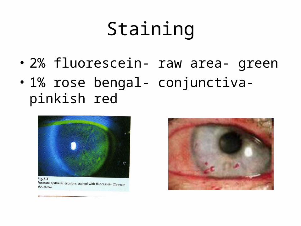
Staining
• 2% fluorescein- raw area- green
• 1% rose bengal- conjunctiva- pinkish red

Sclera
• Colour change • Pigmentation• Protrusion of uveal
tissue• Congestion
• Nodule formation
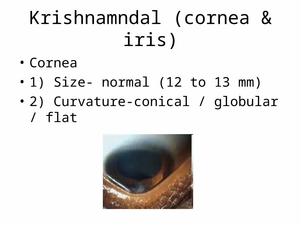
Krishnamndal (cornea & iris)
• Cornea
• 1) Size- normal (12 to 13 mm)
• 2) Curvature-conical / globular / flat
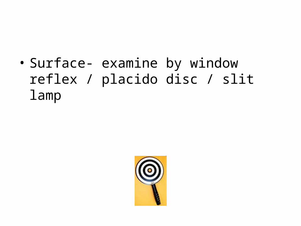
• Surface- examine by window reflex / placido disc / slit lamp
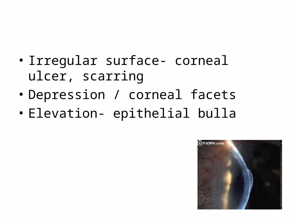
• Irregular surface- corneal ulcer, scarring
• Depression / corneal facets
• Elevation- epithelial bulla
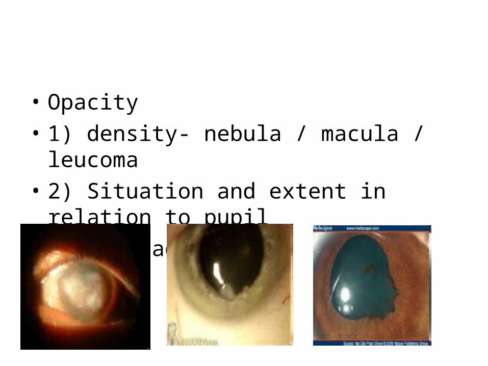
• Opacity
• 1) density- nebula / macula / leucoma
• 2) Situation and extent in relation to pupil
• 3) Iris adhesion
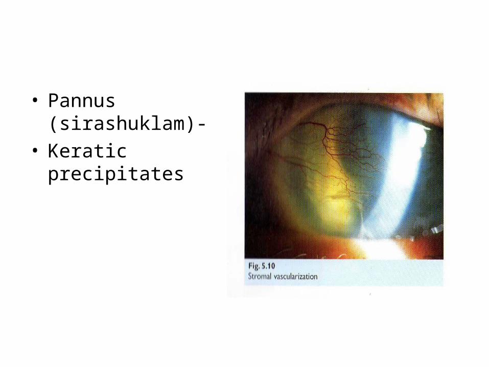
• Pannus (sirashuklam)-
• Keratic precipitates
• Corneal sensationsDiminished sensation- • Herpes• 5th nerve paralysis• ACG and absolute glaucoma• Leprosy• Prolonged use of contact lens• Post surgery• Local anaesthesia
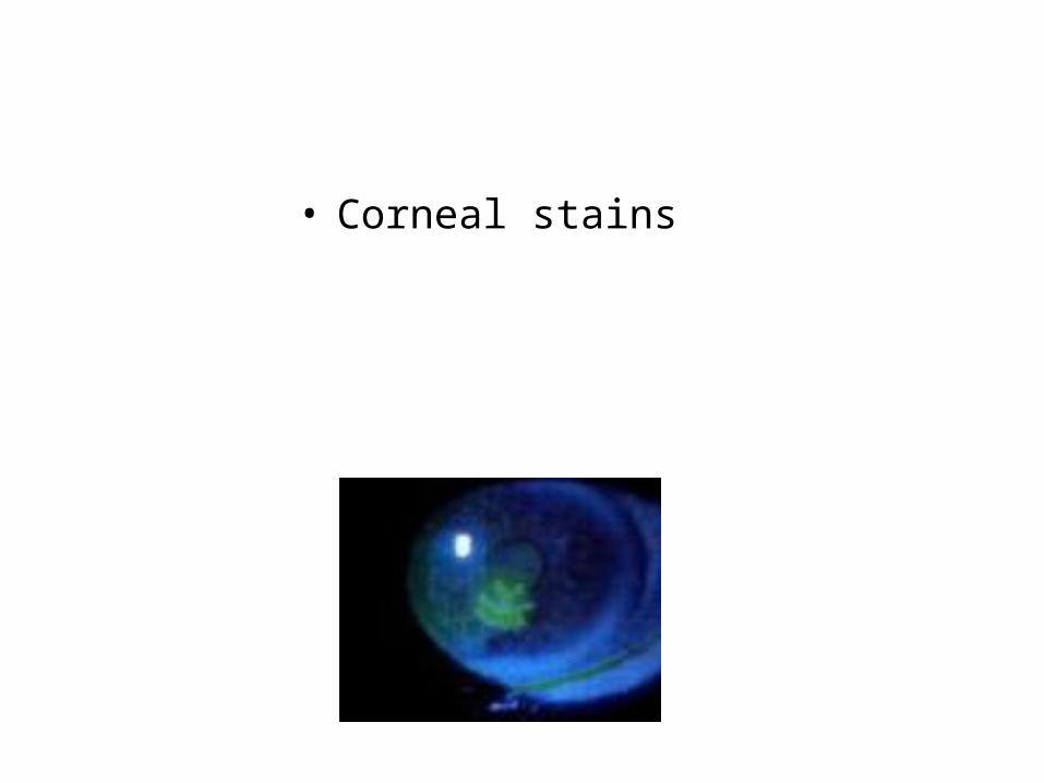
• Corneal stains
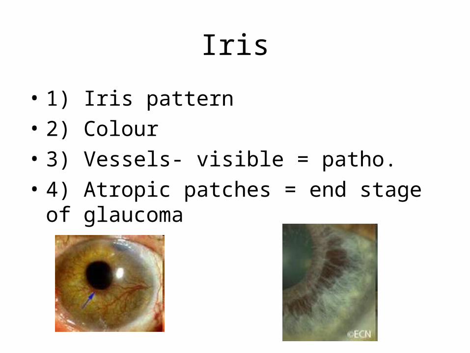
Iris
• 1) Iris pattern
• 2) Colour
• 3) Vessels- visible = patho.
• 4) Atropic patches = end stage of glaucoma
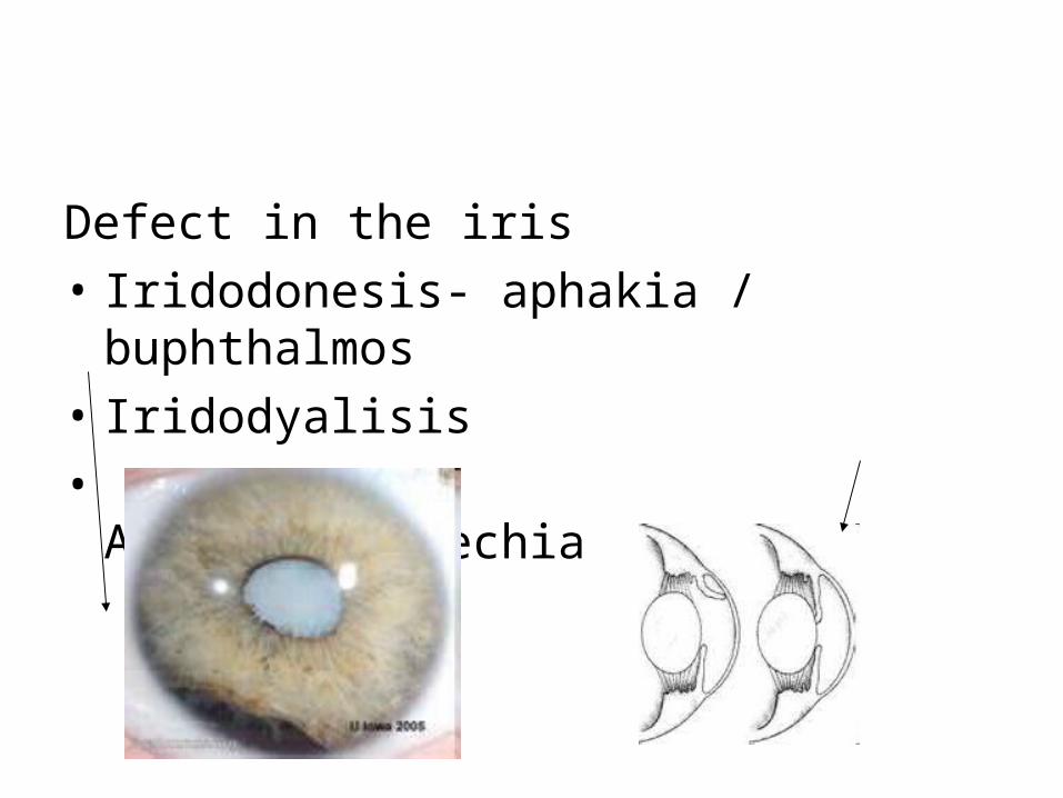
Defect in the iris
• Iridodonesis- aphakia / buphthalmos
• Iridodyalisis
• Anterior synechia
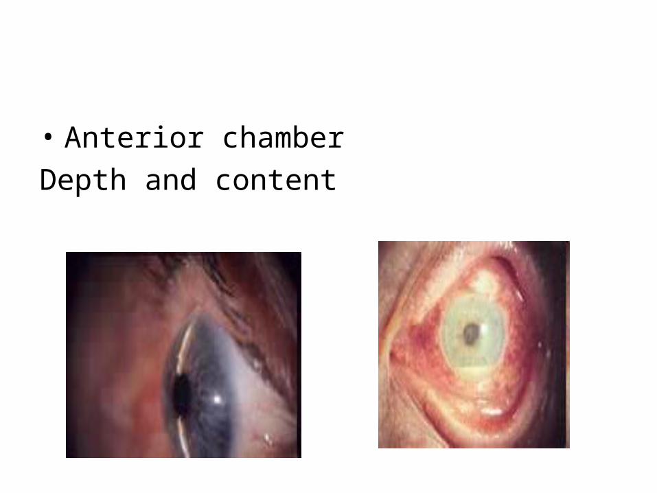
• Anterior chamber
Depth and content
Pupil
• Size- (3 to 4mm)
• Shape
• Position
• Pupillary margin
• Pupillary aperture
• Pupil reaction
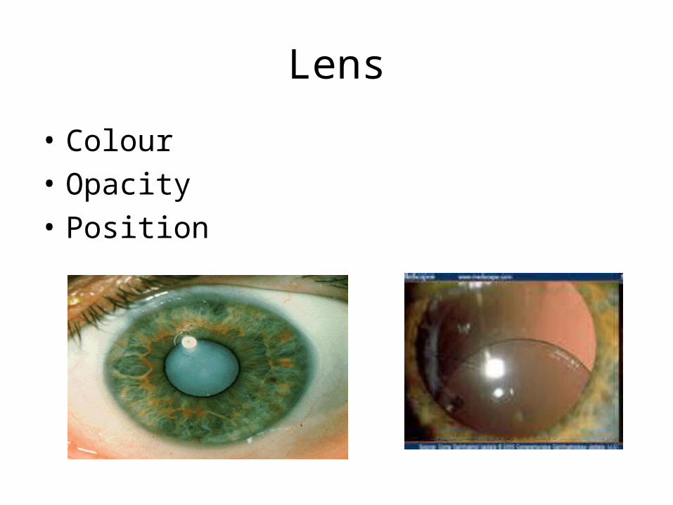
Lens
• Colour
• Opacity
• Position
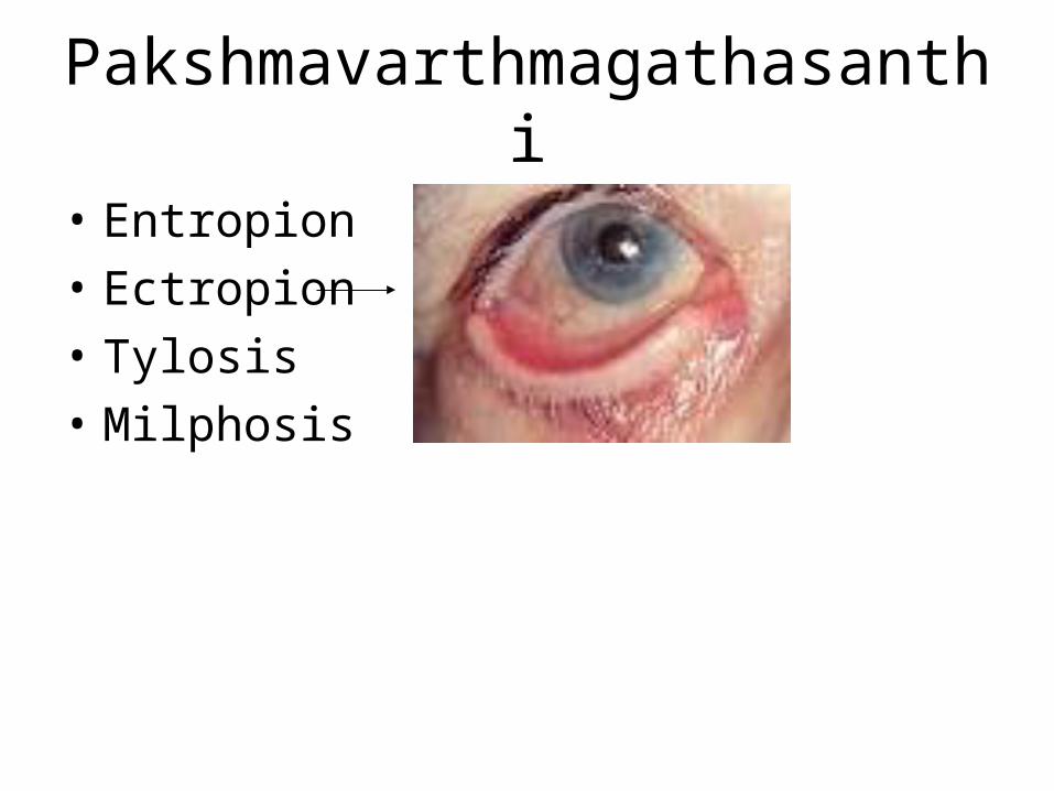
Pakshmavarthmagathasanthi
• Entropion
• Ectropion
• Tylosis
• Milphosis

Kaneenika sandhi
• Lacrimal puncta- eversion / stenosis / absence
• Skin around it
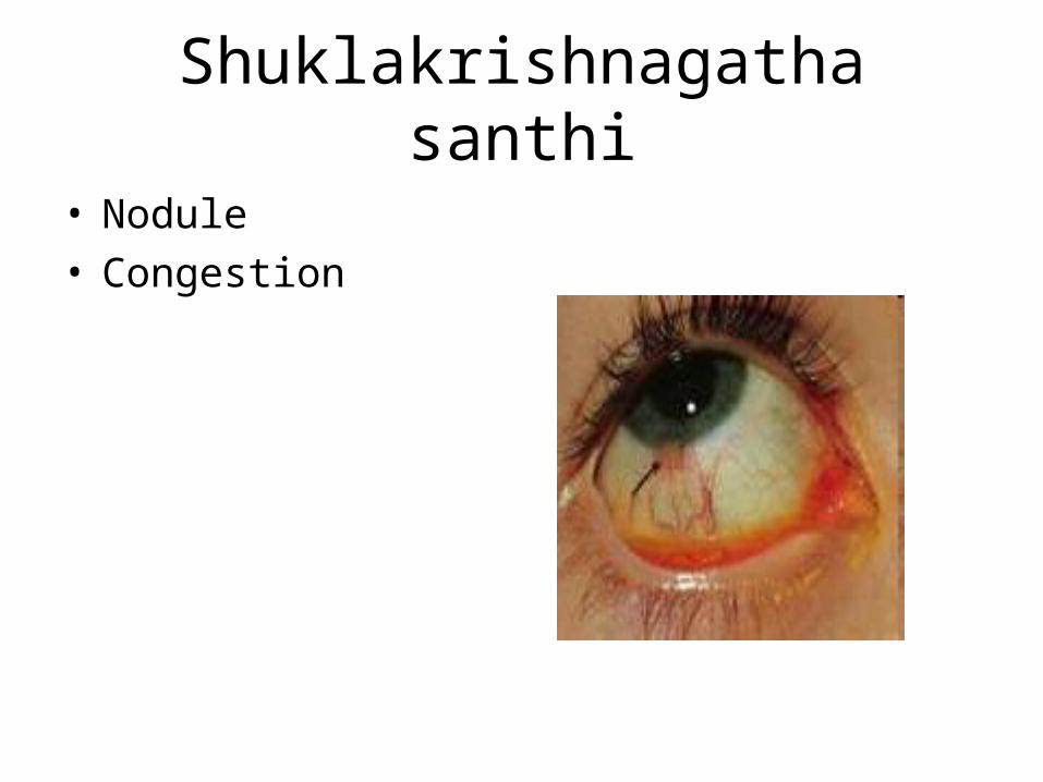
Shuklakrishnagatha santhi
• Nodule• Congestion
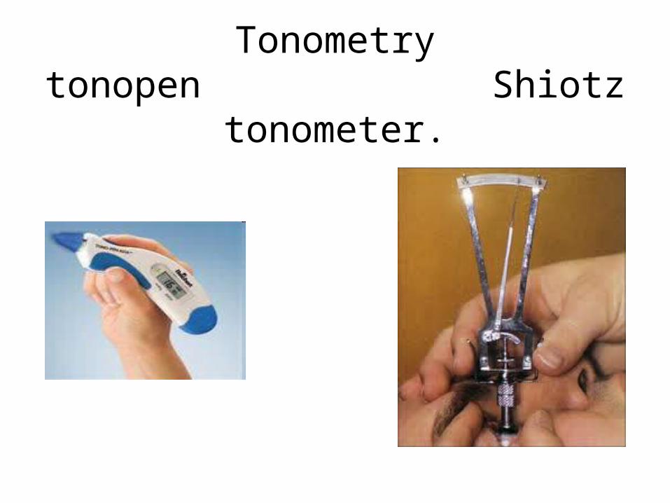
Tonometrytonopen Shiotz tonometer.
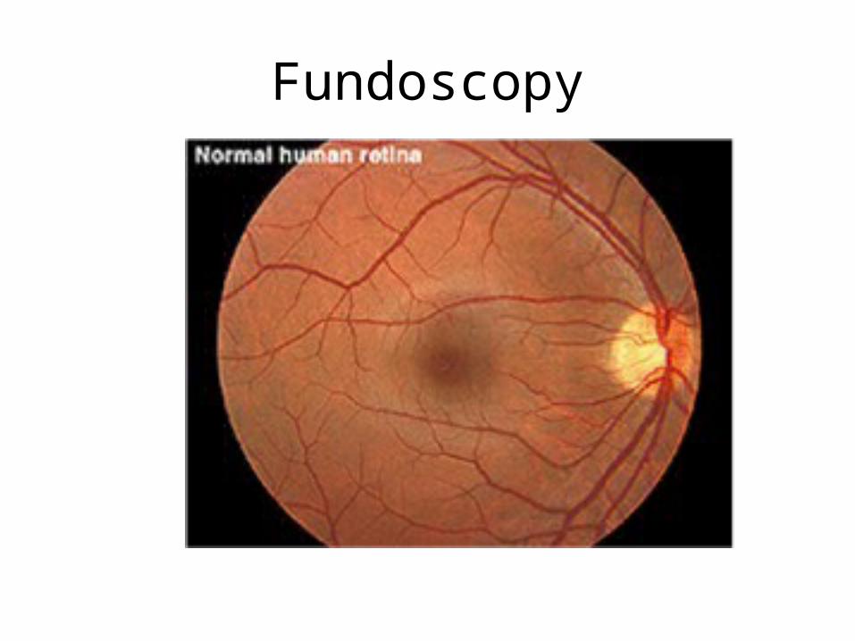
Fundoscopy
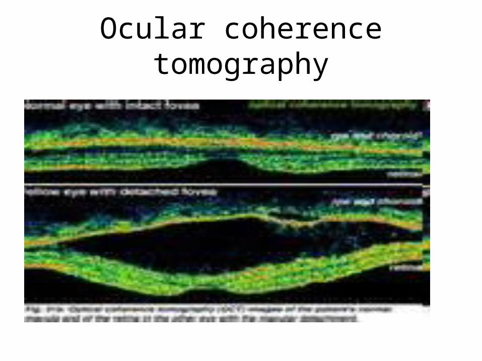
Ocular coherence tomography

• Thank you!