Embed Size (px)
Citation preview
Extraintestinal Manifestations of IBD
Hyun Kim, M.D. San Diego Digestive Disease Consultants
Associate Professor, UCSD School of Medicine
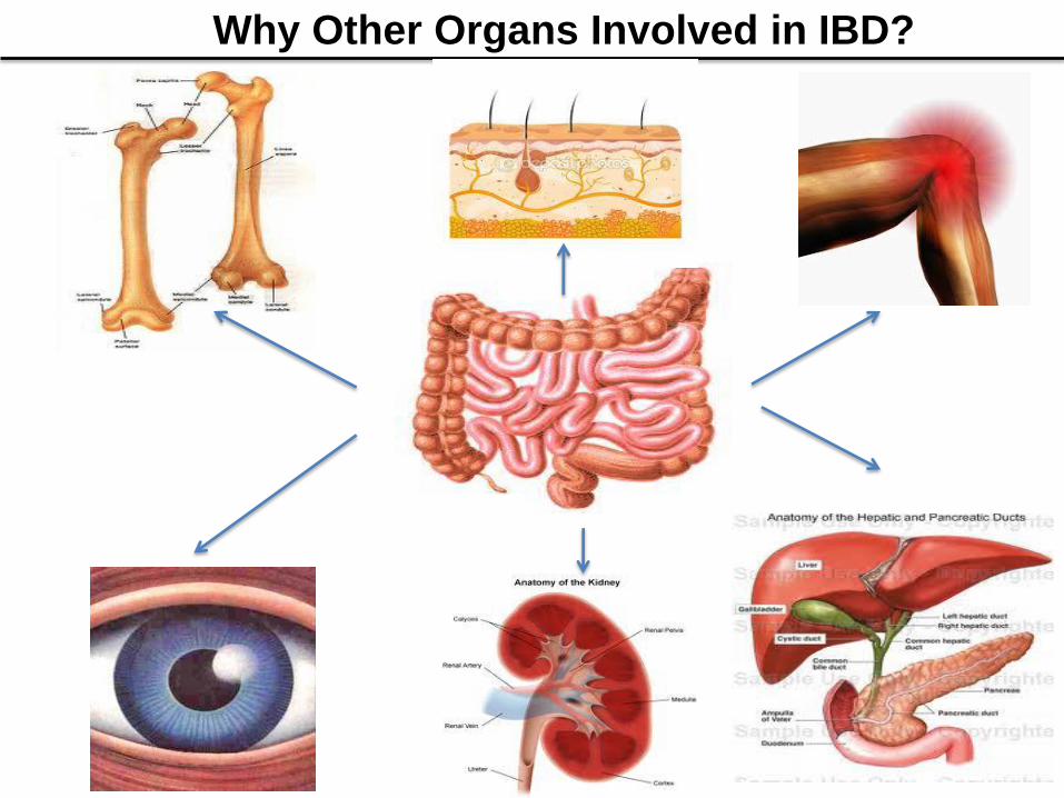
Why Other Organs Involved in IBD?
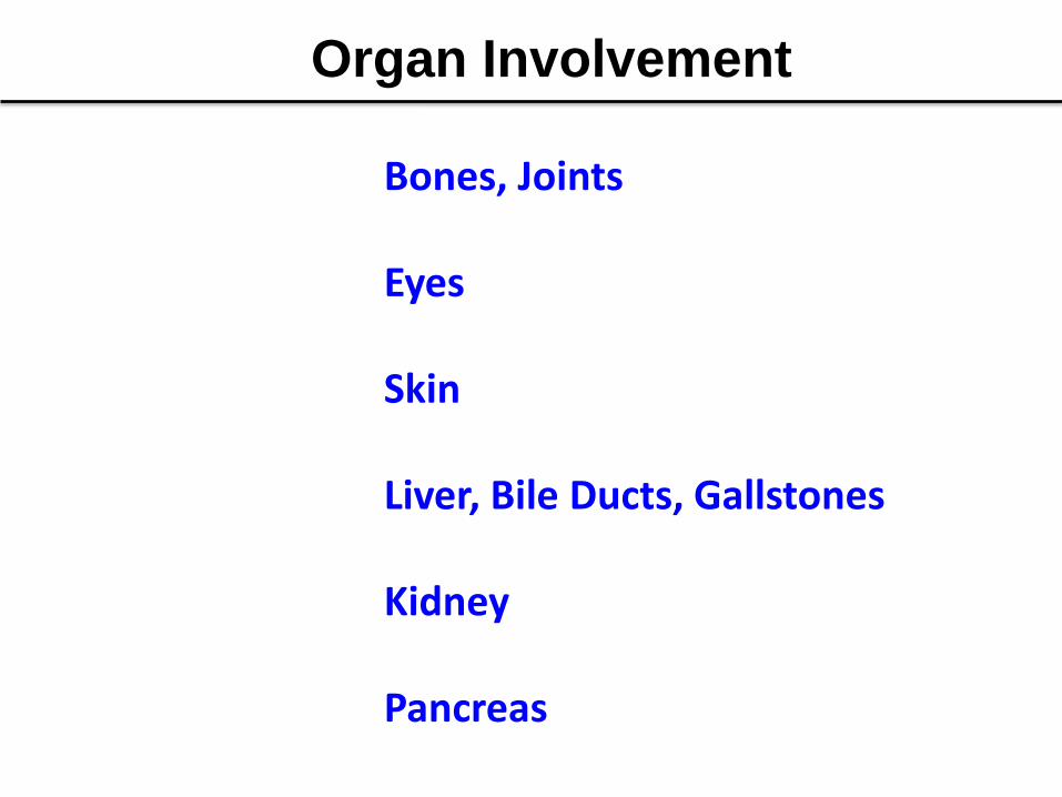
Organ Involvement
Bones, Joints Eyes Skin Liver, Bile Ducts, Gallstones Kidney Pancreas
Bone and Joint
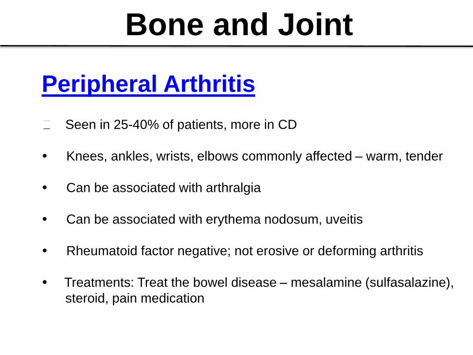
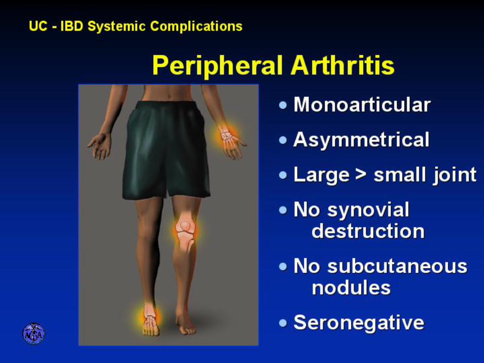
Peripheral Arthritis
Seen in 25-40% of patients, more in CD
Knees, ankles, wrists, elbows commonly affected – warm, tender
Can be associated with arthralgia
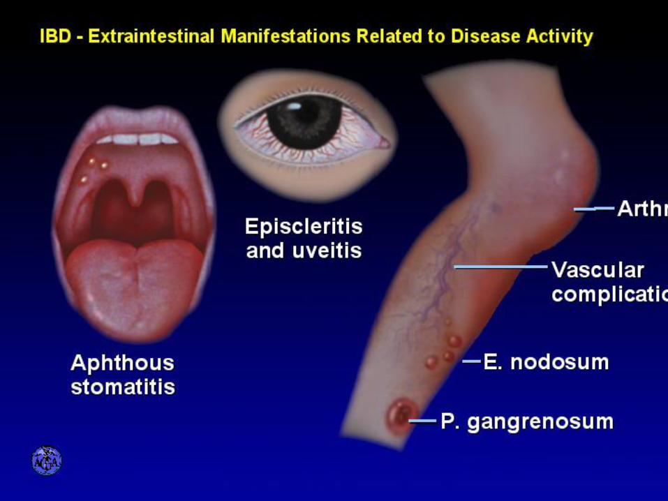
Can be associated with erythema nodosum, uveitis
Rheumatoid factor negative; not erosive or deforming arthritis
Treatments: Treat the bowel disease – mesalamine (sulfasalazine),
steroid, pain medication
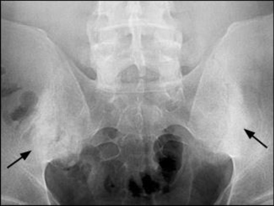
Axial Arthritis
Ankylosing spondylitis, Sacroiliitis – low back pain, pelvic
bone pain, spine pain
HLA-B27 +
Bone inflammation can lead to bone fusion and skeletal
deformity
Usually need aggressive treatment for IBD, including
TNF Ab, pain management, steroid joint injection
May not be associated with bowel inflammation
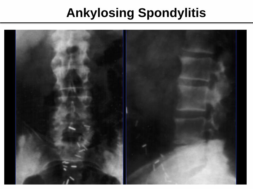
Ankylosing Spondylitis
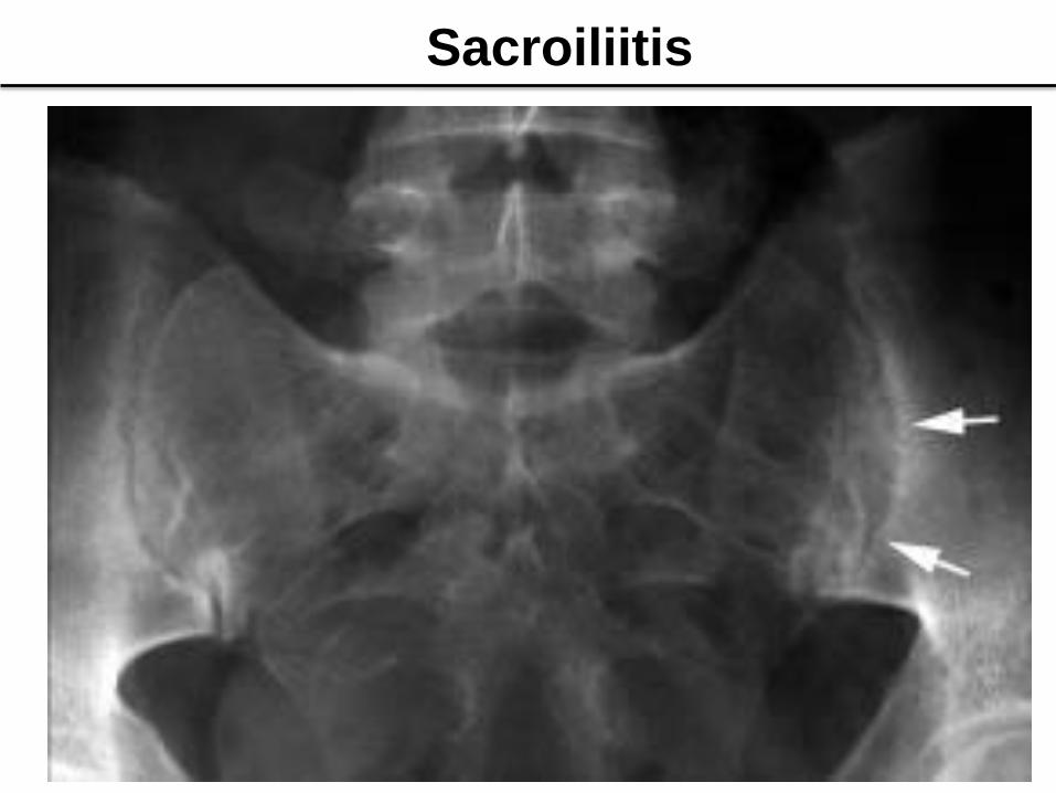
Sacroiliitis
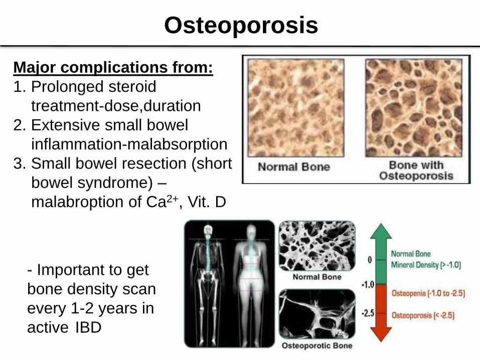
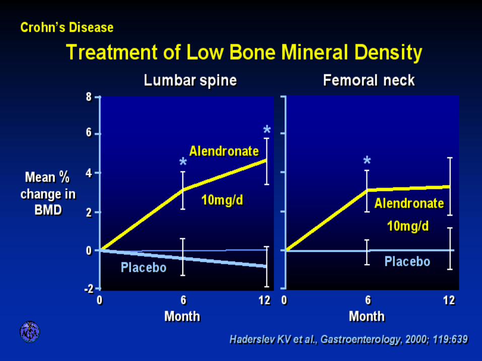
Osteoporosis
Major complications from:
1. Prolonged steroid
treatment-dose,duration
2. Extensive small bowel
inflammation-malabsorption
3. Small bowel resection (short
bowel syndrome) –
malabroption of Ca2+, Vit. D
- Important to get
bone density scan
every 1-2 years in
active IBD
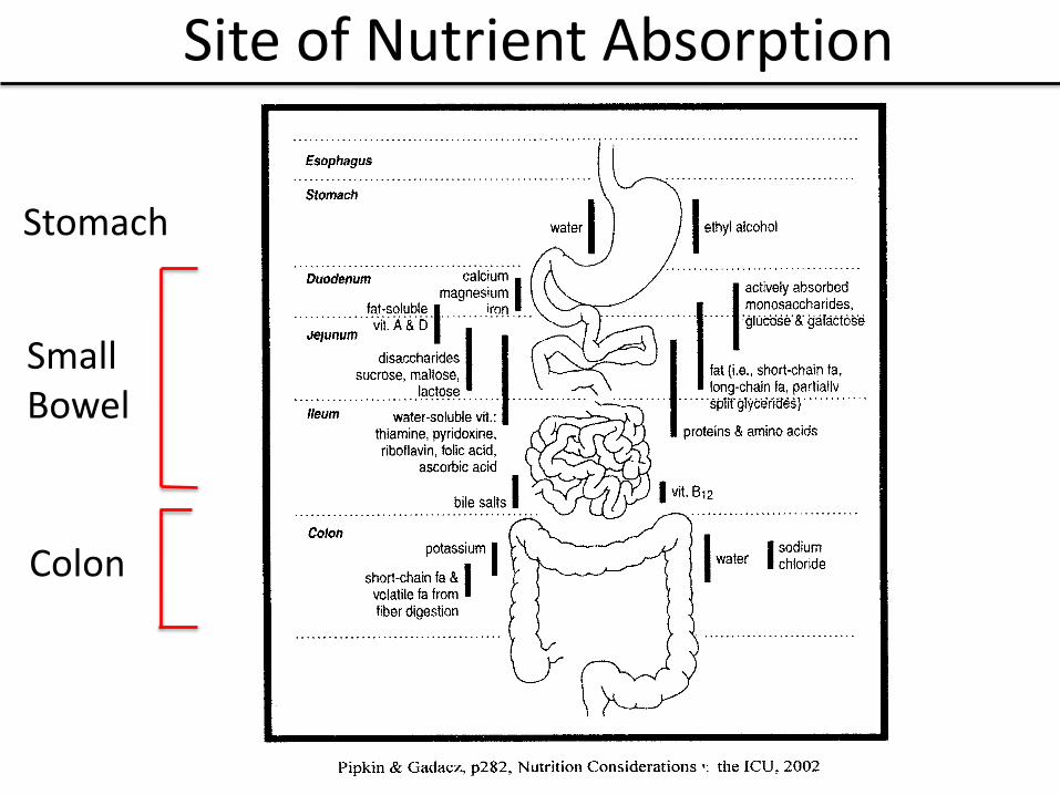
Site of Nutrient Absorption
Small Bowel
Stomach
Colon
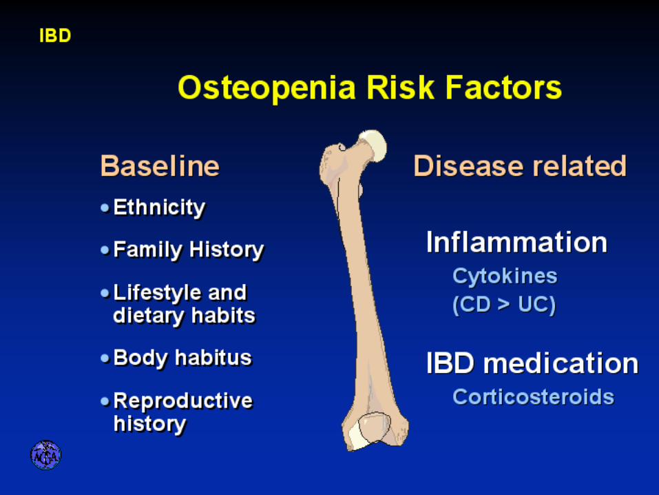
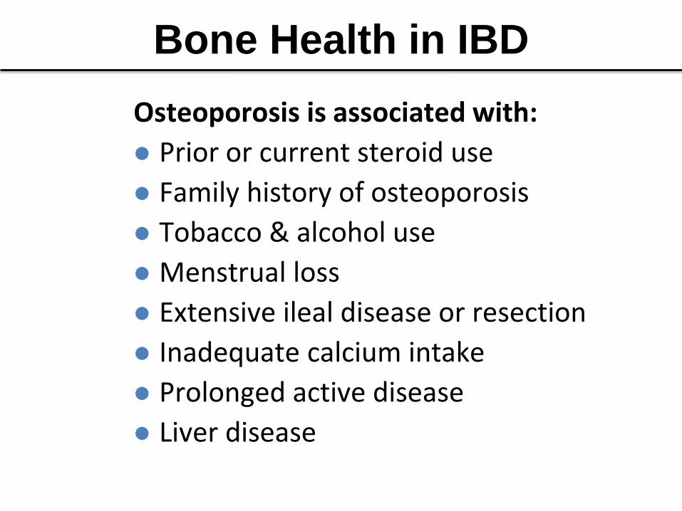
Bone Health in IBD
Osteoporosis is associated with:
Prior or current steroid use
Family history of osteoporosis
Tobacco & alcohol use
Menstrual loss
Extensive ileal disease or resection
Inadequate calcium intake
Prolonged active disease
Liver disease
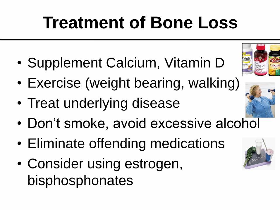
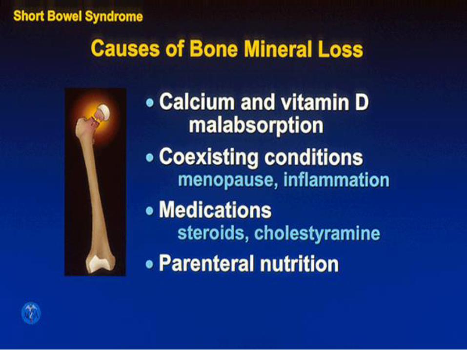
Treatment of Bone Loss
• Supplement Calcium, Vitamin D
• Exercise (weight bearing, walking)
• Treat underlying disease
• Don’t smoke, avoid excessive alcohol
• Eliminate offending medications
• Consider using estrogen,
bisphosphonates
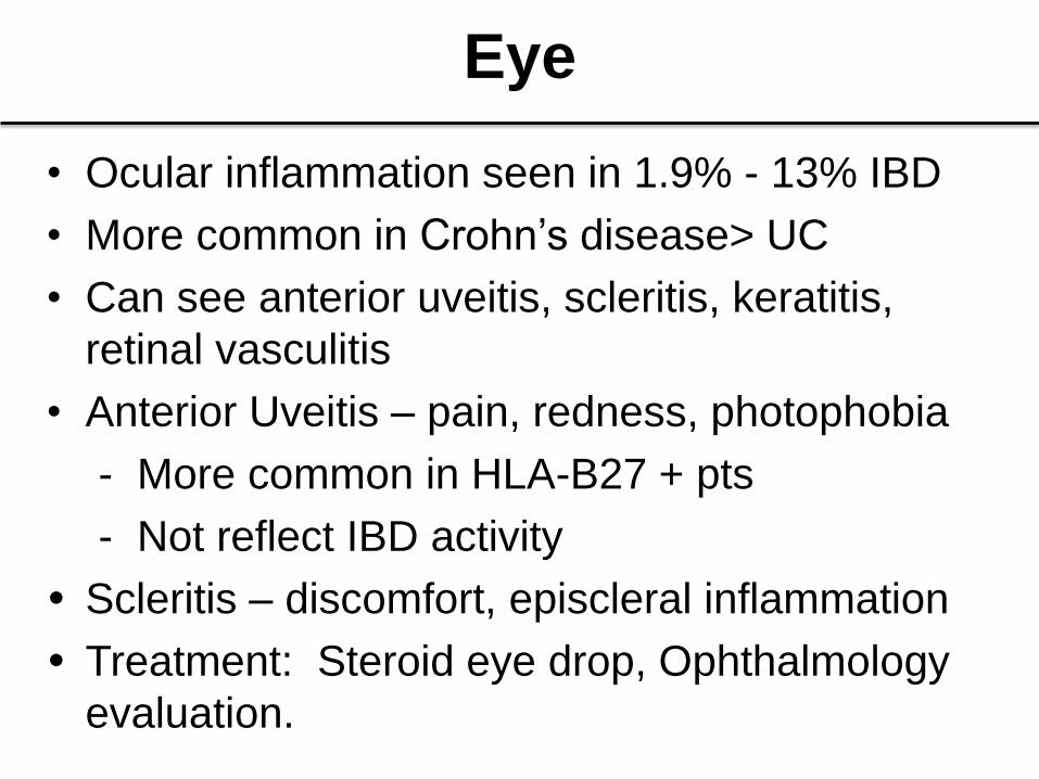
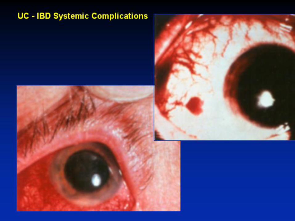
Eye
• Ocular inflammation seen in 1.9% - 13% IBD
• More common in Crohn’s disease> UC
• Can see anterior uveitis, scleritis, keratitis,
retinal vasculitis
• Anterior Uveitis – pain, redness, photophobia
- More common in HLA-B27 + pts
- Not reflect IBD activity
Scleritis – discomfort, episcleral inflammation
Treatment: Steroid eye drop, Ophthalmology
evaluation.
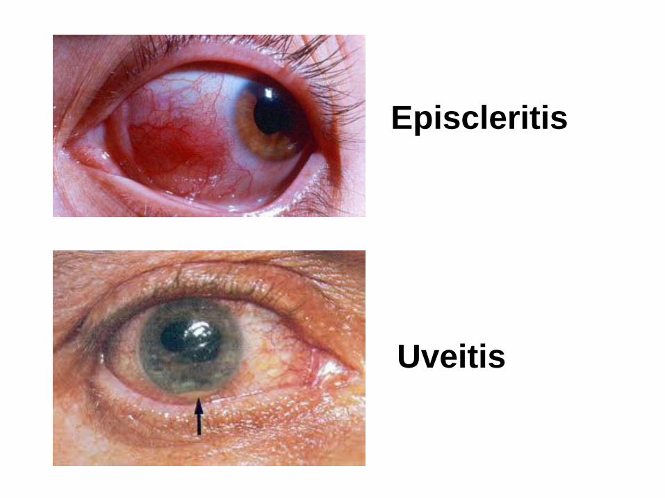
Episcleritis
Uveitis
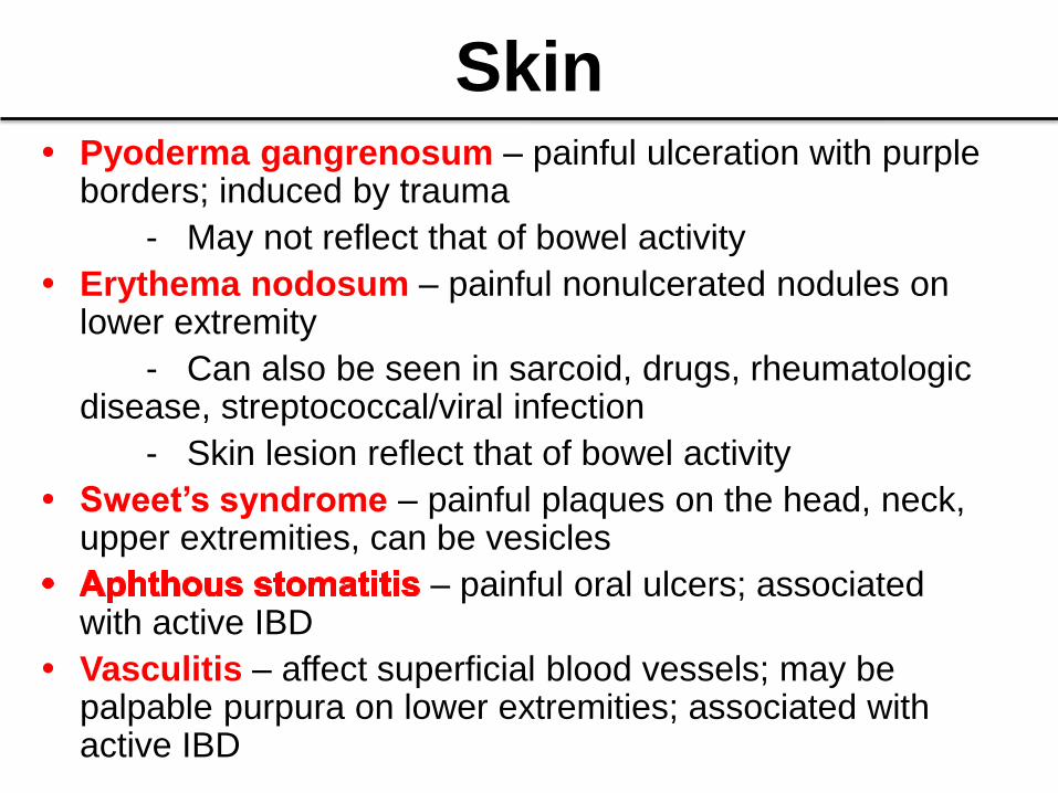
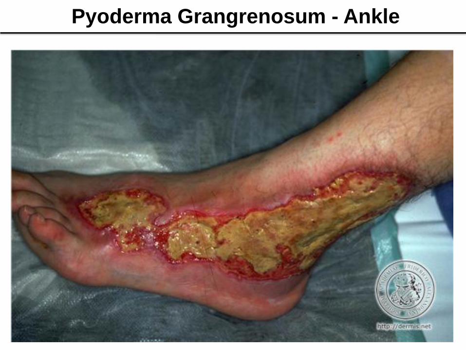
Skin Pyoderma gangrenosum – painful ulceration with purple
borders; induced by trauma
- May not reflect that of bowel activity
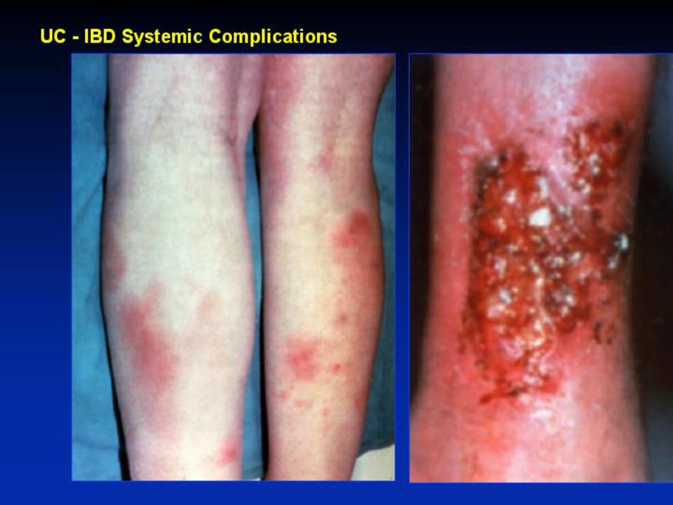
Erythema nodosum – painful nonulcerated nodules on lower extremity
- Can also be seen in sarcoid, drugs, rheumatologic disease, streptococcal/viral infection
- Skin lesion reflect that of bowel activity
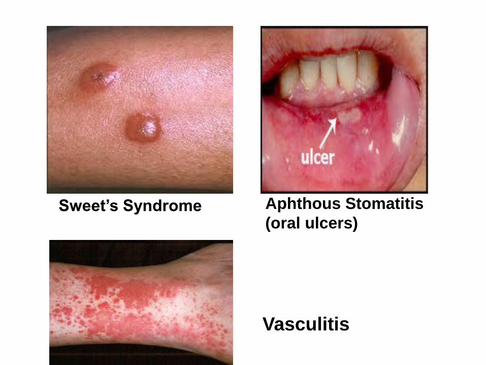
Sweet’s syndrome – painful plaques on the head, neck, upper extremities, can be vesicles
– painful oral ulcers; associated with active IBD
Vasculitis – affect superficial blood vessels; may be palpable purpura on lower extremities; associated with active IBD
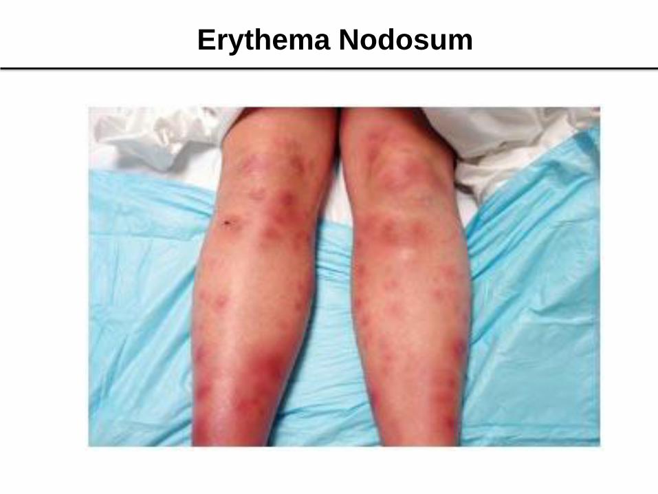
Erythema Nodosum
Pyoderma Grangrenosum - Ankle
Sweet’s Syndrome Aphthous Stomatitis
(oral ulcers)
Vasculitis
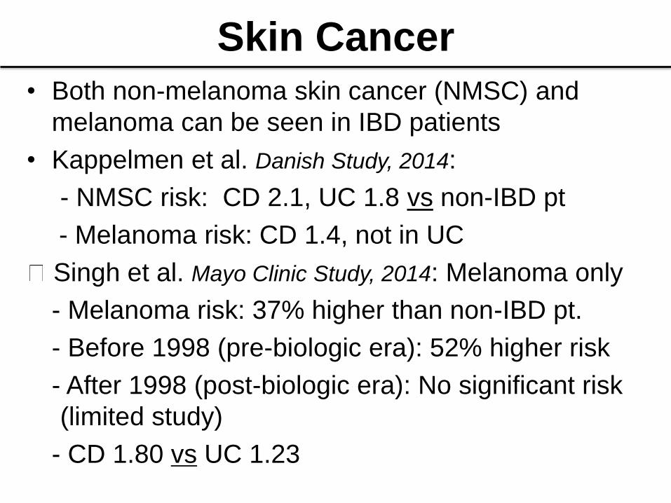
Skin Cancer
• Both non-melanoma skin cancer (NMSC) and
melanoma can be seen in IBD patients
• Kappelmen et al. Danish Study, 2014:
- NMSC risk: CD 2.1, UC 1.8 vs non-IBD pt
- Melanoma risk: CD 1.4, not in UC
Singh et al. Mayo Clinic Study, 2014: Melanoma only
- Melanoma risk: 37% higher than non-IBD pt.
- Before 1998 (pre-biologic era): 52% higher risk
- After 1998 (post-biologic era): No significant risk
(limited study)
- CD 1.80 vs UC 1.23
Skin Cancers
• NMSC risk higher in thiopurine treatment (6-mercaptopurine, azathiopurine) by increasing photosensitivity to ultraviolet A (UVA)
- higher risk in longer treatment duration
Prevention:
1. Sunscreen lotion, sun protection clothing
2. Avoid tanning salon
3. No smoking
4. Get regular dermatology exam, careful self-exam for skin lesion
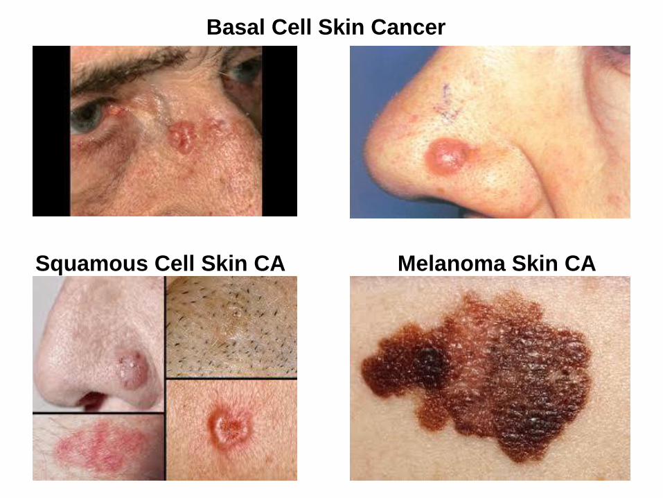
Basal Cell Skin Cancer
Squamous Cell Skin CA Melanoma Skin CA
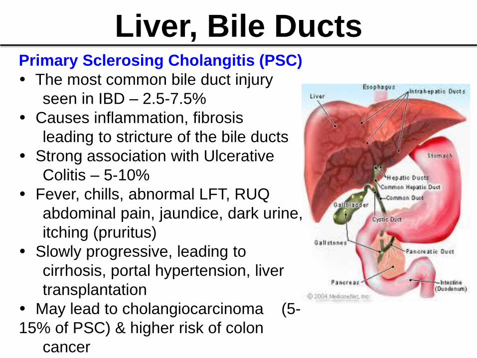
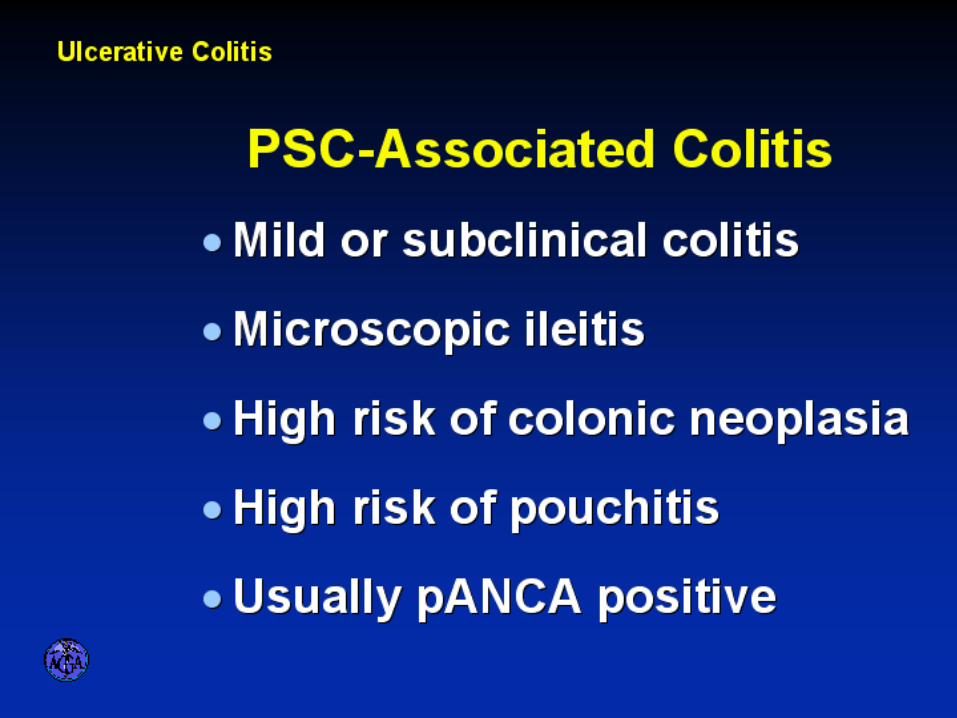
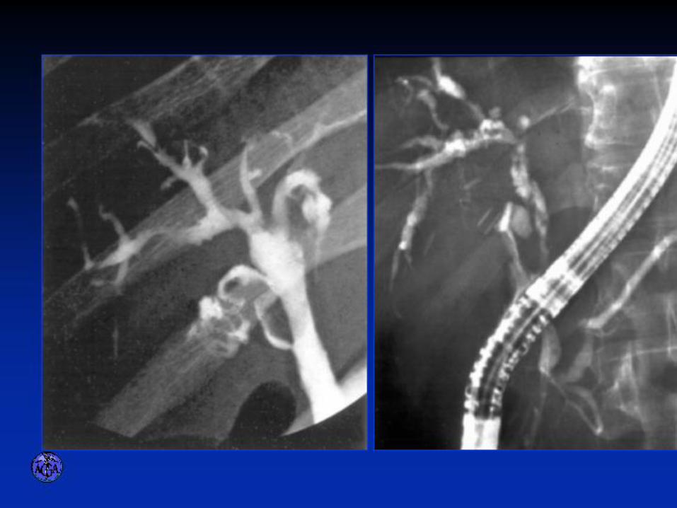
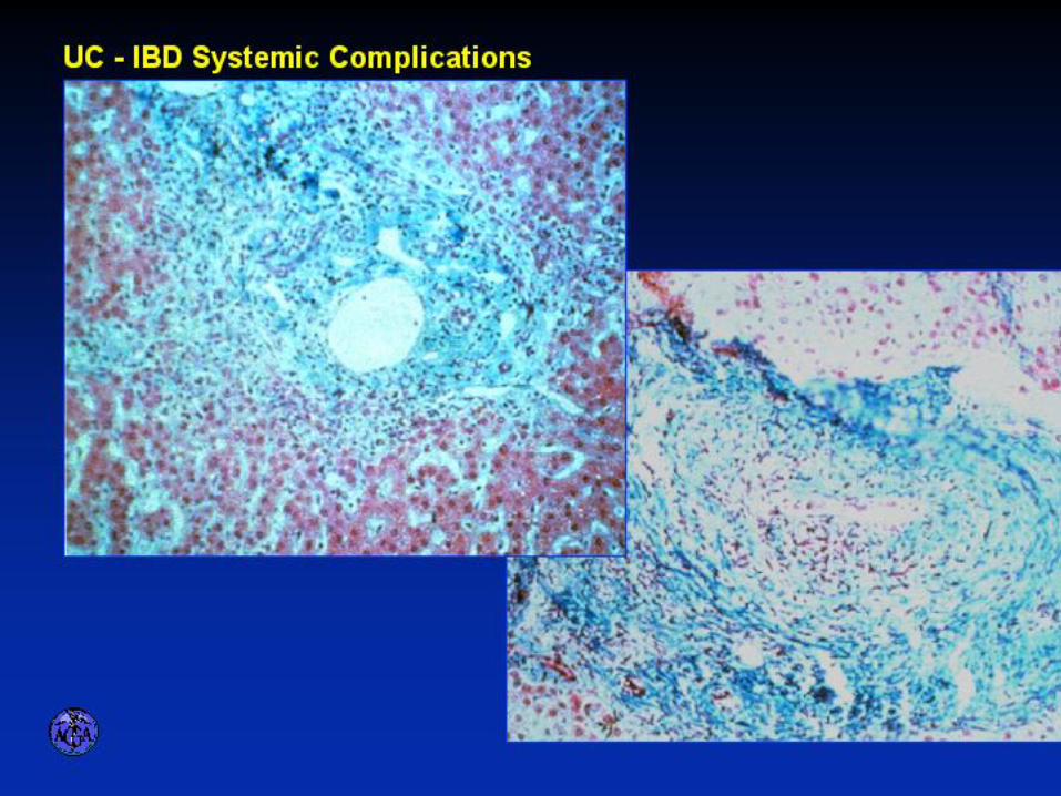
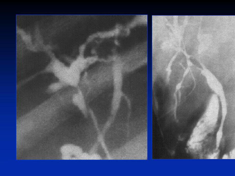
Liver, Bile Ducts Primary Sclerosing Cholangitis (PSC)
The most common bile duct injury
seen in IBD – 2.5-7.5%
Causes inflammation, fibrosis
leading to stricture of the bile ducts
Strong association with Ulcerative
Colitis – 5-10%
Fever, chills, abnormal LFT, RUQ
abdominal pain, jaundice, dark urine,
itching (pruritus)
Slowly progressive, leading to
cirrhosis, portal hypertension, liver
transplantation
May lead to cholangiocarcinoma (5-
15% of PSC) & higher risk of colon
cancer
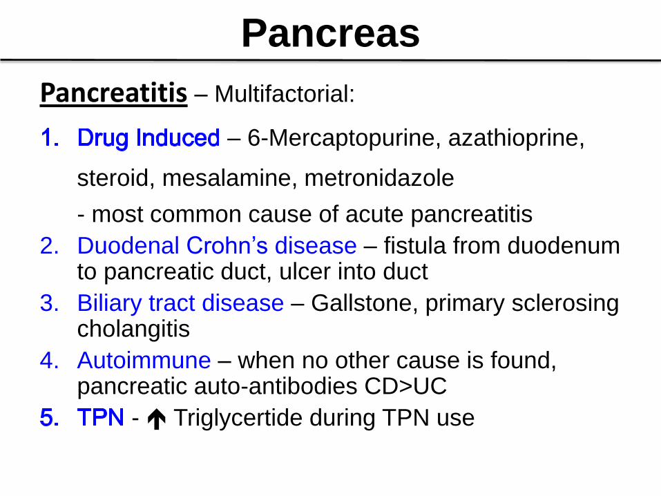
Pancreas
Pancreatitis – Multifactorial:
– 6-Mercaptopurine, azathioprine,
steroid, mesalamine, metronidazole
- most common cause of acute pancreatitis
2. Duodenal Crohn’s disease – fistula from duodenum to pancreatic duct, ulcer into duct
3. Biliary tract disease – Gallstone, primary sclerosing cholangitis
4. Autoimmune – when no other cause is found, pancreatic auto-antibodies CD>UC
- Triglycertide during TPN use
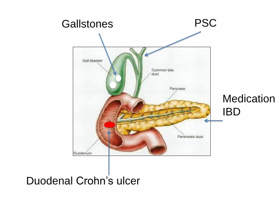
Duodenal Crohn’s ulcer
Gallstones
Medication
IBD
PSC
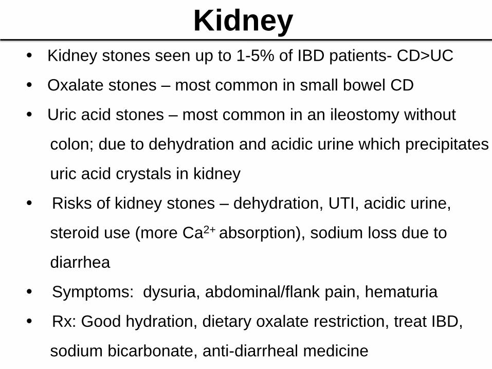
Kidney Kidney stones seen up to 1-5% of IBD patients- CD>UC
Oxalate stones – most common in small bowel CD
Uric acid stones – most common in an ileostomy without
colon; due to dehydration and acidic urine which precipitates
uric acid crystals in kidney
Risks of kidney stones – dehydration, UTI, acidic urine,
steroid use (more Ca2+ absorption), sodium loss due to
diarrhea
Symptoms: dysuria, abdominal/flank pain, hematuria
Rx: Good hydration, dietary oxalate restriction, treat IBD,
sodium bicarbonate, anti-diarrheal medicine
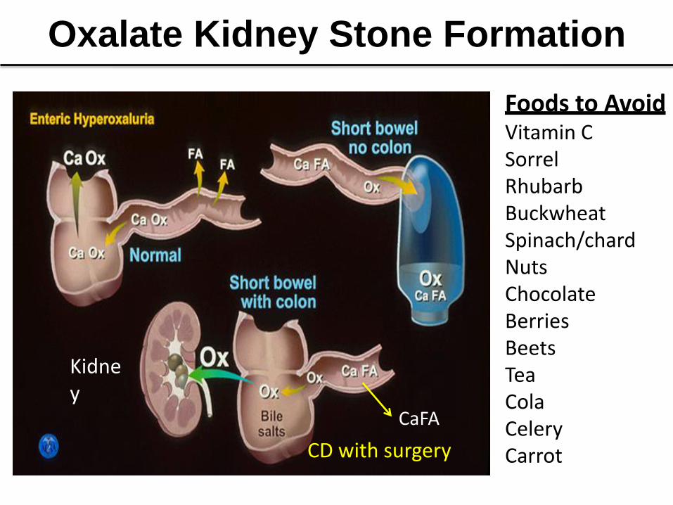
Oxalate Kidney Stone Formation
Foods to Avoid Vitamin C Sorrel Rhubarb Buckwheat Spinach/chard Nuts Chocolate Berries Beets Tea Cola Celery Carrot
CD with surgery
Kidney
CaFA
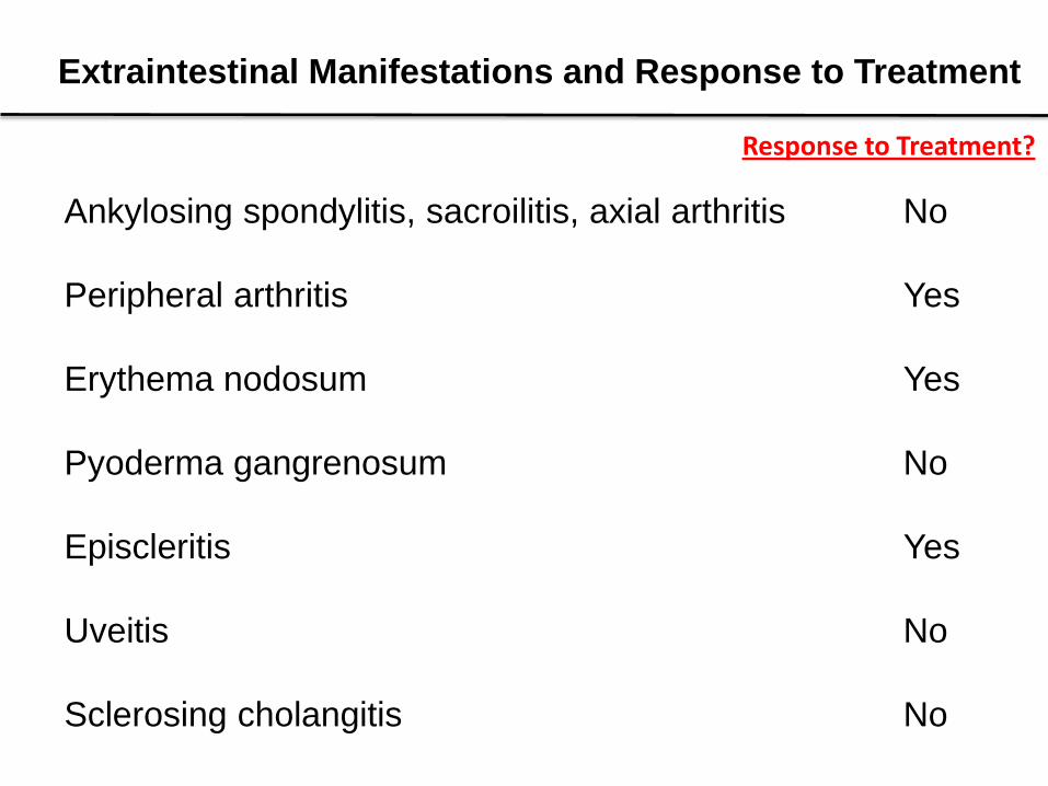
Extraintestinal Manifestations and Response to Treatment
Ankylosing spondylitis, sacroilitis, axial arthritis No
Peripheral arthritis Yes
Erythema nodosum Yes
Pyoderma gangrenosum No
Episcleritis Yes
Uveitis No
Sclerosing cholangitis No
Response to Treatment?